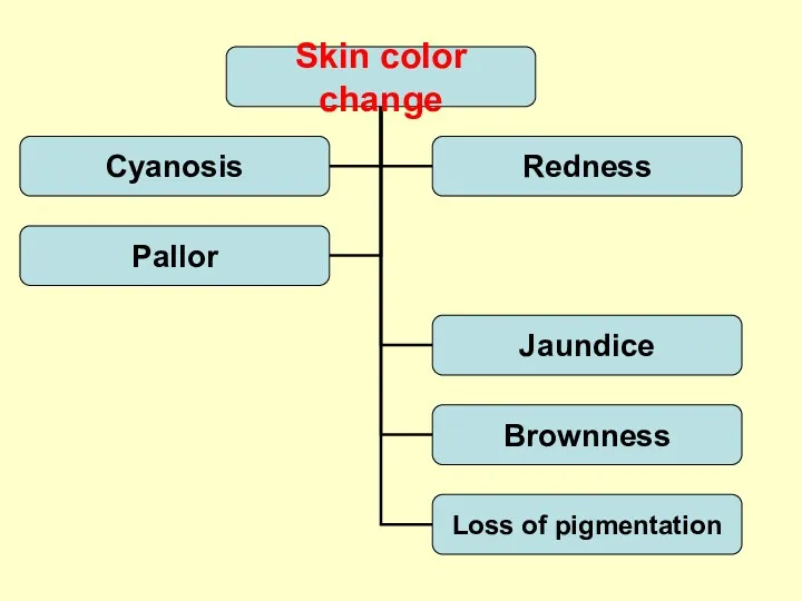
- Functions of the skin

Содержание
- 2. Plan of lecture Functions of the skin. Structure of the skin. The features of the skin
- 3. Purposes of the skin Protection: mechanical barrier; the oily and slightly acid secretions of sebaceous glands
- 4. Structure of the skin Epidermis Dermis Subcutaneous tissue Appendages of the skin: Hair nails sebaceous glands
- 5. Epidermis the outermost cellular membrane of relatively uniform thickness; Diseases of the skin focus mainly on
- 6. Appendages of the skin The types of hair are fetal lanugo, terminal, and vellus. Sebaceous glands:
- 7. Appendages of the skin Eccrine sweat glands are distributed over the entire body surface; respond to
- 8. The skin of the infant far more susceptible to superficial bacterial infection more likely to have
- 9. Newborns more often show blistering (bullous) reactions caused by the poor adherence between epidermis and dermis
- 10. Evaluation of the skin: inspection and palpation Skin is assessed for colour, turgor, texture, temperature, and
- 11. Factors influencing assessment of color Genetic: From a milky-white and rosy color to a more deep-hued
- 12. The skin of the newborn is velvety smooth and puffy At birth the skin is covered
- 13. The skin of the newborn Milia, distended sebaceous glands, appear as tiny white papules on the
- 14. The skin of the newborn Acrocyanosis Cutis marmorata Erythema toxicum Harlequin color change
- 15. The skin of the newborn Mongolian spots - irregular areas of deep blue pigmentation, usually in
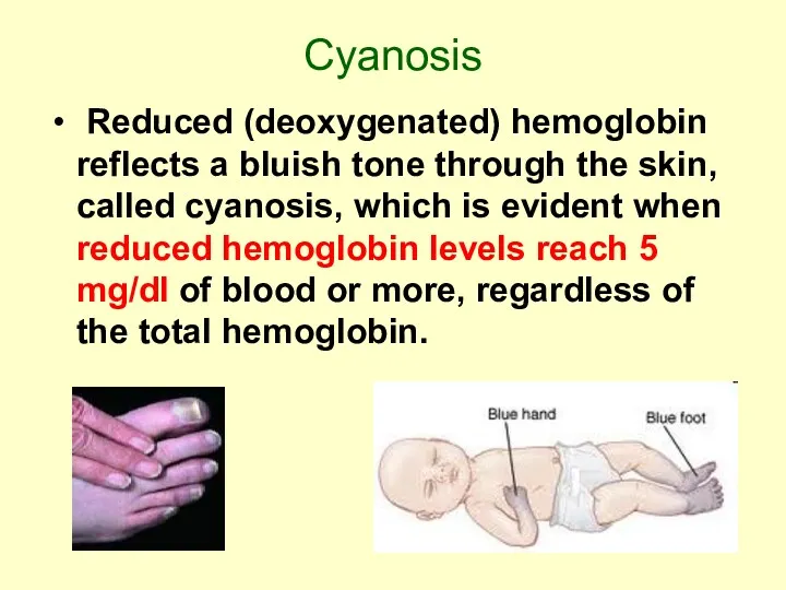
- 17. Cyanosis Reduced (deoxygenated) hemoglobin reflects a bluish tone through the skin, called cyanosis, which is evident
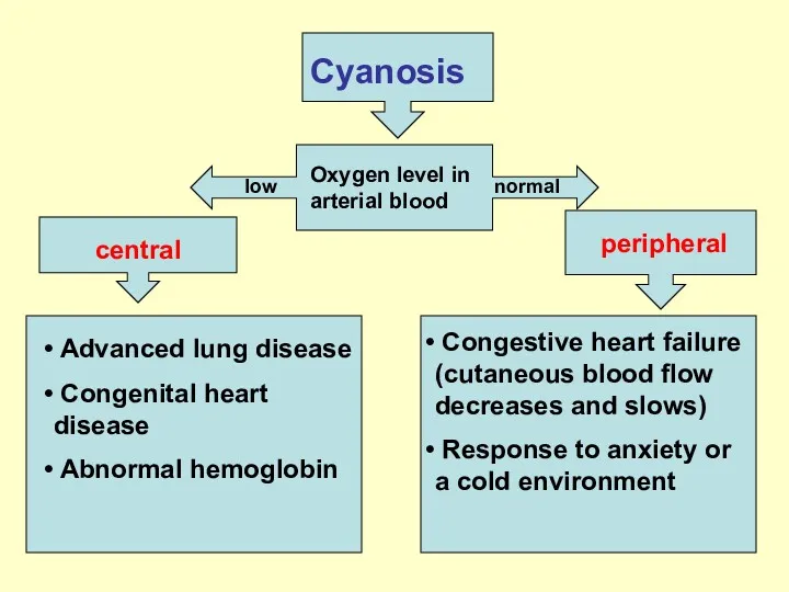
- 18. Cyanosis Oxygen level in arterial blood low normal central peripheral Congestive heart failure (cutaneous blood flow
- 19. Pallor Pallor, or paleness, is evident as a loss of the rosy glow in light-skinned individuals,
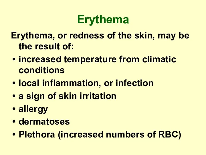
- 20. Erythema Erythema, or redness of the skin, may be the result of: increased temperature from climatic
- 21. Erythema Dermatomyositis Erythema nodosum
- 22. Jaundice, a yellow staining of the skin, usually caused by bile pigments
- 23. Jaundice Causes: Physiologic in newborn Excessive hemolysis of RBC (hemolytic disease of the newborn) Liver disease
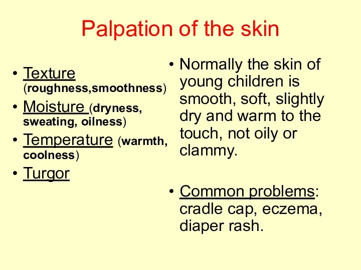
- 24. Palpation of the skin Texture (roughness,smoothness) Moisture (dryness, sweating, oilness) Temperature (warmth, coolness) Turgor Normally the
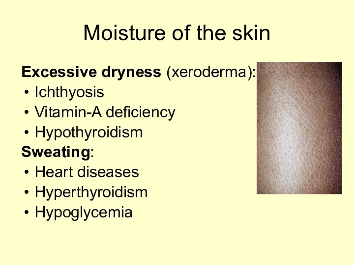
- 25. Moisture of the skin Excessive dryness (xeroderma): Ichthyosis Vitamin-A deficiency Hypothyroidism Sweating: Heart diseases Hyperthyroidism Hypoglycemia
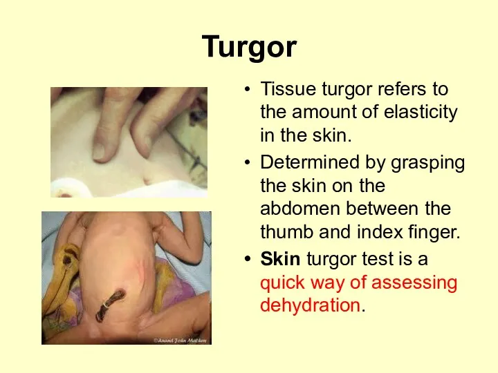
- 26. Turgor Tissue turgor refers to the amount of elasticity in the skin. Determined by grasping the
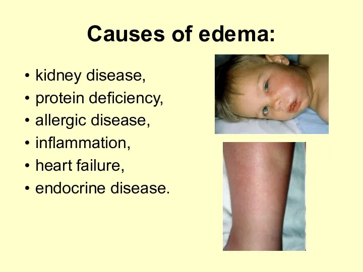
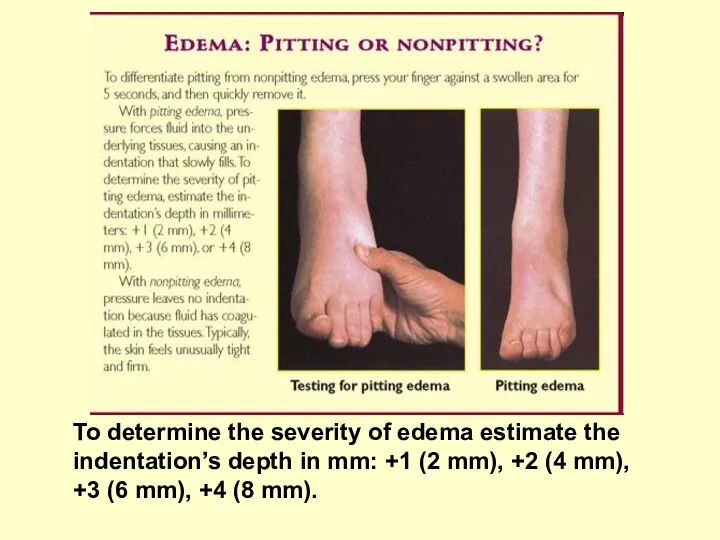
- 27. Causes of edema: kidney disease, protein deficiency, allergic disease, inflammation, heart failure, endocrine disease.
- 28. To determine the severity of edema estimate the indentation’s depth in mm: +1 (2 mm), +2
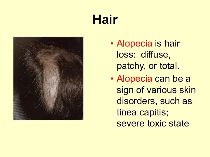
- 29. Hair Alopecia is hair loss: diffuse, patchy, or total. Alopecia can be a sign of various
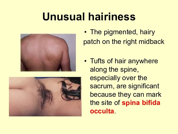
- 30. Unusual hairiness The pigmented, hairy patch on the right midback Tufts of hair anywhere along the
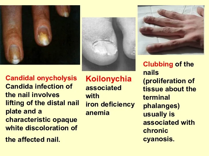
- 31. Candidal onycholysis Candida infection of the nail involves lifting of the distal nail plate and a
- 32. Lesions of the skin In general skin lesions originate from contact with injurious agents such as
- 33. Observe any lesions of the skin, noting their characteristics: Their anatomic location and distribution over the
- 34. The type(s) of skin lesions: Primary lesions are skin changes produced by some causative factor; may
- 36. Primary lesions A macule represents an alteration in skin color but cannot be felt. When larger
- 37. Primary lesions Vesicles are raised, fluid-filled lesions less than 0.5 cm in diameter; when larger, they
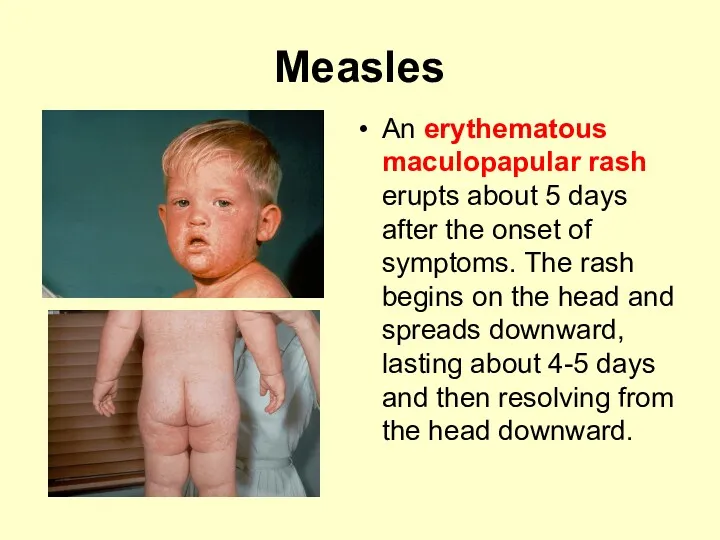
- 38. Measles An erythematous maculopapular rash erupts about 5 days after the onset of symptoms. The rash
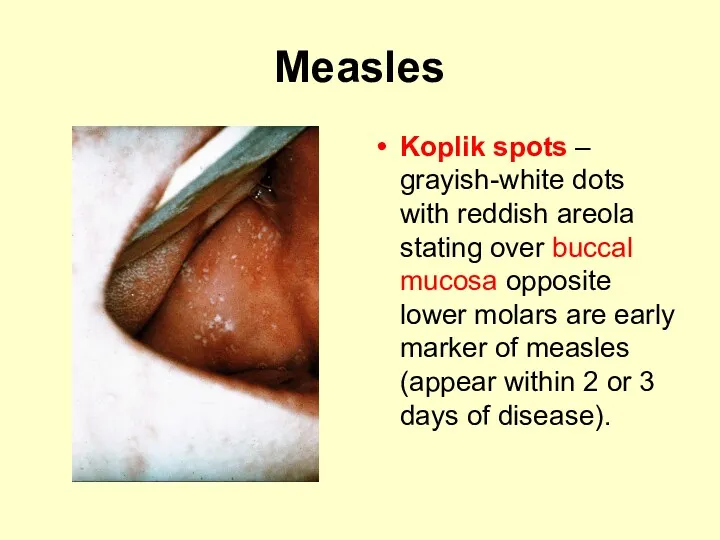
- 39. Measles Koplik spots – grayish-white dots with reddish areola stating over buccal mucosa opposite lower molars
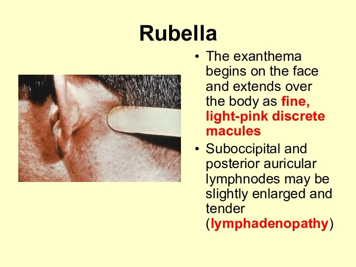
- 40. Rubella The exanthema begins on the face and extends over the body as fine, light-pink discrete
- 41. Scarlet fever The rash is erythematous, finely punctuate, it appears on the trunk and becomes generalized
- 42. Scarlet fever The skin rash fades over 1 week followed by desquamation, which may last for
- 43. Chickenpox The varying stages of development (macules, papules, and vesicles) present at the same time
- 44. Vesicular eruption Zoster - vesicles confined to a dermatome area. Herpes - vesicles are located in
- 45. Hives (urticaria) is allergic disorder; the lesions are transient, well-circumscribed erythematous wheals of varying size affecting
- 46. Hemorrhagic rash Petechiae - small, distinct pinpoint hemorrhages 2 mm or less in size Purpura –
- 47. Henoch-Schonlein purpura The characterictic purpuric rash is slightly raised above the surface, the rash is distributed
- 48. Hemorrhagic rash Thrombocytopenic Purpura: Patients with this disease present with a diffuse, nonpalpable petechial/purpuric rash. Hemophilia
- 49. Menigococcemia- infection with Neisseria meningitidis typical irregular purpuric and gangrenous skin rash over the trunk, legs,
- 51. Secondary lesions Scales consist of compressed layers of stratum corneum cells that are retained on the
- 52. Secondary lesions Fissures are caused by splitting or cracking; they occur usually in diseased skin. Scars
- 53. Secondary lesions Crusts consist of matted, retained accumulations of blood, serum, pus, and epithelial debris on
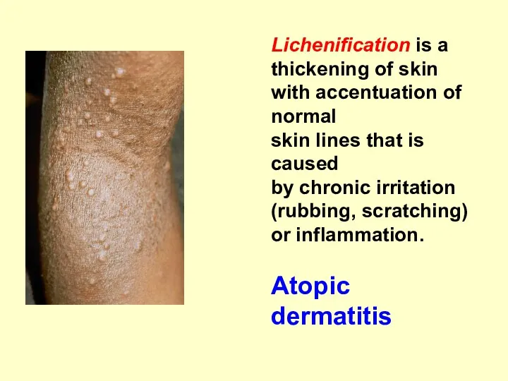
- 54. Lichenification is a thickening of skin with accentuation of normal skin lines that is caused by
- 55. Distribution The pattern is a useful aid in diagnosis. It may be: generalized or localized; widespread,
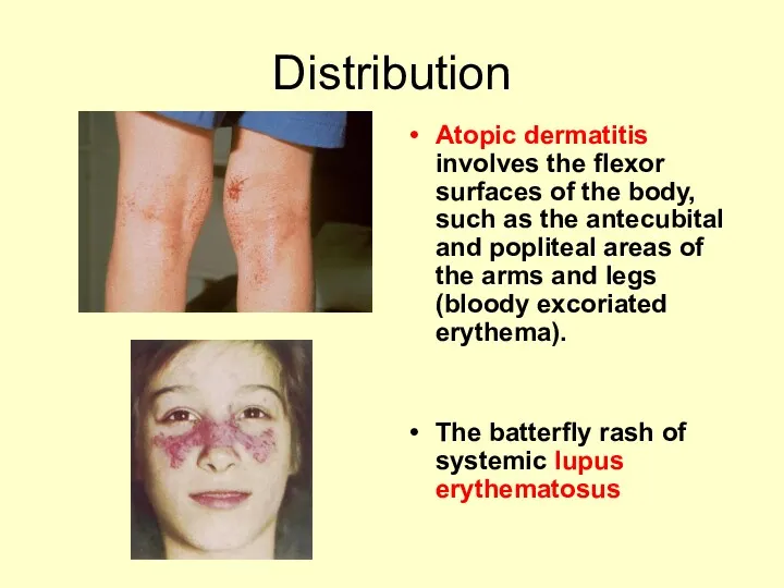
- 56. Distribution Atopic dermatitis involves the flexor surfaces of the body, such as the antecubital and popliteal
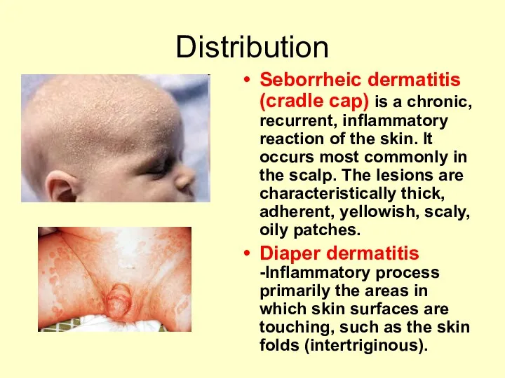
- 57. Distribution Seborrheic dermatitis (cradle cap) is a chronic, recurrent, inflammatory reaction of the skin. It occurs
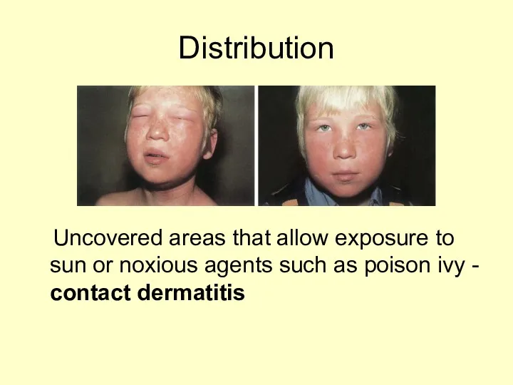
- 58. Distribution Uncovered areas that allow exposure to sun or noxious agents such as poison ivy -
- 59. Arrangement Clustered (grouped) - herpes Annular (in a ring) - vascular reactions such as urticaria Arciform
- 60. Subjective symptoms Itching Pain or tenderness Alterations in local feeling or sensation: - absence of sensation
- 62. Скачать презентацию
Plan of lecture
Functions of the skin.
Structure of the skin.
The features of
Plan of lecture
Functions of the skin.
Structure of the skin.
The features of

Purposes of the skin
Protection: mechanical barrier; the oily and slightly acid
Purposes of the skin
Protection: mechanical barrier; the oily and slightly acid

Structure of the skin
Epidermis
Dermis
Subcutaneous tissue
Appendages
of the skin:
Hair
nails
Structure of the skin
Epidermis
Dermis
Subcutaneous tissue
Appendages
of the skin:
Hair
nails
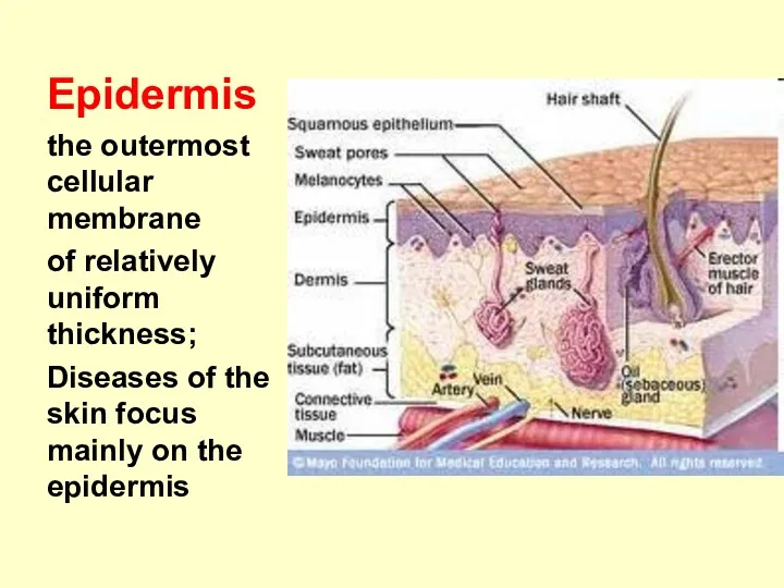
Epidermis
the outermost cellular membrane
of relatively uniform thickness;
Diseases of the skin
Epidermis
the outermost cellular membrane
of relatively uniform thickness;
Diseases of the skin
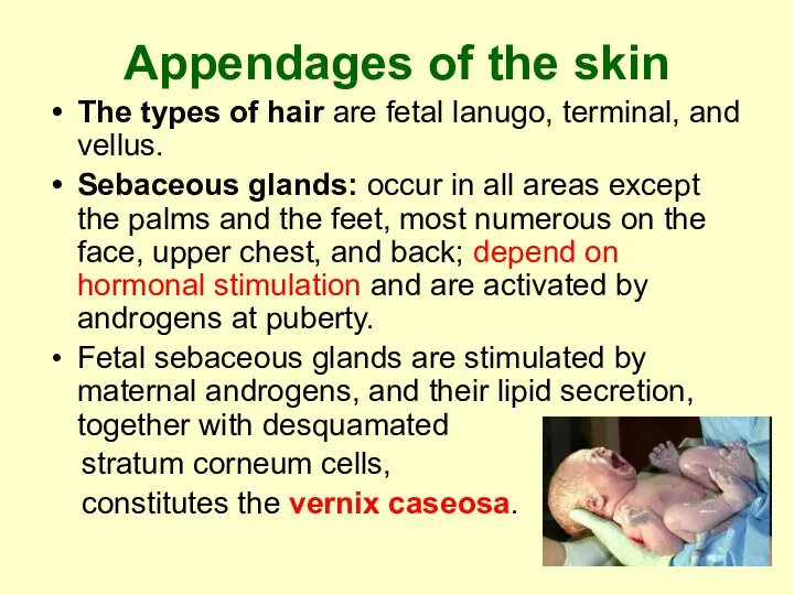
Appendages of the skin
The types of hair are fetal lanugo, terminal,
Appendages of the skin
The types of hair are fetal lanugo, terminal,
Appendages of the skin
Eccrine sweat glands are distributed over the entire
Appendages of the skin
Eccrine sweat glands are distributed over the entire
The skin of the infant
far more susceptible to superficial bacterial infection
The skin of the infant
far more susceptible to superficial bacterial infection
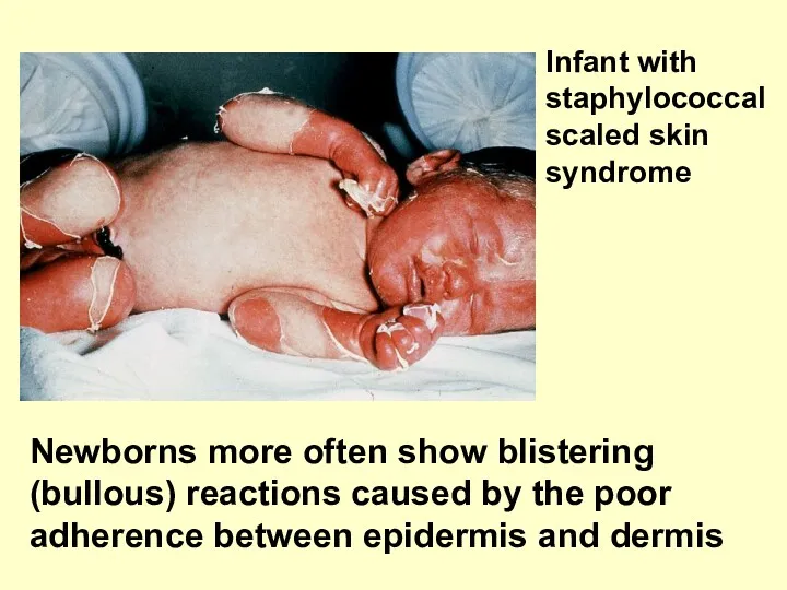
Newborns more often show blistering (bullous) reactions caused by the poor
Newborns more often show blistering (bullous) reactions caused by the poor
Evaluation of the skin:
inspection and palpation
Skin is assessed for colour,
Evaluation of the skin:
inspection and palpation
Skin is assessed for colour,
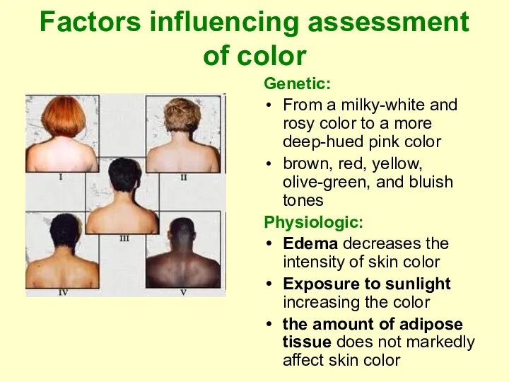
Factors influencing assessment of color
Genetic:
From a milky-white and rosy color
Factors influencing assessment of color
Genetic:
From a milky-white and rosy color
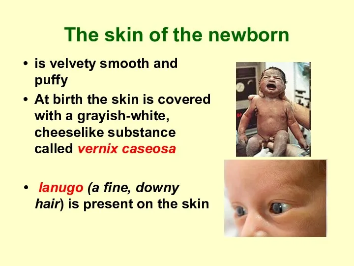
The skin of the newborn
is velvety smooth and puffy
At
The skin of the newborn
is velvety smooth and puffy
At
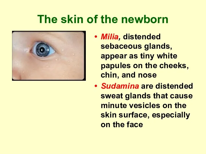
The skin of the newborn
Milia, distended sebaceous glands, appear as tiny
The skin of the newborn
Milia, distended sebaceous glands, appear as tiny
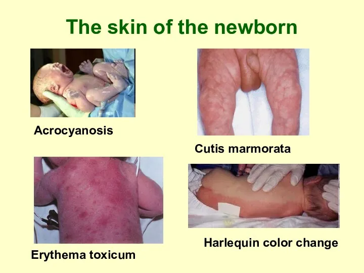
The skin of the newborn
Acrocyanosis
Cutis marmorata
Erythema toxicum
Harlequin color change
The skin of the newborn
Acrocyanosis
Cutis marmorata
Erythema toxicum
Harlequin color change
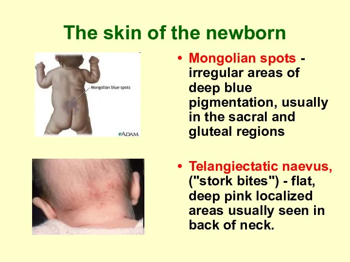
The skin of the newborn
Mongolian spots - irregular areas of deep
The skin of the newborn
Mongolian spots - irregular areas of deep
Cyanosis
Reduced (deoxygenated) hemoglobin reflects a bluish tone through the skin,
Cyanosis
Reduced (deoxygenated) hemoglobin reflects a bluish tone through the skin,
Cyanosis
Oxygen level in arterial blood
low
normal
central
peripheral
Congestive heart failure (cutaneous blood flow
Cyanosis
Oxygen level in arterial blood
low
normal
central
peripheral
Congestive heart failure (cutaneous blood flow
Pallor
Pallor, or paleness, is evident as a loss of the rosy
Pallor
Pallor, or paleness, is evident as a loss of the rosy
Erythema
Erythema, or redness of the skin, may be the result of:
Erythema
Erythema, or redness of the skin, may be the result of:
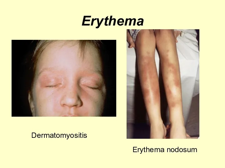
Erythema
Dermatomyositis
Erythema nodosum
Erythema
Dermatomyositis
Erythema nodosum
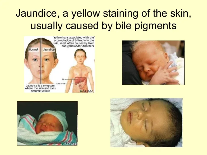
Jaundice, a yellow staining of the skin, usually caused by bile
Jaundice, a yellow staining of the skin, usually caused by bile
Jaundice
Causes:
Physiologic in newborn
Excessive hemolysis of RBC (hemolytic disease of the newborn)
Liver
Jaundice
Causes:
Physiologic in newborn
Excessive hemolysis of RBC (hemolytic disease of the newborn)
Liver
Palpation of the skin
Texture (roughness,smoothness)
Moisture (dryness, sweating, oilness)
Temperature (warmth, coolness)
Turgor
Normally
Palpation of the skin
Texture (roughness,smoothness)
Moisture (dryness, sweating, oilness)
Temperature (warmth, coolness)
Turgor
Normally
Moisture of the skin
Excessive dryness (xeroderma):
Ichthyosis
Vitamin-A deficiency
Hypothyroidism
Sweating:
Heart diseases
Hyperthyroidism
Hypoglycemia
Moisture of the skin
Excessive dryness (xeroderma):
Ichthyosis
Vitamin-A deficiency
Hypothyroidism
Sweating:
Heart diseases
Hyperthyroidism
Hypoglycemia
Turgor
Tissue turgor refers to the amount of elasticity in the skin.
Turgor
Tissue turgor refers to the amount of elasticity in the skin.
Causes of edema:
kidney disease,
protein deficiency,
allergic disease,
inflammation,
heart failure,
endocrine disease.
Causes of edema:
kidney disease,
protein deficiency,
allergic disease,
inflammation,
heart failure,
endocrine disease.
To determine the severity of edema estimate the indentation’s depth in
To determine the severity of edema estimate the indentation’s depth in
Hair
Alopecia is hair loss: diffuse, patchy, or total.
Alopecia can be
Hair
Alopecia is hair loss: diffuse, patchy, or total.
Alopecia can be
Unusual hairiness
The pigmented, hairy
patch on the right midback
Tufts of hair
Unusual hairiness
The pigmented, hairy
patch on the right midback
Tufts of hair
Candidal onycholysis Candida infection of the nail involves lifting of the
Candidal onycholysis Candida infection of the nail involves lifting of the
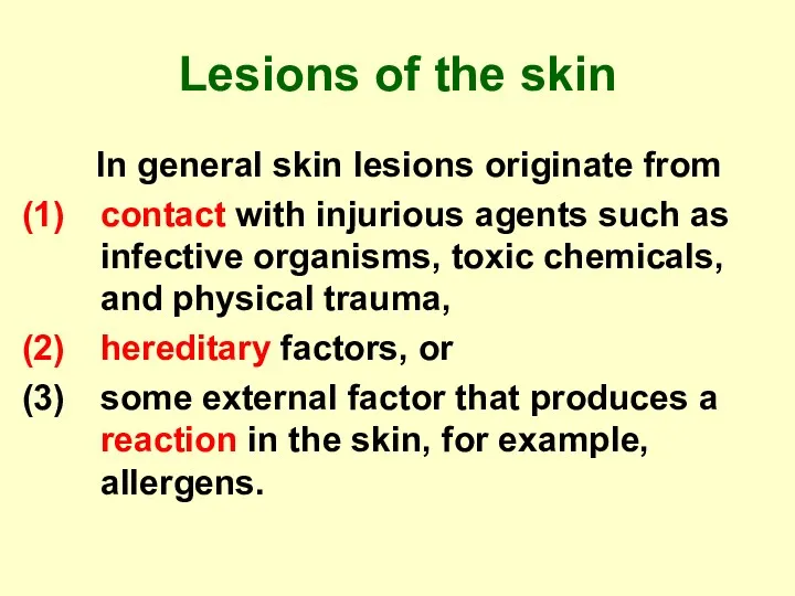
Lesions of the skin
In general skin lesions originate from
contact
Lesions of the skin
In general skin lesions originate from
contact
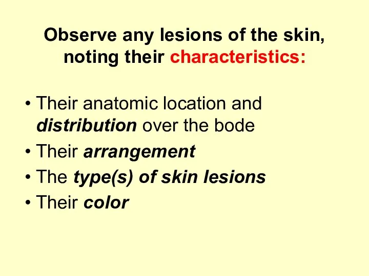
Observe any lesions of the skin, noting their characteristics:
Their anatomic location
Observe any lesions of the skin, noting their characteristics:
Their anatomic location
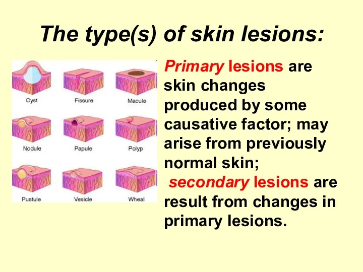
The type(s) of skin lesions:
Primary lesions are skin changes produced by
The type(s) of skin lesions:
Primary lesions are skin changes produced by

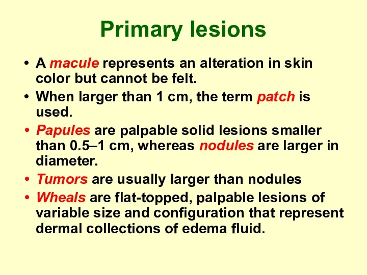
Primary lesions
A macule represents an alteration in skin color but cannot
Primary lesions
A macule represents an alteration in skin color but cannot
Primary lesions
Vesicles are raised, fluid-filled lesions less than 0.5 cm in
Primary lesions
Vesicles are raised, fluid-filled lesions less than 0.5 cm in
Measles
An erythematous maculopapular rash erupts about 5 days after the onset
Measles
An erythematous maculopapular rash erupts about 5 days after the onset
Measles
Koplik spots – grayish-white dots with reddish areola stating over buccal
Measles
Koplik spots – grayish-white dots with reddish areola stating over buccal
Rubella
The exanthema begins on the face and extends over the body
Rubella
The exanthema begins on the face and extends over the body
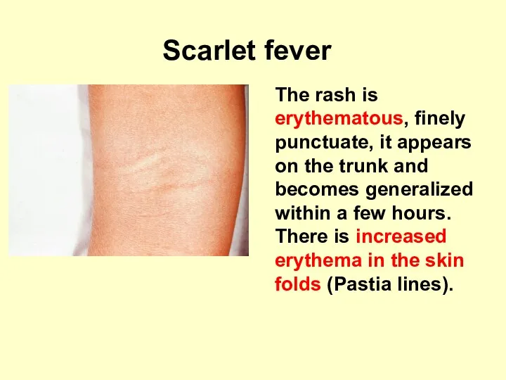
Scarlet fever
The rash is erythematous, finely punctuate, it appears on
Scarlet fever
The rash is erythematous, finely punctuate, it appears on
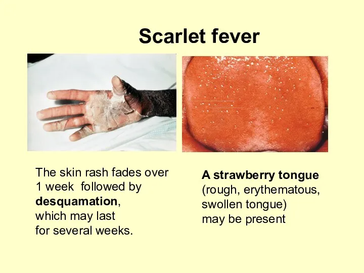
Scarlet fever
The skin rash fades over
1 week followed by
Scarlet fever
The skin rash fades over
1 week followed by
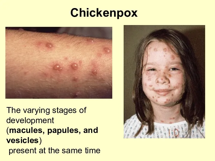
Chickenpox
The varying stages of development
(macules, papules, and vesicles)
present at the
Chickenpox
The varying stages of development
(macules, papules, and vesicles)
present at the
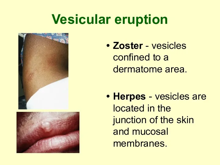
Vesicular eruption
Zoster - vesicles confined to a dermatome area.
Herpes - vesicles
Vesicular eruption
Zoster - vesicles confined to a dermatome area.
Herpes - vesicles
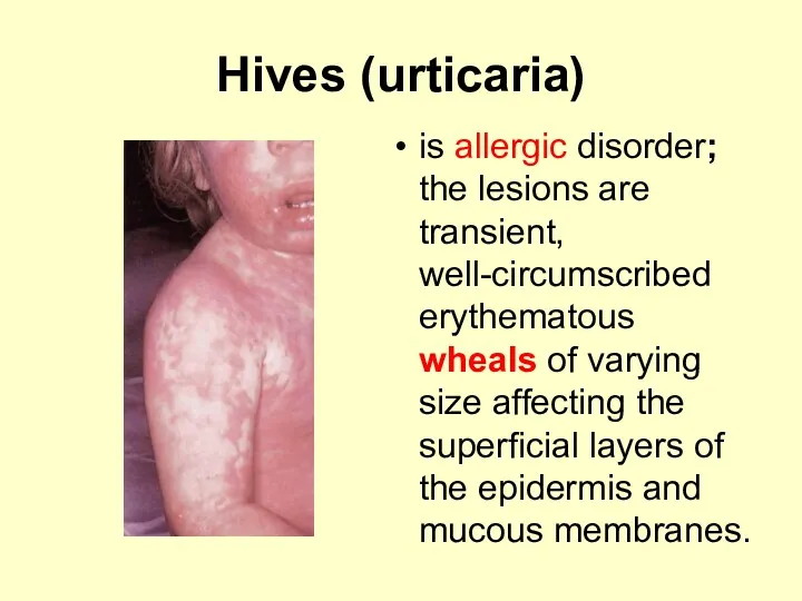
Hives (urticaria)
is allergic disorder; the lesions are transient, well-circumscribed erythematous wheals
Hives (urticaria)
is allergic disorder; the lesions are transient, well-circumscribed erythematous wheals
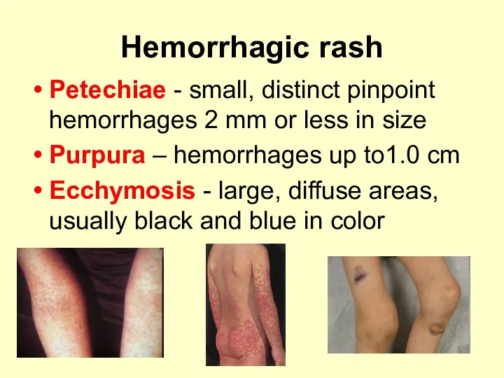
Hemorrhagic rash
Petechiae - small, distinct pinpoint hemorrhages 2 mm or
Hemorrhagic rash
Petechiae - small, distinct pinpoint hemorrhages 2 mm or
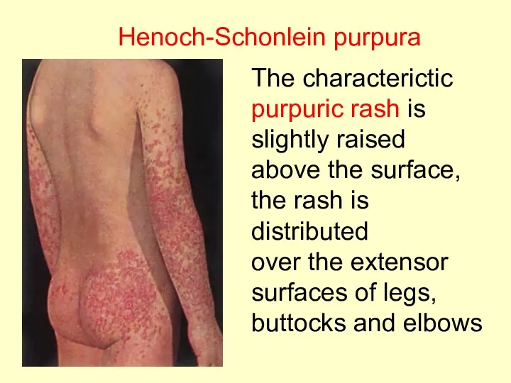
Henoch-Schonlein purpura
The characterictic
purpuric rash is slightly raised
above the surface,
Henoch-Schonlein purpura
The characterictic
purpuric rash is slightly raised
above the surface,
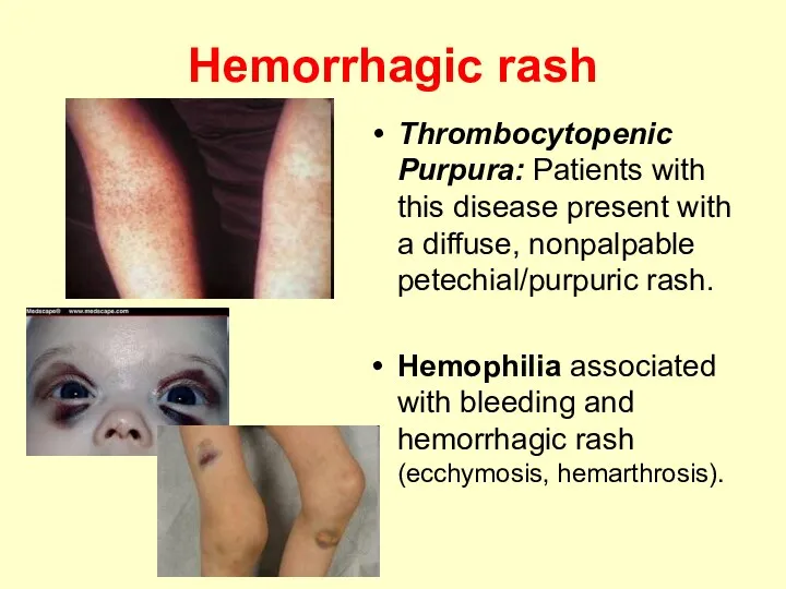
Hemorrhagic rash
Thrombocytopenic Purpura: Patients with this disease present with a diffuse,
Hemorrhagic rash
Thrombocytopenic Purpura: Patients with this disease present with a diffuse,
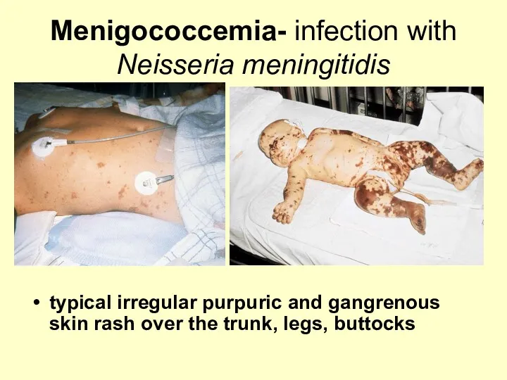
Menigococcemia- infection with Neisseria meningitidis
typical irregular purpuric and gangrenous skin
Menigococcemia- infection with Neisseria meningitidis
typical irregular purpuric and gangrenous skin

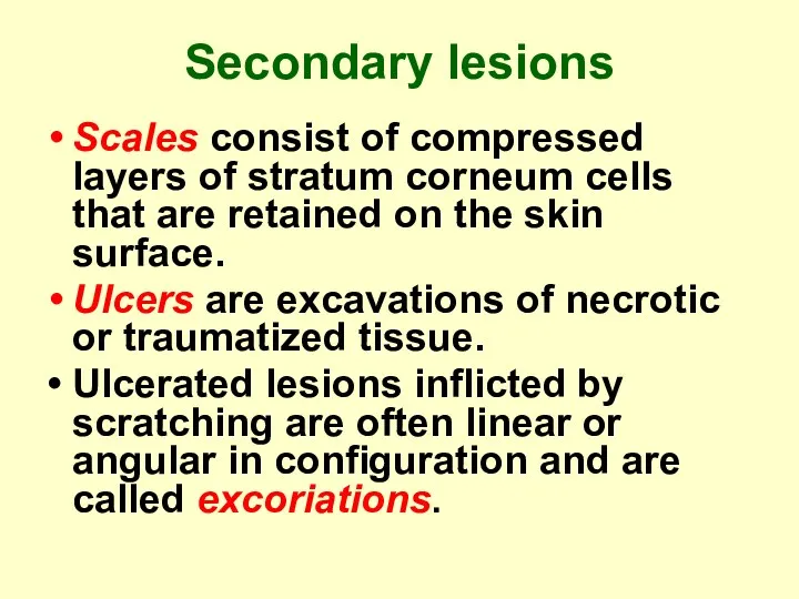
Secondary lesions
Scales consist of compressed layers of stratum corneum cells that
Secondary lesions
Scales consist of compressed layers of stratum corneum cells that
Secondary lesions
Fissures are caused by splitting or cracking; they occur usually
Secondary lesions
Fissures are caused by splitting or cracking; they occur usually
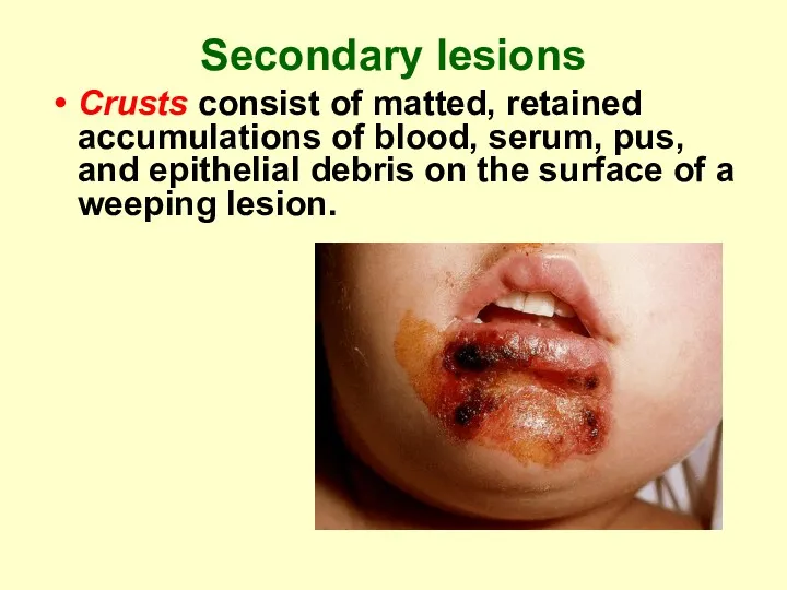
Secondary lesions
Crusts consist of matted, retained accumulations of blood, serum, pus,
Secondary lesions
Crusts consist of matted, retained accumulations of blood, serum, pus,
Lichenification is a thickening of skin with accentuation of normal
skin
Lichenification is a thickening of skin with accentuation of normal
skin
Distribution
The pattern is a useful aid in diagnosis. It may be:
Distribution
The pattern is a useful aid in diagnosis. It may be:
Distribution
Atopic dermatitis involves the flexor surfaces of the body, such as
Distribution
Atopic dermatitis involves the flexor surfaces of the body, such as
Distribution
Seborrheic dermatitis (cradle cap) is a chronic, recurrent, inflammatory reaction of
Distribution
Seborrheic dermatitis (cradle cap) is a chronic, recurrent, inflammatory reaction of
Distribution
Uncovered areas that allow exposure to sun or noxious
Distribution
Uncovered areas that allow exposure to sun or noxious
Arrangement
Clustered (grouped) - herpes
Annular (in a ring) - vascular
Arrangement
Clustered (grouped) - herpes
Annular (in a ring) - vascular
Subjective symptoms
Itching
Pain or tenderness
Alterations in local feeling or
Subjective symptoms
Itching
Pain or tenderness
Alterations in local feeling or
 Анатомия опорно-двигательного аппарата
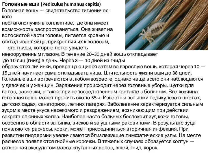 Головные вши (Pediculus humanus capitis)
 Острые респираторные заболевания у детей
 Нефротический синдром
 Медицинские аэрозольные баллоны
 Антибиотки: лечат или калечат?
 Абсцессы и флегмоны скуловой области
 Оперативная хирургия. Кожный шов
 Контроль качества в здравоохранении
 Эндоцервикоз шейки матки
 Звук, природа звука. Анатомия и физиология слуховой системы. Методы исследования состояния слуха
 Патология воли и влечений
 Лазерний скальпель
 Внегоспитальные пневмонии тяжелого течения
 Компоненты анестезии и клиническая фармакология препаратов
 Клеткалық цикл және митоз. Клетканың тіршілік
 Личная и общественная гигиена. Гигиенические основы физических упражнений
 Врожденные пороки развития лица и челюстей
 Гнойно-воспалительные заболевания новорожденных. Этиология, классификация, клиника, терапия. Сепсис новорожденных
 Некробактеріоз
 Механические повреждения
 Қан тамырларының мүшелік ерекшеліктері және тамырлардың жасқа қарай өзгерілуі
 Факторы риска развития рака яичников у женщин молодого возраста
 Международная статистическая классификация болезней и проблем, связанных со здоровьем 10 пересмотра
 Тік ішек геморройы және анус сызаты
 Ранний детский аутизм (РДА)
 Визуальный план [Автосохраненный]
 Использование бесприборных тест-систем Иммунокомб. Памятка для заводчиков и владельцев питомников кошек