- Histology of gallbladder and biliary tract

Содержание
- 2. Plan: Biliary tract Histology of biliary tract Histology of gallbladder Age changes Regeneration Conclusion
- 3. Biliary tract— a channel of a liver for a bile conclusion, part of a gastrointestinal tract
- 5. The wall of biliary tract consists of 3 layer: Mucous-single-layer (high) prismatic epithelium Muscular –bundles of
- 6. Epitelial cells are rich with lysosomes and mitochondria which concentrate mainly in their apical part. The
- 7. Generally the muscular layer is expressed better and presented to the item by two layers —
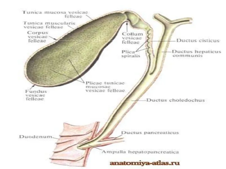
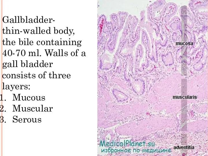
- 8. Gallbladder- thin-walled body, the bile containing 40-70 ml. Walls of a gall bladder consists of three
- 9. The mucous membrane forms numerous folds. It is covered by the high prismatic epithelial cells having
- 10. The muscular layer of a gall bladder consists of bunches of the smooth myocytes located a
- 11. Age changes In hepatocytes the quantity of a lipofusin which paints cells in brown color increases.
- 12. Regeneration The liver possesses high ability to physiological and reparative regeneration. At animals during removal from
- 14. Скачать презентацию
Plan:
Biliary tract
Histology of biliary tract
Histology of gallbladder
Age changes
Regeneration
Conclusion
Plan:
Biliary tract
Histology of biliary tract
Histology of gallbladder
Age changes
Regeneration
Conclusion
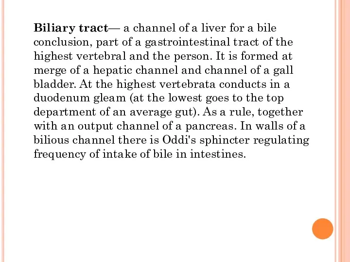
Biliary tract— a channel of a liver for a bile conclusion,
Biliary tract— a channel of a liver for a bile conclusion,
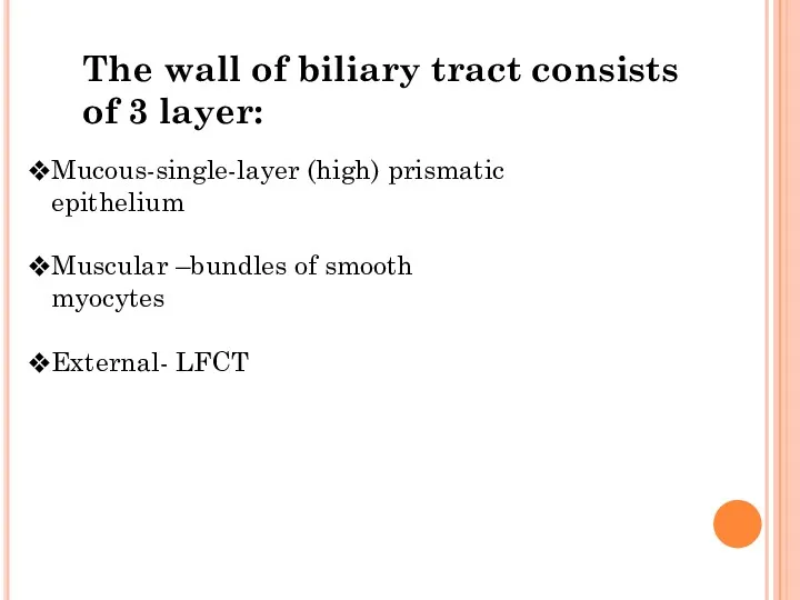
The wall of biliary tract consists of 3 layer:
Mucous-single-layer (high) prismatic
The wall of biliary tract consists of 3 layer:
Mucous-single-layer (high) prismatic
Epitelial cells are rich with lysosomes and mitochondria which concentrate mainly
Epitelial cells are rich with lysosomes and mitochondria which concentrate mainly
Generally the muscular layer is expressed better and presented to the
Generally the muscular layer is expressed better and presented to the
Gallbladder- thin-walled body, the bile containing 40-70 ml. Walls of a
Gallbladder- thin-walled body, the bile containing 40-70 ml. Walls of a
The mucous membrane forms numerous folds. It is covered by the
The mucous membrane forms numerous folds. It is covered by the
The muscular layer of a gall bladder consists of bunches of
The muscular layer of a gall bladder consists of bunches of
Age changes
In hepatocytes the quantity of a lipofusin which paints
Age changes
In hepatocytes the quantity of a lipofusin which paints
Regeneration
The liver possesses high ability to physiological and reparative regeneration. At
Regeneration
The liver possesses high ability to physiological and reparative regeneration. At
 Leptospira interrogans – возбудитель лептоспироза
 Искусство видеть. КМО (синдром ирвина-гасса)
 Организация деятельности акушерского стационара, задачи, роль акушерки
 Рахит у детей
 Организмнің биоритміне байланысты дәрілік заттардың көрсететін әсерлері
 Профилактика синдрома эмоционального выгорания педагога
 Острый коронарный синдром без подъема с. St
 Физические факторы в лечении ишемических невропатий и невритов лицевого нерва
 Раны
Раны Факторы риска развития рака яичников у женщин молодого возраста
 Cad\cam системы в ортопедической стоматологии
 Средства, влияющие на систему крови
 Супратенториальные опухоли
 Кровотечения. Первая помощь при кровотечениях
 Бауыр және өт жолдары, ұйқы безі ауруларын тағаммен емдеу және емдік дене шынықтыру
 Раны и раневой процесс
 Эндовидеохирургические инструменты
 Артикуляционно-акустическая дисграфия
 Заболевания органов пищеварения у пожилых людей
 Рентгенография
 Бағаналық жасушалар. Генетика негіздері. Тұқым қуалаушылықты зерттеудің гибридологиялық әдісі. Тұқым қуалау заңдары
 Применение препаратов гиалуроновой кислоты в комплексном лечении гонартроза
 Функциональная анатомия мышц головы, шеи и туловища
 Реабилитация постинсультных психических расстройств
 Приобретенные (вторичные) иммунодефициты
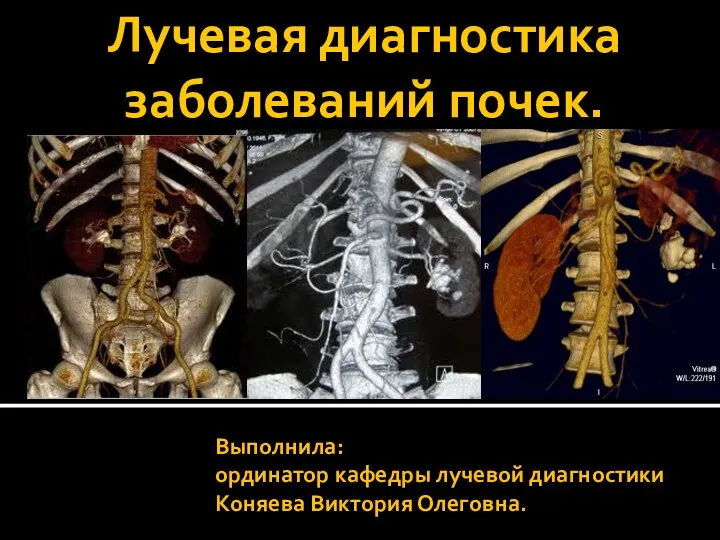 Лучевая диагностика заболеваний почек
 Побочные действия антибиотиков
 Обструкция дыхательных путей