- Immunology of Dermatophytes and Dermatophytosis

Содержание
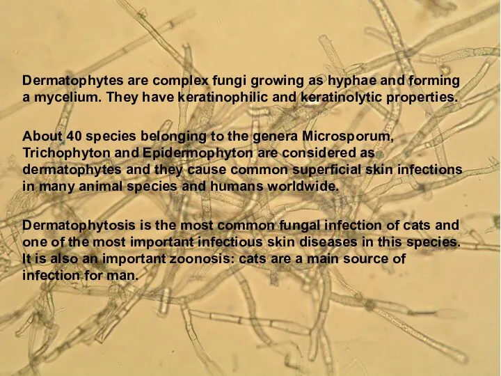
- 2. Agent Properties Dermatophytes are complex fungi growing as hyphae and forming a mycelium. They have keratinophilic
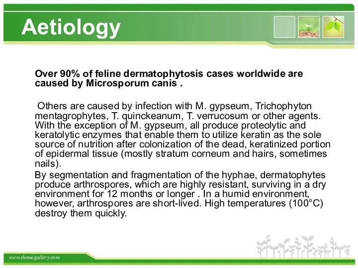
- 3. Aetiology Over 90% of feline dermatophytosis cases worldwide are caused by Microsporum canis . Others are
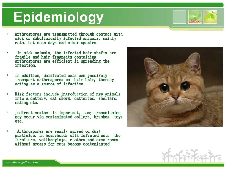
- 4. Epidemiology Arthrospores are transmitted through contact with sick or subclinically infected animals, mainly cats, but also
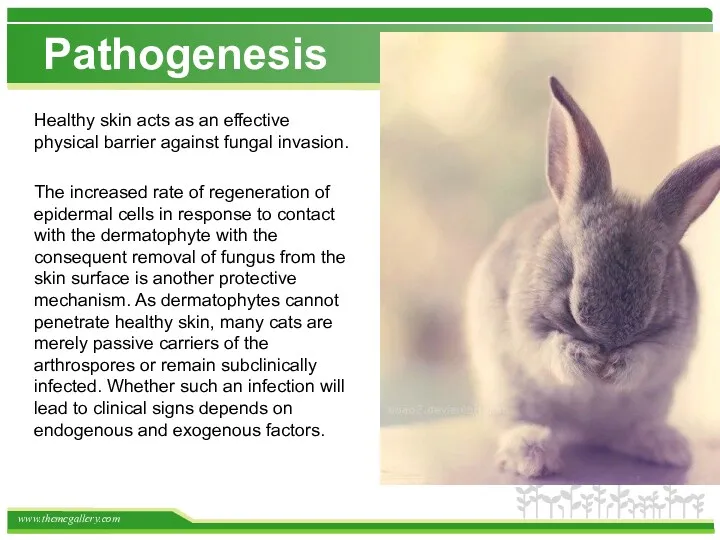
- 5. Pathogenesis Healthy skin acts as an effective physical barrier against fungal invasion. The increased rate of
- 6. The incubation period of ringworm caused by M. canis is 1 to 3 weeks. During this
- 7. Predisposing factors Young age (first 2 years of life), immunosuppression (including immunosuppressive treatment), other diseases, nutritional
- 8. In general, the zoophilic species cause more inflammatory infections which may heal spontaneously and result in
- 9. Immunity Ringworm rarely recurs, suggesting an effective and long-lasting immunity. Experimental studies confirm that animals express
- 10. Although dermatophyte infection is confined to the superficial keratinised tissues, humoral and cellular immune responses are
- 11. Antibodies In spite of the superficial nature of most infections, circulating antibodies have been demonstrated in
- 12. Citron was the first to report the presence of circulating antibodies to dermatophytes in sera of
- 13. Several investigators have demonstrated the production of agglutinins, precipitins, and complement-fixing antibodies by immunization of animals,
- 14. Investigations showed that inoculation of living or killed dermatophyte cells by routes other than the cutaneous
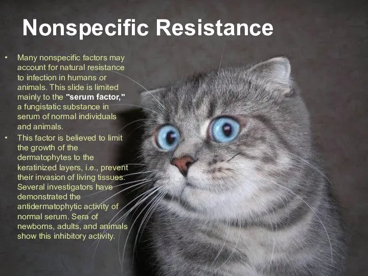
- 15. Nonspecific Resistance Many nonspecific factors may account for natural resistance to infection in humans or animals.
- 16. The dermatophytes do not usually grow in internal organs of the living body. However, they can
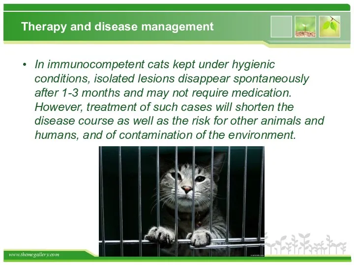
- 17. Therapy and disease management In immunocompetent cats kept under hygienic conditions, isolated lesions disappear spontaneously after
- 18. Topical treatment is less effective in cats compared to humans due to poor penetration of the
- 19. Systemic therapy Griseofulvin In some countries, the fungistatic drug griseofulvin is still used. It is administered
- 20. Вакцина содержит антиген из штамма Microsporum camS ВГНКИ N 2293, и антиген из штамма Trichophyton mentagrophytes
- 23. Скачать презентацию
Agent Properties
Dermatophytes are complex fungi growing as hyphae and forming
Agent Properties
Dermatophytes are complex fungi growing as hyphae and forming
Aetiology
Over 90% of feline dermatophytosis cases worldwide are caused by
Aetiology
Over 90% of feline dermatophytosis cases worldwide are caused by
Epidemiology
Arthrospores are transmitted through contact with sick or subclinically infected
Epidemiology
Arthrospores are transmitted through contact with sick or subclinically infected
Pathogenesis
Healthy skin acts as an effective physical barrier against fungal invasion.
Pathogenesis
Healthy skin acts as an effective physical barrier against fungal invasion.
The incubation period of ringworm caused by M. canis is 1
The incubation period of ringworm caused by M. canis is 1
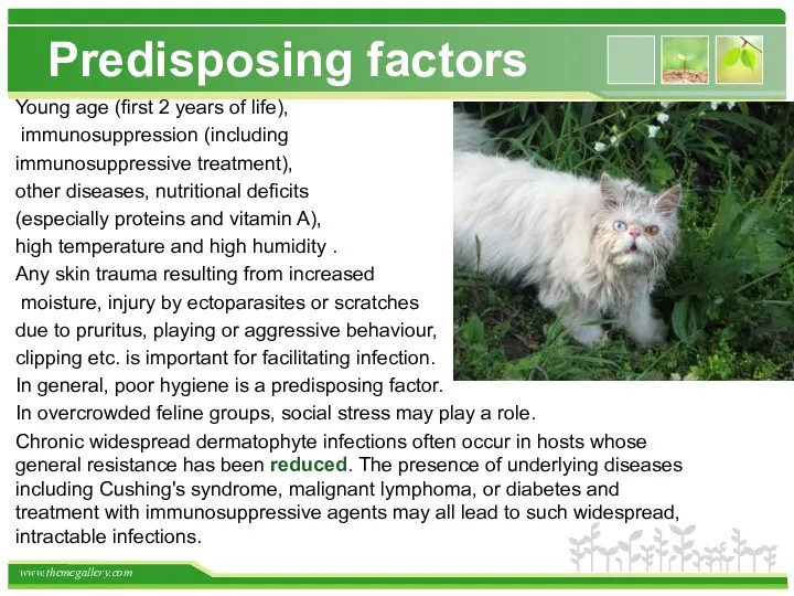
Predisposing factors
Young age (first 2 years of life),
immunosuppression (including
Predisposing factors
Young age (first 2 years of life),
immunosuppression (including
In general, the zoophilic species cause more inflammatory infections which may
In general, the zoophilic species cause more inflammatory infections which may
Immunity
Ringworm rarely recurs, suggesting an effective and long-lasting immunity. Experimental
Immunity
Ringworm rarely recurs, suggesting an effective and long-lasting immunity. Experimental
Although dermatophyte infection is confined to the superficial keratinised tissues, humoral
Although dermatophyte infection is confined to the superficial keratinised tissues, humoral
Antibodies
In spite of the superficial nature of most infections, circulating antibodies
Antibodies
In spite of the superficial nature of most infections, circulating antibodies
Citron was the first to report the presence of circulating antibodies
Citron was the first to report the presence of circulating antibodies
Several investigators have demonstrated the production of agglutinins, precipitins, and complement-fixing
Several investigators have demonstrated the production of agglutinins, precipitins, and complement-fixing
Investigations showed that inoculation of living or killed dermatophyte cells by
Investigations showed that inoculation of living or killed dermatophyte cells by
Nonspecific Resistance
Many nonspecific factors may account for natural resistance to infection
Nonspecific Resistance
Many nonspecific factors may account for natural resistance to infection
The dermatophytes do not usually grow in internal organs of the
The dermatophytes do not usually grow in internal organs of the
Therapy and disease management
In immunocompetent cats kept under hygienic conditions,
Therapy and disease management
In immunocompetent cats kept under hygienic conditions,
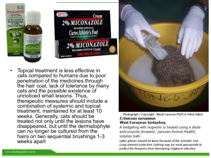
Topical treatment is less effective in cats compared to humans due
Topical treatment is less effective in cats compared to humans due
Systemic therapy
Griseofulvin In some countries, the fungistatic drug griseofulvin is still
Systemic therapy
Griseofulvin In some countries, the fungistatic drug griseofulvin is still
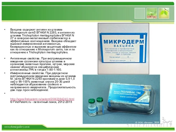
Вакцина содержит антиген из штамма Microsporum camS ВГНКИ N 2293, и
Вакцина содержит антиген из штамма Microsporum camS ВГНКИ N 2293, и

 Кровь. Состав крови
 Сапонины. Строение сапонинов
 Зондирование
 Тісжегімен және пародонттың қабыну ауруларының патогенетикалық даму механизмі
 Воспалительные заболевания придатков матки: диагностика и лечение
 Коррекция кальций-фосфорного обмена у пациентов на гемодиализе
 Иммунологическое бесплодие
 Diplomnaya_prezentatsia
 Иммунодепрессивные болезни птиц
 Лазеры в медицине. История изобретения
 Роль физической культуры и спорта в профилактике заболеваний и укреплении здоровья
 Эпилепсия. Эпилептическая реакция
 Гис будасының тармақшалар блокадасы
 Анатомо-морфологические особенности формирующейся зубочелюстной системы у детей
 Дихання. Анатомо-функціональні особливості дихальної системи
 Дегенерация сетчатки глаза
 Этиология, определение понятия. Причины, условия и факторы болезни. Определение понятий. Виды
 Лабораторная диагностика паразитарных болезней
 Чума. Источники инфекции
 Естественное вскармливание ребенка первого года жизни
 Клиническая фармакология лекарственных средств, применяемых для лечения гастро-дуоденальной патологии
 Рак гортани. Злокачественное новообразование гортани
 Транспортировка пострадавших. Транспортные положения
 Абдоминальное кесарево сечение
 Здоровый образ жизни
 Туберкулезге қарсы іс шаралар
 Грипп и его профилактика
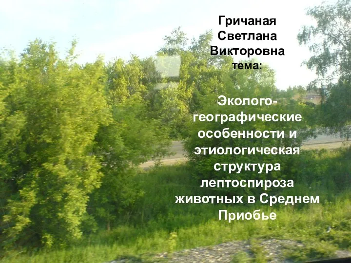 Эколого-географические особенности и этиологическая структура лептоспироза животных в Среднем Приобье