- Immunophysiology of reproductive system

Содержание
- 2. Over 50 years ago, there was the assumption that the placenta is an allograft expressing paternal
- 3. Integrational view of the immune system during pregnancy. The old model conceives the maternal immune system
- 4. Role of the placenta as a modulator of fetal and maternal responses. Inflammation at the placenta
- 7. Trophoblasts are specialized cells of the placenta that play an important role in embryo implantation and
- 8. During normal pregnancy, the human decidua contains a high number of immune cells, such as macrophages,
- 10. Comparison and contrast between a cellular response to a skin allograft and to a semi-allogeneic fetus
- 11. The interaction between the trophoblast HLA molecules and the KIR receptors of the uNK cells of
- 12. Cytokines produced by uNK cells at the human fetal-maternal interface include interleukin (IL) 8, interferon-inducible-protein-10 (IP-10),
- 13. Figure 6. Schematic illustration of the mechanism by which dNK cells promote trophoblastic cells differentiation. The
- 14. During gestation, uNK (uterine NK cells) are in intimate contact with placental cells and mediate trophic
- 16. Скачать презентацию
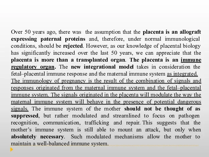
Over 50 years ago, there was the assumption that the placenta
Over 50 years ago, there was the assumption that the placenta
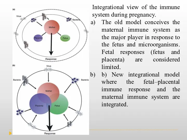
Integrational view of the immune system during pregnancy.
The old model
Integrational view of the immune system during pregnancy.
The old model
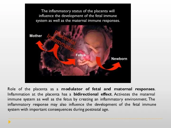
Role of the placenta as a modulator of fetal and maternal
Role of the placenta as a modulator of fetal and maternal
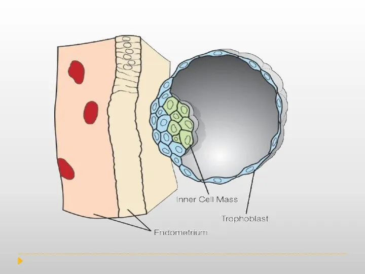
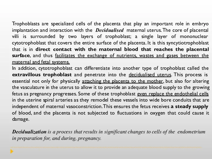
Trophoblasts are specialized cells of the placenta that play an important
Trophoblasts are specialized cells of the placenta that play an important
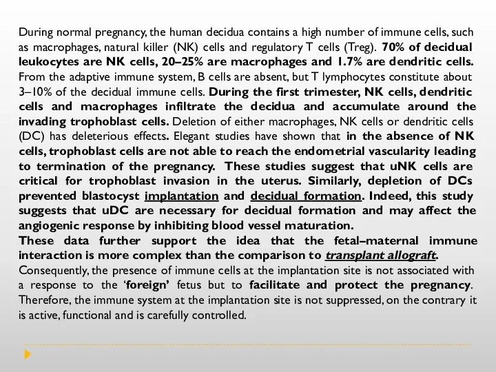
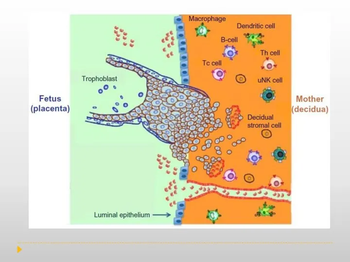
During normal pregnancy, the human decidua contains a high number of
During normal pregnancy, the human decidua contains a high number of
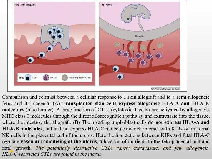
Comparison and contrast between a cellular response to a skin allograft
Comparison and contrast between a cellular response to a skin allograft
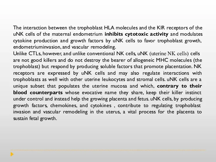
The interaction between the trophoblast HLA molecules and the KIR receptors
The interaction between the trophoblast HLA molecules and the KIR receptors
Cytokines produced by uNK cells at the human fetal-maternal interface include
Cytokines produced by uNK cells at the human fetal-maternal interface include
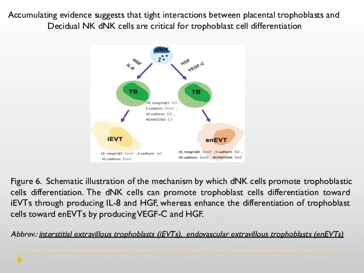
Figure 6. Schematic illustration of the mechanism by which dNK cells
Figure 6. Schematic illustration of the mechanism by which dNK cells
During gestation, uNK (uterine NK cells) are in intimate contact with
During gestation, uNK (uterine NK cells) are in intimate contact with
 Опухолевые маркеры: роль в клинической практике
 Кровь, её состав и функции. Группы крови
 Биопсия почек. Показания, методика проведения
 Анестезиология и реаниматология. Введение в дисциплину
 Endodontics includes a treatment of root canals inside the tooth
 Влияние материала ИОЛ и величины передне-задней оси глаза при миопии на развитие вторичной катаракты в послеоперационный период
 Постуральный контроль
 Тауарлар қорының құрылымы және классификациясы
 Отработка. Особенности анатомического строения зубов боковой группы : премоляров , моляров верхней , нижней челюстей
 Медицинская визуализация мочевыделительной системы. Часть 1. Рентгенанатомия. Методы диагностики
 Паровая стерилизация на фармацевтическом производстве. Компания Миллаб
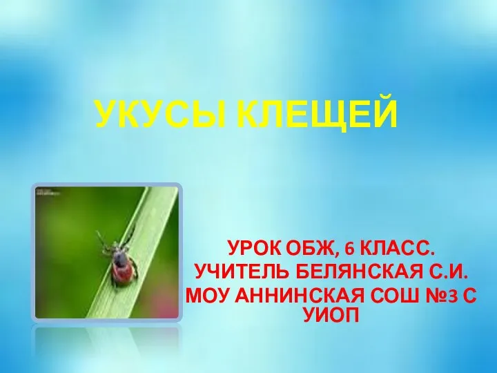 Укусы клещей
 Технология определения хим.свойств мочи
 Оперативные доступы к органам брюшной полости
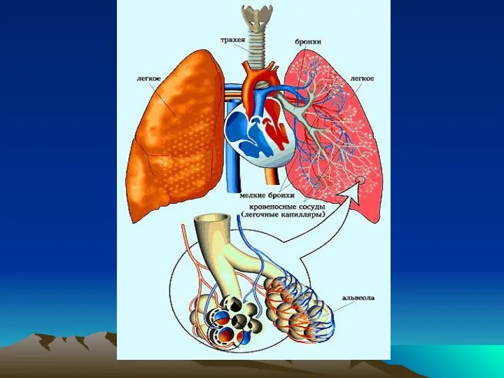 Инструментальные методы исследования. Синдромы при заболеваниях органов дыхания
 Моторні функції мозочка. Моторні функції великих півкуль і базальних гангліїв
 Рекомендации ESC по артериальной гипертензии
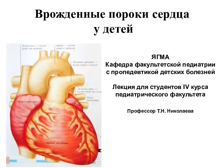 Врожденные пороки сердца у детей
 Учение об инфекции патогенность и вирулентность микробов
 Предоперационный период. Операция. Послеоперационный период
 Алкоголизм. Наркомания. Токсикомания
 Реабилитация пациентов в гинекологии. Лекция №21
 Ингаляционные методы анестезии у детей
 Менингит. Менингококкты инфекция
 Новый продукт
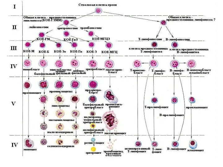 Эритрон. Показатели эритропоэза
 Новые подходы в организации и проведении предварительных и периодических медицинских осмотров
 Сестринский процесс при уходе за пациентами с сахарным диабетом второго типа