- Inflammaione. (Subject 4)

Содержание
- 2. Causes of inflammation Exogenous Infectious factors Exogenous Non-infectious factors: physical chemical biological Endogenous products of tissue
- 3. Inflammation – local manifestation of the organism general reaction to the tissue injury Inflammation events Alteration
- 4. Signs of inflammation Local: Calor - heat Rubor - redness Dolor - pain Tumor - swelling
- 5. Alteration Primary alteration - direct action of pathogenic factor (functional and structural injury of the cells)
- 6. Metabolism changes Prevalence of catabolic processes in the early stages High speed of metabolic reaction (heat)
- 7. Inflammatory mediators
- 8. Arachidonic acid metabolites Cell membrane phospholipids Phospholipases Arachidonic Acid Lipooxygenase Cyclooxygenase Leukotrienes Thromboxane Prostacycline Prostoglandins -
- 9. Arachidonic acids metabolites Thromboxane A2 - platelet aggregator and vasoconstrictor Prostacyclin - ↓ platelet aggregation and
- 10. Cellular mediators Active oxygen radicals: endothelial cells damage (? vessels permeability) other cells injury Platelet activating
- 11. Cellular mediators Lysosomal enzymes: mediate tissue injury activate bradykinine synthesis mast cells degranulation chemotaxis Nitric oxide:
- 12. Plasma mediators Clotting system mobilization of molecules of adherence activation of cyclooxygenase production of NO and
- 13. The summary of inflammatory mediators’ activity Vasodilation ↑ of blood vessels permeability Leukocyte adhesion Chemotaxis Fever
- 14. Changes in vascular flow Arterioles constriction (activation of sympathetic nerves, mediators influence) -localization of injuring agent
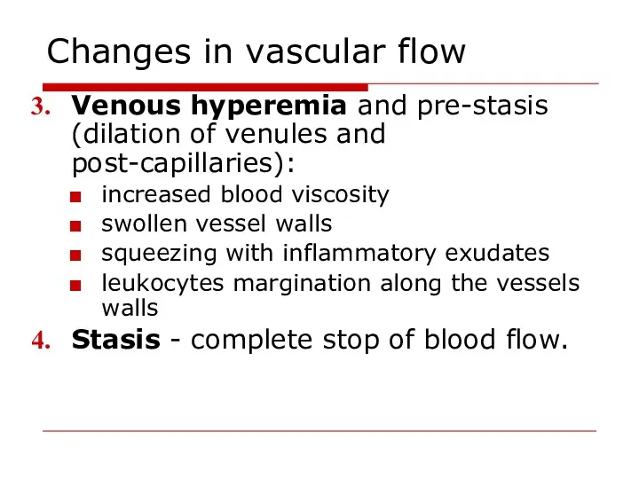
- 15. Changes in vascular flow Venous hyperemia and pre-stasis (dilation of venules and post-capillaries): increased blood viscosity
- 16. Venous hyperemia and stasis prevent the spreading of the damage to surrounding tissues. Arterial and venous
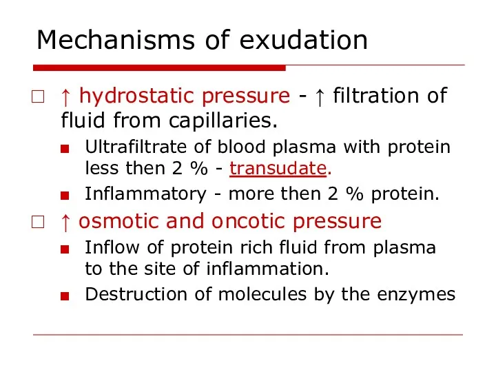
- 17. Mechanisms of exudation ↑ vascular permeability (vascular leakage). ↑ intravascular hydrostatic pressure ↑ osmotic and oncotic
- 18. Increase of vascular permeability Endothelial cells contraction histamine, bradykinin occurs rapidly after exposure to mediator reversible
- 19. Mechanisms of exudation ↑ hydrostatic pressure - ↑ filtration of fluid from capillaries. Ultrafiltrate of blood
- 20. The role of exudation Negative squeezing of tissues and organs exudate outflow to body cavities and
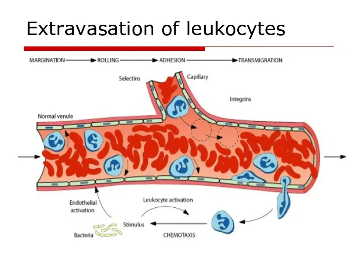
- 21. Extravasation of leukocytes
- 22. Leukocytes migration Move pseudopods into the junctions between the endothelial cells Squeeze through interendothelial junctions Release
- 23. Chemotaxis Chemotactic agents: bacterial membrane lipopolysaccharides components of the complement (3b, 5a,5b,6,7 leukotrienes products of tissue
- 24. Leukocytes role in inflammation Protective function – phagocytosis. Synthesis and secretion of inflammatory mediators. Processing and
- 25. Stages of phagocytosis Chemotaxis 2. Adherence (opsonins - IgM, IgG, C3b) 3. Phagosome formation 4. Killing
- 26. Two mechanisms of bacterial killing Oxygen-dependent mechanism reactive oxygen species – superoxide anion, hydroxyl ion, hydroperoxide
- 27. Proliferation in inflammation Regeneration - replacement of dead cells with new ones; the function is restored.
- 28. The steps of repair Phagocytosis Proliferation of endothelial cells and fibroblasts in the damaged area. The
- 29. Factors influencing proliferation Local: Persisting infection, foreign material Inadequate blood supply Excessive movement Irradiation Systemic: Age
- 30. Classification of inflammation Classification based on the cause of inflammation: Infectious: non-specific (cocci) and specific (tuberculosis,
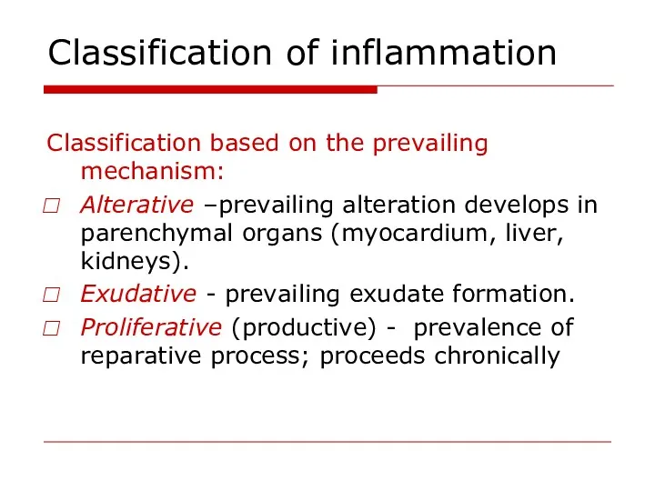
- 31. Classification of inflammation Classification based on the prevailing mechanism: Alterative –prevailing alteration develops in parenchymal organs
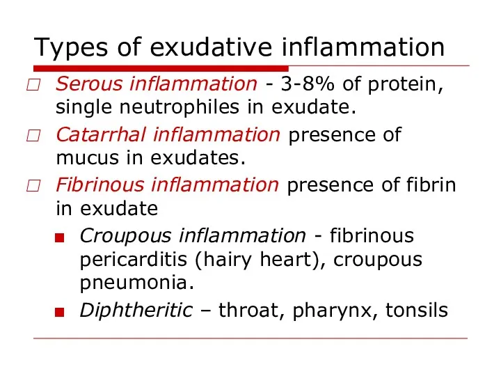
- 32. Types of exudative inflammation Serous inflammation - 3-8% of protein, single neutrophiles in exudate. Catarrhal inflammation
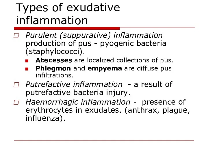
- 33. Types of exudative inflammation Purulent (suppurative) inflammation production of pus - pyogenic bacteria (staphylococci). Abscesses are
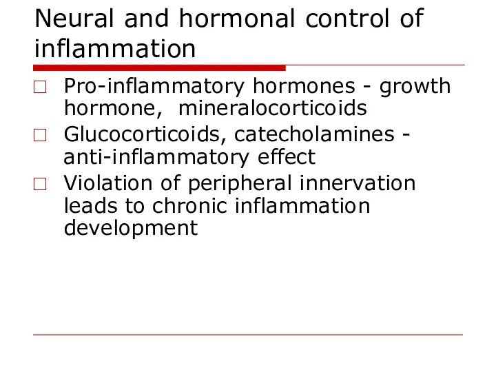
- 34. Neural and hormonal control of inflammation Pro-inflammatory hormones - growth hormone, mineralocorticoids Glucocorticoids, catecholamines - anti-inflammatory
- 35. Inflammation outcomes Complete resolution - the injury is limited Healing by scarring – impossibility of regeneration
- 37. Скачать презентацию
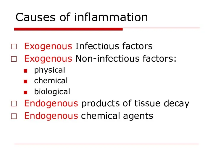
Causes of inflammation
Exogenous Infectious factors
Exogenous Non-infectious factors:
physical
chemical
biological
Endogenous products
Causes of inflammation
Exogenous Infectious factors
Exogenous Non-infectious factors:
physical
chemical
biological
Endogenous products
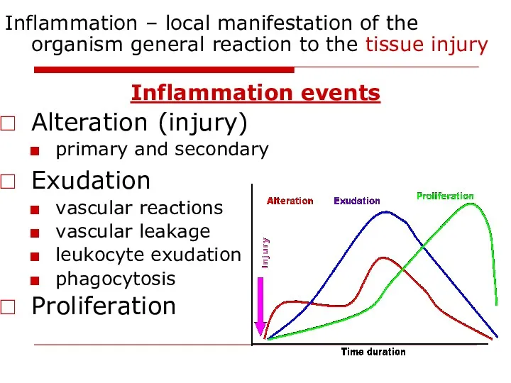
Inflammation – local manifestation of the organism general reaction to the
Inflammation – local manifestation of the organism general reaction to the
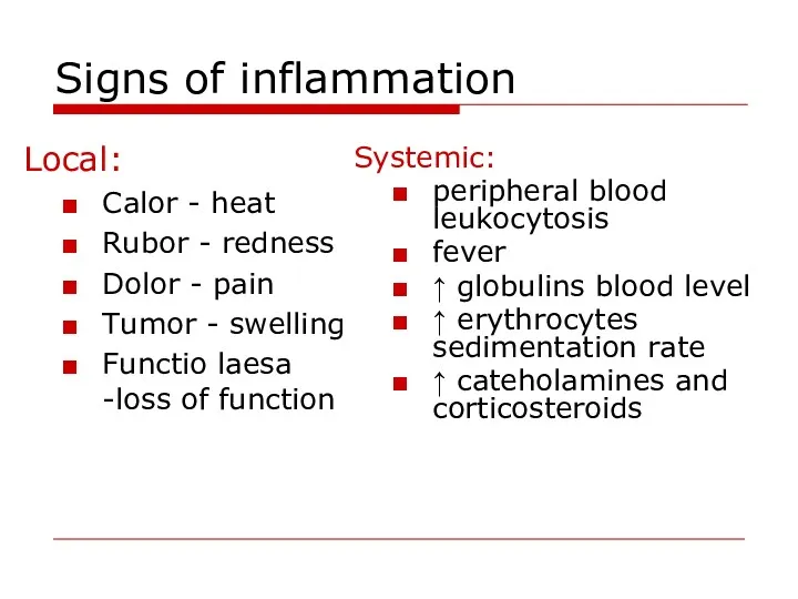
Signs of inflammation
Local:
Calor - heat
Rubor - redness
Dolor - pain
Signs of inflammation
Local:
Calor - heat
Rubor - redness
Dolor - pain
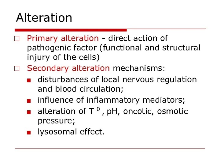
Alteration
Primary alteration - direct action of pathogenic factor (functional and
Alteration
Primary alteration - direct action of pathogenic factor (functional and
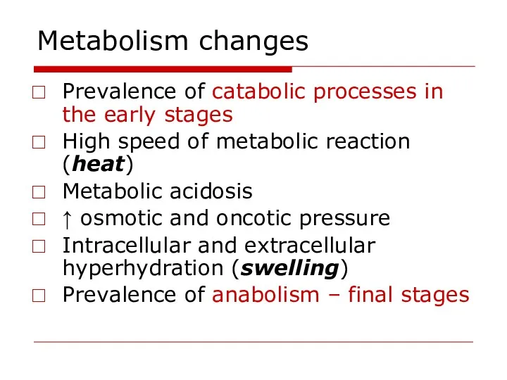
Metabolism changes
Prevalence of catabolic processes in the early stages
High speed
Metabolism changes
Prevalence of catabolic processes in the early stages
High speed
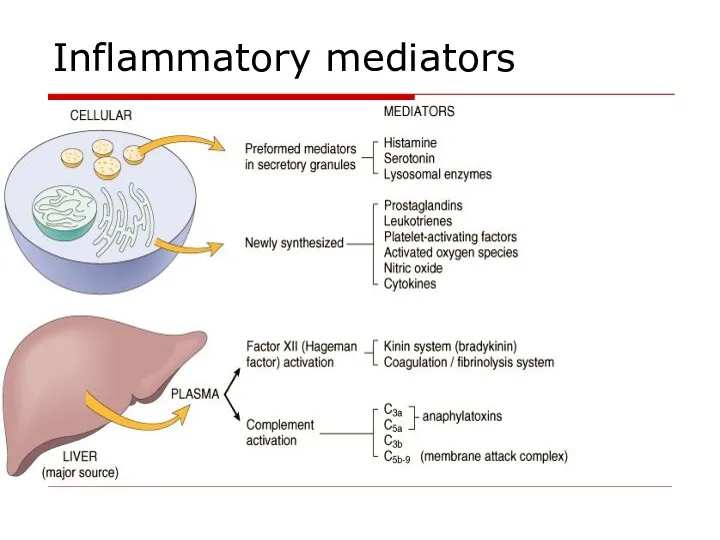
Inflammatory mediators
Inflammatory mediators
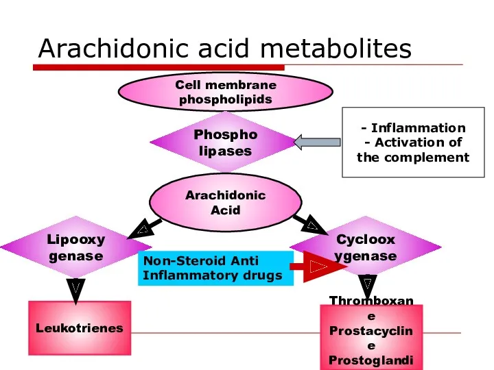
Arachidonic acid metabolites
Cell membrane phospholipids
Phospholipases
Arachidonic Acid
Lipooxygenase
Cyclooxygenase
Leukotrienes
Thromboxane
Prostacycline
Prostoglandins
- Inflammation
- Activation of
the complement
Arachidonic acid metabolites
Cell membrane phospholipids
Phospholipases
Arachidonic Acid
Lipooxygenase
Cyclooxygenase
Leukotrienes
Thromboxane
Prostacycline
Prostoglandins
- Inflammation
- Activation of
the complement
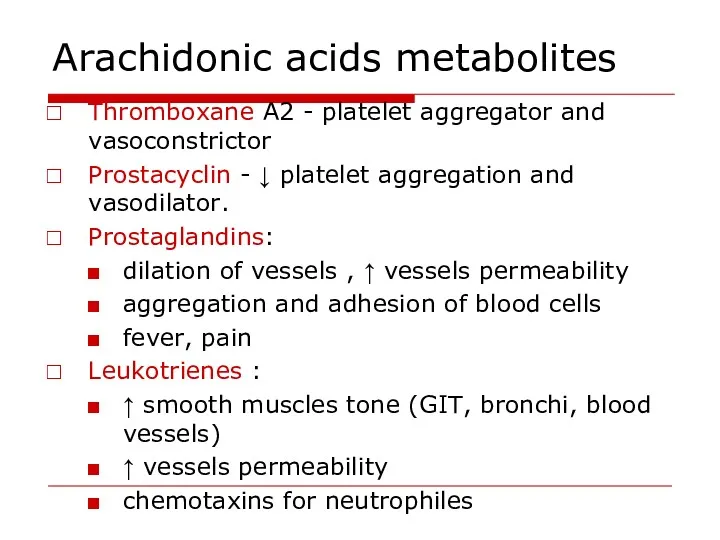
Arachidonic acids metabolites
Thromboxane A2 - platelet aggregator and vasoconstrictor
Prostacyclin -
Arachidonic acids metabolites
Thromboxane A2 - platelet aggregator and vasoconstrictor
Prostacyclin -
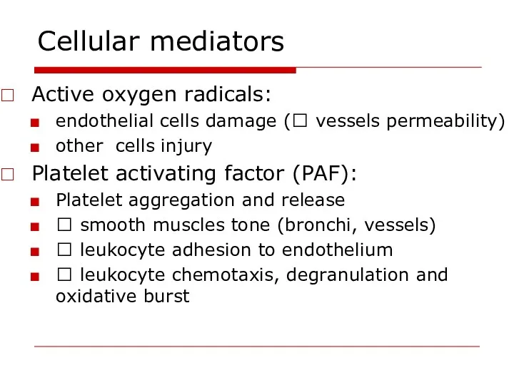
Cellular mediators
Active oxygen radicals:
endothelial cells damage (? vessels permeability)
other cells
Cellular mediators
Active oxygen radicals:
endothelial cells damage (? vessels permeability)
other cells
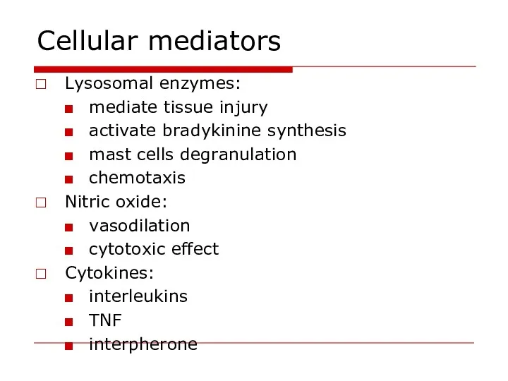
Cellular mediators
Lysosomal enzymes:
mediate tissue injury
activate bradykinine synthesis
mast cells degranulation
chemotaxis
Nitric oxide:
vasodilation
cytotoxic
Cellular mediators
Lysosomal enzymes:
mediate tissue injury
activate bradykinine synthesis
mast cells degranulation
chemotaxis
Nitric oxide:
vasodilation
cytotoxic
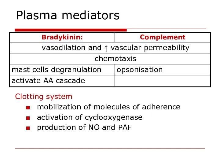
Plasma mediators
Clotting system
mobilization of molecules of adherence
activation of cyclooxygenase
production of
Plasma mediators
Clotting system
mobilization of molecules of adherence
activation of cyclooxygenase
production of
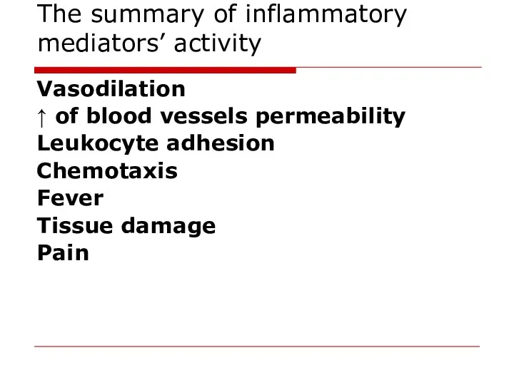
The summary of inflammatory mediators’ activity
Vasodilation
↑ of blood vessels permeability
Leukocyte
The summary of inflammatory mediators’ activity
Vasodilation
↑ of blood vessels permeability
Leukocyte
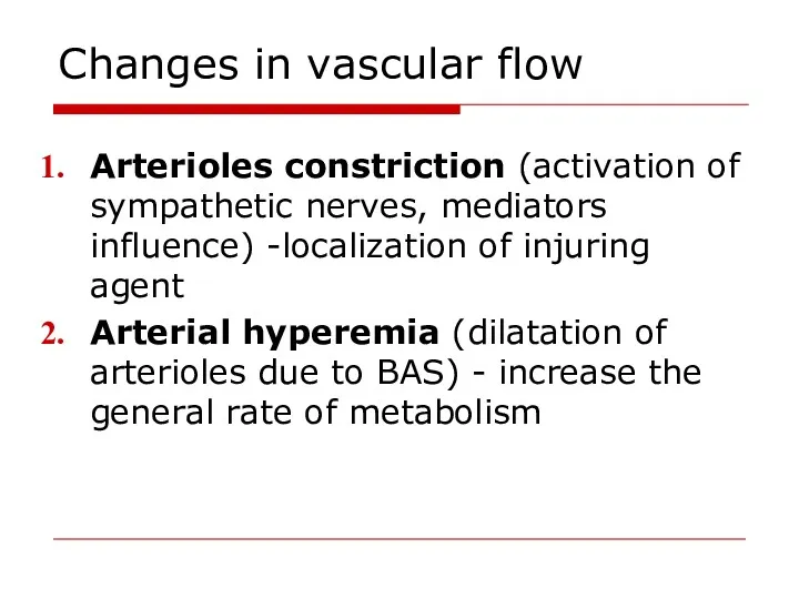
Changes in vascular flow
Arterioles constriction (activation of sympathetic nerves, mediators
Changes in vascular flow
Arterioles constriction (activation of sympathetic nerves, mediators
Changes in vascular flow
Venous hyperemia and pre-stasis (dilation of venules and
Changes in vascular flow
Venous hyperemia and pre-stasis (dilation of venules and
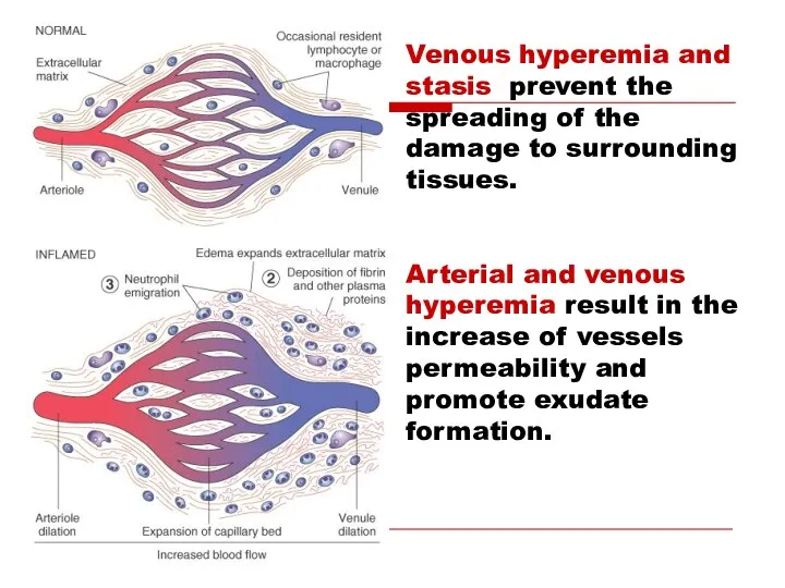
Venous hyperemia and stasis prevent the spreading of the damage to
Venous hyperemia and stasis prevent the spreading of the damage to
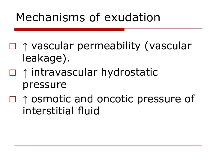
Mechanisms of exudation
↑ vascular permeability (vascular leakage).
↑ intravascular hydrostatic pressure
↑
Mechanisms of exudation
↑ vascular permeability (vascular leakage).
↑ intravascular hydrostatic pressure
↑
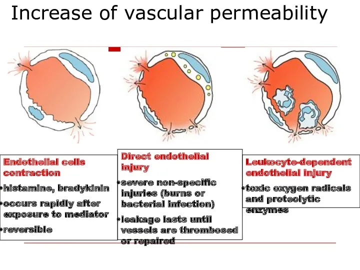
Increase of vascular permeability
Endothelial cells contraction
histamine, bradykinin
occurs rapidly after exposure
Increase of vascular permeability
Endothelial cells contraction
histamine, bradykinin
occurs rapidly after exposure
Mechanisms of exudation
↑ hydrostatic pressure - ↑ filtration of fluid from
Mechanisms of exudation
↑ hydrostatic pressure - ↑ filtration of fluid from
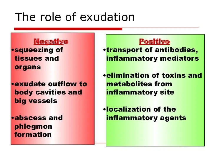
The role of exudation
Negative
squeezing of tissues and organs
exudate outflow
The role of exudation
Negative
squeezing of tissues and organs
exudate outflow
Extravasation of leukocytes
Extravasation of leukocytes
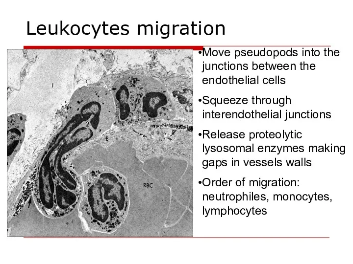
Leukocytes migration
Move pseudopods into the junctions between the endothelial cells
Squeeze through
Leukocytes migration
Move pseudopods into the junctions between the endothelial cells
Squeeze through
Chemotaxis
Chemotactic agents:
bacterial membrane lipopolysaccharides
components of the complement (3b, 5a,5b,6,7
leukotrienes
products of
Chemotaxis
Chemotactic agents:
bacterial membrane lipopolysaccharides
components of the complement (3b, 5a,5b,6,7
leukotrienes
products of
Leukocytes role in inflammation
Protective function – phagocytosis.
Synthesis and secretion of
Leukocytes role in inflammation
Protective function – phagocytosis.
Synthesis and secretion of
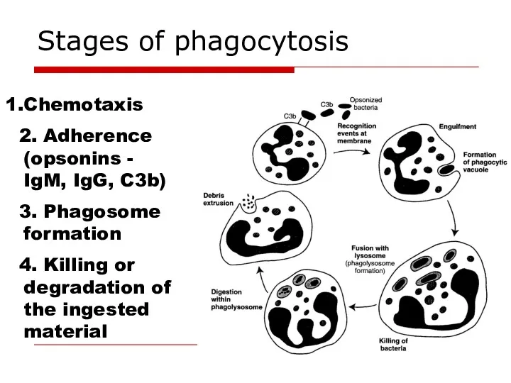
Stages of phagocytosis
Chemotaxis
2. Adherence (opsonins - IgM, IgG, C3b)
3. Phagosome
Stages of phagocytosis
Chemotaxis
2. Adherence (opsonins - IgM, IgG, C3b)
3. Phagosome
Two mechanisms of bacterial killing
Oxygen-dependent mechanism reactive oxygen species – superoxide
Two mechanisms of bacterial killing
Oxygen-dependent mechanism reactive oxygen species – superoxide
Proliferation in inflammation
Regeneration - replacement of dead cells with new ones;
Proliferation in inflammation
Regeneration - replacement of dead cells with new ones;
The steps of repair
Phagocytosis
Proliferation of endothelial cells and fibroblasts
The steps of repair
Phagocytosis
Proliferation of endothelial cells and fibroblasts
Factors influencing proliferation
Local:
Persisting infection, foreign material
Inadequate blood supply
Factors influencing proliferation
Local:
Persisting infection, foreign material
Inadequate blood supply

Classification of inflammation
Classification based on the cause of inflammation:
Infectious: non-specific
Classification of inflammation
Classification based on the cause of inflammation:
Infectious: non-specific
Classification of inflammation
Classification based on the prevailing mechanism:
Alterative –prevailing alteration develops
Classification of inflammation
Classification based on the prevailing mechanism:
Alterative –prevailing alteration develops
Types of exudative inflammation
Serous inflammation - 3-8% of protein, single neutrophiles
Types of exudative inflammation
Serous inflammation - 3-8% of protein, single neutrophiles
Types of exudative inflammation
Purulent (suppurative) inflammation production of pus - pyogenic
Types of exudative inflammation
Purulent (suppurative) inflammation production of pus - pyogenic
Neural and hormonal control of inflammation
Pro-inflammatory hormones - growth hormone, mineralocorticoids
Neural and hormonal control of inflammation
Pro-inflammatory hormones - growth hormone, mineralocorticoids
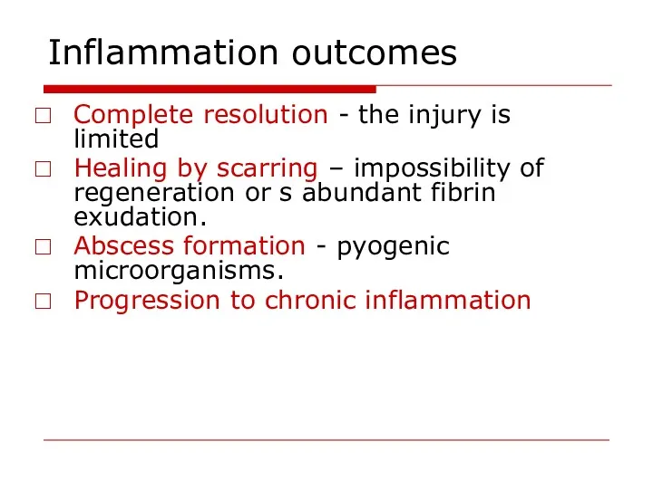
Inflammation outcomes
Complete resolution - the injury is limited
Healing by scarring –
Inflammation outcomes
Complete resolution - the injury is limited
Healing by scarring –
 Пневмония. Пневмония жіктелуі
 Основные принципы современной химиотерапии злокачественных опухолей
 Эпидемиялық процестің әртүрлі халық топтары арасындағы көрінуінің қазіргі кездегі сипаттамасы
 Гиподинамия как фактор риска развития заболеваний
 Лабораторная диагностика сахарного диабета
 Доброкачественные и злокачественные опухоли радужной оболочки и цилиарного тела
 Комплексний порівняльний аналіз стану громадського здоров’я та діяльності системи охорони здоров’я України та Румунії
 Методы контрацепции
 Лазеры в медицине
 Соединительно-тканный массаж
 Түрлі мүшелер электрлік белсеңділігін зерттеу әдістері
 Осторожно! Клещи
 Грипп и ОРВИ: Обоснованные подходы к лечению и профилактике
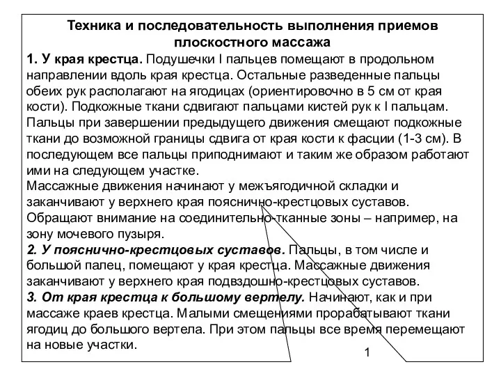 Техника и последовательность выполнения приемов плоскостного массажа
 Болезнь Виллебранда
 Вступ в курс інфектології. Поняття про інфекційні хвороби. Принципи діагностики, лікування, профілактики
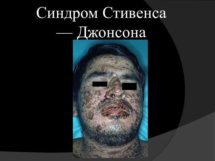 Синдром Стивенса — Джонсона
 Инфекционно-токсический шок. Критические состояния и факторы риска их развития у инфекционных больных
 Долихосигма. Этиология
 Гормоны
 Интенсивная терапия и анестезия при кровопотере в акушерстве
 Дезинфекционные технологии и оборудование для обеззараживания воздуха
 Обучение по оказанию первой помощи пострадавшим на производстве
 Рентгеновская и позитронная томография
 Анестезиологическое обеспечение экстренных хирургических вмешательств у детей. Оценка риска и безопасность
 Физкультура в семье. Здоровье родителей и детей
 Обучение в сестринском деле
 Возрастные особенности формирования и развития эндокринной системы (гипо- и гиперфункция эндокринных желез)