- Классификация опухолей ЦНС. ВОЗ 2016

Содержание
- 2. Классификации ВОЗ опухолей ЦНС 1 издание,1979 2 издание,1993 3 издание, 2000 4 издание, 2007 Диагнозы, классификация,
- 3. После 2007 года
- 4. «I believe that we have reached the tipping point, the point at which we know enough
- 5. ISN-Haarlem Guidelines 2014 “A critical question with major practical consequences has therefore arisen: how should clinically
- 6. ISN-Haarlem Guidelines 2014 – “integrated diagnosis”
- 7. В 2016 году выходит пересмотр 4 издания классификации опухолей ЦНС ВОЗ Над ним работают 122 автора
- 8. Структура и номенклатура
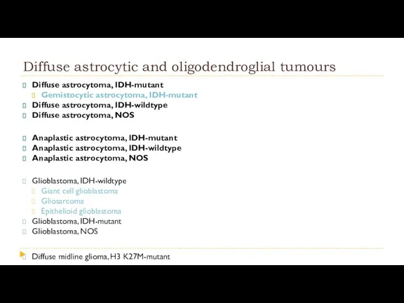
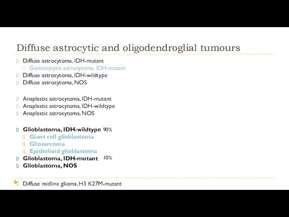
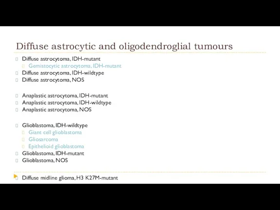
- 10. Diffuse astrocytic and oligodendroglial tumours Diffuse astrocytoma, IDH-mutant Gemistocytic astrocytoma, IDH-mutant Diffuse astrocytoma, IDH-wildtype Diffuse astrocytoma,
- 11. Как формулируется диагноз? Diffuse astrocytoma, IDH-mutant WHO Grade II
- 12. Варианты обозначения молекулярного профиля опухоли
- 13. WHO Grade I WHO Grade II WHO Grade III WHO Grade IV Степень злокачественности
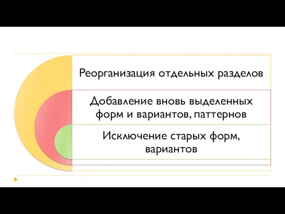
- 14. Что нового?
- 16. Почему нельзя основываться только на молекулярном анализе?
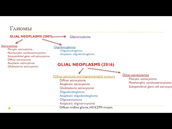
- 18. Глиомы GLIAL NEOPLASMS (2007) Astrocytomas Pilocytic astrocytoma Pleomorphic xanthoastrocytoma Subependimal giant cell astrocytoma Diffuse astrocytoma Anaplastic
- 19. Глиомы GLIAL NEOPLASMS (2007) Astrocytomas Pilocytic astrocytoma Pleomorphic xanthoastrocytoma Subependimal giant cell astrocytoma Diffuse astrocytoma Anaplastic
- 20. Диффузные глиомы. Зависимость прогноза от молекулярного типа опухоли
- 21. Clinical Outcomes According to Histologic Class and Grade According to Molecular Subtype Comprehensive, Integrative Genomic Analysis
- 22. Диффузные глиомы. Зависимость прогноза от молекулярных свойств опухоли Прогноз у пациентов с «IDH-mutant» типом лучше, чем
- 23. Molecular taxonomy of IDH-mutant glioma progression Hiroaki Wakimoto et al. Targetable Signaling Pathway Mutations Are Associated
- 24. A novel view of the genetic landscape of glioblastomas Parsons DW et al. An integrated genomic
- 25. Гены IDH IDH1 – 2q34 IDH2 – 15q26.1
- 26. Изомеры белка IDH (изоцитратдегидрогеназа)
- 27. Соотношение различных мутаций в генах IDH1 и IDH2
- 28. Мутантный белок R132H Lenny Dang et al. Cancer-associated IDH1 mutations produce 2-hydroxyglutarate. Nature, 2010;465(7300):966. Strong reaction
- 29. Нормальные ферменты IDH vs Мутантные формы IDH Andrew R. Mullen and Ralph J. DeBerardinis. Genetically-defined metabolic
- 30. Роль R2-HG в развитии опухолевого процесса Michael Heuser et al. Enigmas of IDH mutations in hematology/oncology.
- 31. Diffuse astrocytic and oligodendroglial tumours Diffuse astrocytoma, IDH-mutant Gemistocytic astrocytoma, IDH-mutant Diffuse astrocytoma, IDH-wildtype Diffuse astrocytoma,
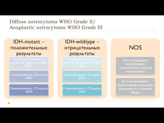
- 32. Diffuse astrocytoma WHO Grade II/ Anaplastic astrocytoma WHO Grade III
- 33. IDH-mutant IDH-mutant Секвенирование IDH1 и IDH2 Секвенирование IDH1 и IDH2 Мутация найдена IDH-mutant Мутация не найдена
- 34. Diffuse astrocytoma, IDH-mutant WHO Grade II
- 35. Gemistocytic astrocytoma, IDH-mutant WHO Grade II Гемистоциты характеризуются насыщенной эозинофильной цитоплазмой и эксцентрично расположенным ядром Количество
- 36. Diffuse astrocytic and oligodendroglial tumours Diffuse astrocytoma, IDH-mutant Gemistocytic astrocytoma, IDH-mutant Diffuse astrocytoma, IDH-wildtype Diffuse astrocytoma,
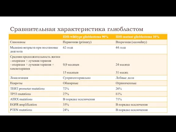
- 37. Сравнительная характеристика глиобластом
- 38. Glioblastoma WHO Grade IV
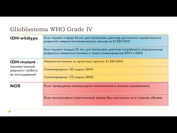
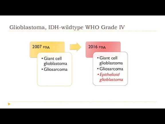
- 39. Glioblastoma, IDH-wildtype WHO Grade IV
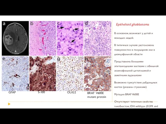
- 40. Epithelioid glioblastoma В основном, возникает у детей и молодых людей. В типичных случаях расположена поверхностно в
- 41. Glioblastoma/Astrocytoma patterns
- 42. Glioblastoma with primitive neuronal component Обычно содержит клетки астроцитарного ряда и состоит из хорошо отделенных узлов,
- 43. Diffuse astrocytic and oligodendroglial tumours Diffuse astrocytoma, IDH-mutant Gemistocytic astrocytoma, IDH-mutant Diffuse astrocytoma, IDH-wildtype Diffuse astrocytoma,
- 44. Oligodendroglioma WHO Grade II/ Anaplastic oligodendroglioma WHO Grade III
- 45. 1p19q co-deletion 1p19q co-deletion in gliomas occurs through an unbalanced whole-arm translocation between chromosomes 1 and
- 46. 1p19q codeletion in oligodendrogliomas via fluorescence in situ hybridization (FISH) Craig Horbinski et al. Gone FISHing:
- 47. Oligodendroglioma WHO Grade II/ Anaplastic oligodendroglioma WHO Grade III
- 48. Diffuse astrocytic and oligodendroglial tumours Diffuse astrocytoma, IDH-mutant Gemistocytic astrocytoma, IDH-mutant Diffuse astrocytoma, IDH-wildtype Diffuse astrocytoma,
- 49. Oligoastrocytoma, NOS WHO Grade II/ Anaplastic oligoastrocytoma, NOS WHO Grade III Из 43 случаев олигоастроцитом после
- 50. Diffuse astrocytic and oligodendroglial tumours Diffuse astrocytoma, IDH-mutant Gemistocytic astrocytoma, IDH-mutant Diffuse astrocytoma, IDH-wildtype Diffuse astrocytoma,
- 51. Diffuse midline glioma, H3 K27M-mutant WHO Grade IV Встречается, в основном, среди детей Мутация гистона H3
- 52. Глиоматоз головного мозга Исключен из классификации ВОЗ 2016 года как отдельный вид опухоли На настоящий момент
- 53. Diffuse astrocytic and oligodendroglial tumours Алгоритм постановки диагноза.
- 54. Other astrocytic tumours Pilocytic astrocytoma Pilomyxoid astrocytoma Subependymal giant cell astrocytoma Pleomorphic xanthoastrocytoma Anaplastic pleomorphic xanthoastrocytoma
- 55. Anaplastic pleomorphic xanthoastrocytoma WHO Grade III Предыдущее название: плейоморфная ксантоастроцитома с анапластическим компонентом Гистологические критерии: Пять
- 56. Ependymal tumours Subependymoma Myxopapillary ependymoma Ependymoma Papillary ependymoma Clear cell ependymoma Tanycytic ependymoma Cellular ependymoma Ependymoma,
- 57. Ependymoma, RELA fusion—positive WHO Grade II or III Большая часть супратенториальных опухолей детей Экспрессия L1CAM обсуждается
- 58. Neuronal and mixed neuronal-glial tumours Dysembryoplastic neuroepithelial tumour Gangliocytoma Ganglioglioma Anaplastic ganglioglioma Dysplastic cerebellar gangliocytoma (Lhermitte-Duclos
- 59. Diffuse leptomeningeal glioneuronal tumour Чаще встречается у детей или подростков Гистологическая картина сходна с олигодендроглиомами: мономорфные
- 60. Multinodular and vacuolated pattern Множественные узлы опухоли с заметной вакуолизацией Дифференцировка клеток по типу глии и/или
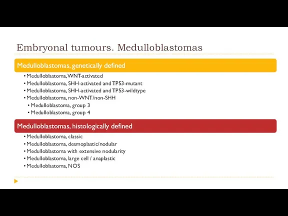
- 61. Embryonal tumours. Medulloblastomas
- 62. Embryonal tumours. Medulloblastomas Taylor MD, Northcott PA, Korshunov A, et al. Molecular subgroups of medulloblastoma: the
- 63. Embryonal tumours. Medulloblastomas
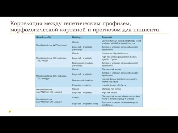
- 64. Корреляция между генетическим профилем, морфологической картиной и прогнозом для пациента.
- 65. Other Embryonal tumours Embryonal tumour with multilayered rosettes, C19MC-aItered Embryonal tumour with multilayered rosettes, NOS Medulloepithelioma
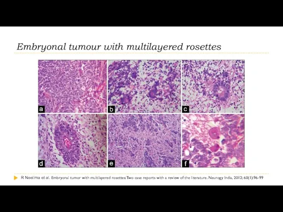
- 66. Embryonal tumour with multilayered rosettes R Neelima et al. Embryonal tumor with multilayered rosettes: Two case
- 67. Embryonal tumour with multilayered rosettes
- 68. Other Embryonal tumours Embryonal tumour with multilayered rosettes, C19MC-aItered Embryonal tumour with multilayered rosettes, NOS Medulloepithelioma
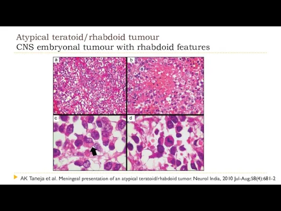
- 69. Atypical teratoid/rhabdoid tumour CNS embryonal tumour with rhabdoid features AK Taneja et al. Meningeal presentation of
- 70. Atypical teratoid/rhabdoid tumour CNS embryonal tumour with rhabdoid features
- 71. Tumours of the cranial and paraspinal nerves Schwannoma Cellular schwannoma Plexiform schwannoma Melanotic schwannoma Neurofibroma Atypical
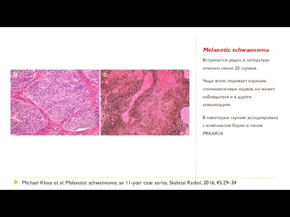
- 72. Melanotic schwannoma Встречается редко, в литературе описано около 20 случаев. Чаще всего поражает корешки спинномозговых нервов,
- 73. Tumours of the cranial and paraspinal nerves Schwannoma Cellular schwannoma Plexiform schwannoma Melanotic schwannoma Neurofibroma Atypical
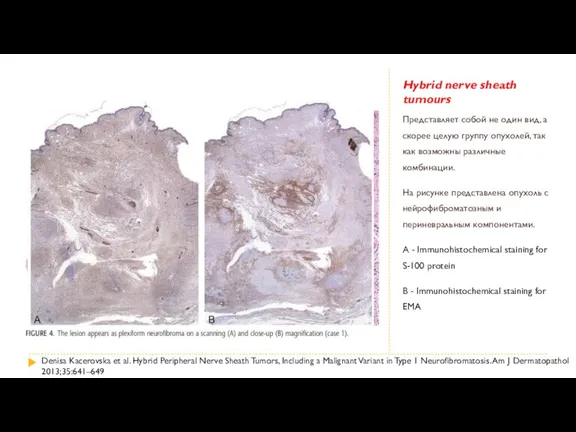
- 74. Hybrid nerve sheath tumours Представляет собой не один вид, а скорее целую группу опухолей, так как
- 75. Tumours of the cranial and paraspinal nerves Schwannoma Cellular schwannoma Plexiform schwannoma Melanotic schwannoma Neurofibroma Atypical
- 76. Malignant peripheral nerve sheath tumour variants Epithelioid MPNST MPNST with perineurial differentiation Vickie Y. Jo et
- 77. Meningiomas Meningioma Meningothelial meningioma Fibrous meningioma Transitional meningioma Psammomatous meningioma Angiomatous meningioma Microcystic meningioma Secretory meningioma
- 78. Atypical (WHO grade II) meningiomas are characterised histologically by
- 79. Mesenchymal, non-meningothelial tumours Solitary fibrous turnour / haemangiopericytoma Grade 1 Grade 2 Grade 3 Haemangioblastoma Haemangiorna
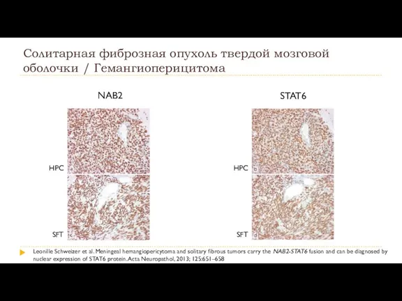
- 80. Солитарная фиброзная опухоль твердой мозговой оболочки / Гемангиоперицитома
- 81. Солитарная фиброзная опухоль твердой мозговой оболочки / Гемангиоперицитома Leonille Schweizer et al. Meningeal hemangiopericytoma and solitary
- 82. Солитарная фиброзная опухоль твердой мозговой оболочки / Гемангиоперицитома NAB2 STAT6 HPC SFT HPC SFT Leonille Schweizer
- 83. Ключевые моменты 1. Мы перешли в эру постановки диагноза с помощью молекулярной генетики 2. Обязательно определение
- 86. Скачать презентацию

Классификации ВОЗ опухолей ЦНС
1 издание,1979
2 издание,1993
3 издание, 2000
4
Классификации ВОЗ опухолей ЦНС
1 издание,1979
2 издание,1993
3 издание, 2000
4
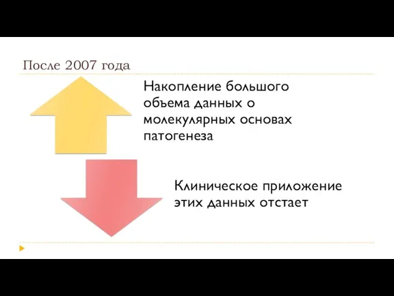
После 2007 года
После 2007 года
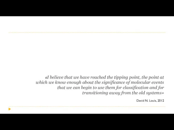
«I believe that we have reached the tipping point, the point
«I believe that we have reached the tipping point, the point
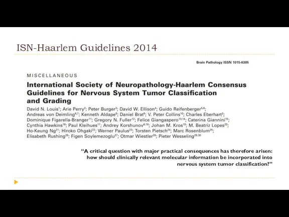
ISN-Haarlem Guidelines 2014
“A critical question with major practical consequences has therefore
ISN-Haarlem Guidelines 2014
“A critical question with major practical consequences has therefore
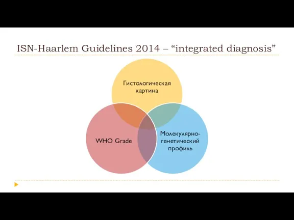
ISN-Haarlem Guidelines 2014 – “integrated diagnosis”
ISN-Haarlem Guidelines 2014 – “integrated diagnosis”
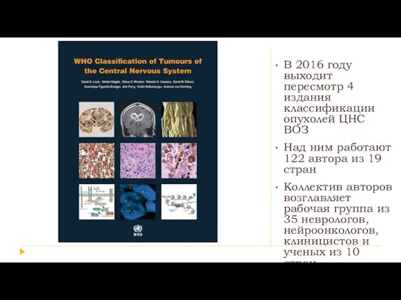
В 2016 году выходит пересмотр 4 издания классификации опухолей ЦНС ВОЗ
Над
В 2016 году выходит пересмотр 4 издания классификации опухолей ЦНС ВОЗ
Над
Структура и номенклатура
Структура и номенклатура

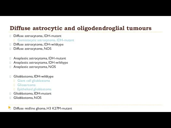
Diffuse astrocytic and oligodendroglial tumours
Diffuse astrocytoma, IDH-mutant
Gemistocytic astrocytoma, IDH-mutant
Diffuse astrocytic and oligodendroglial tumours
Diffuse astrocytoma, IDH-mutant
Gemistocytic astrocytoma, IDH-mutant
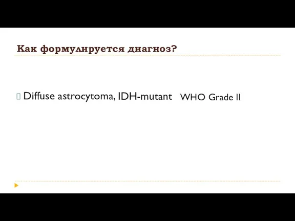
Как формулируется диагноз?
Diffuse astrocytoma,
IDH-mutant
WHO Grade II
Как формулируется диагноз?
Diffuse astrocytoma,
IDH-mutant
WHO Grade II
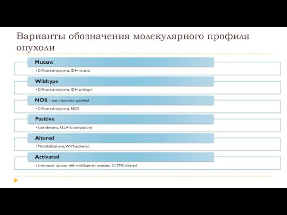
Варианты обозначения молекулярного профиля опухоли
Варианты обозначения молекулярного профиля опухоли
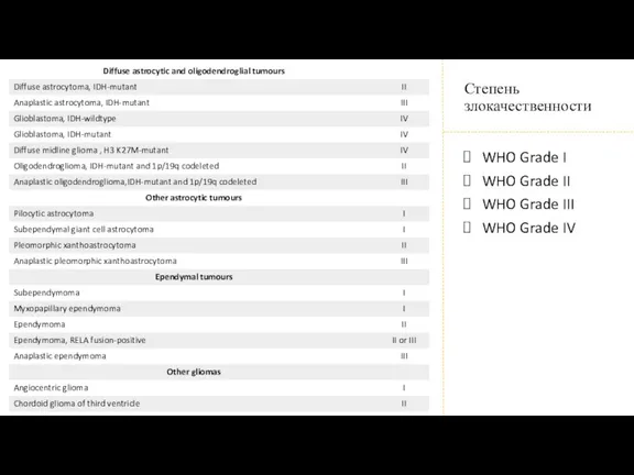
WHO Grade I
WHO Grade II
WHO Grade III
WHO Grade IV
Степень злокачественности
WHO Grade II
WHO Grade III
WHO Grade IV
Степень злокачественности
Что нового?
Что нового?

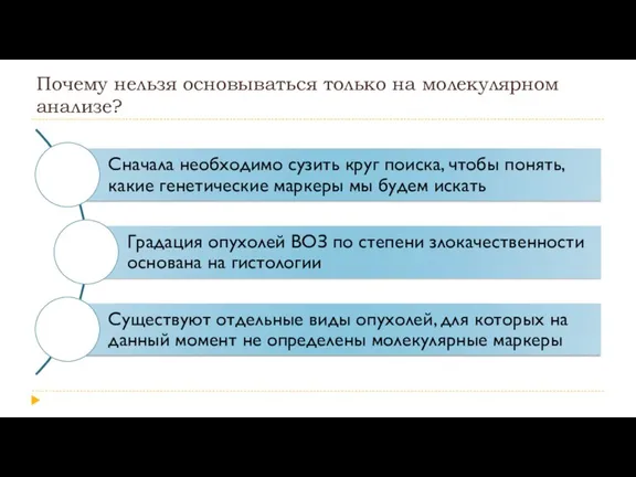
Почему нельзя основываться только на молекулярном анализе?
Почему нельзя основываться только на молекулярном анализе?
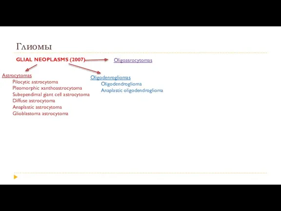
Глиомы
GLIAL NEOPLASMS (2007)
Astrocytomas
Pilocytic astrocytoma
Pleomorphic xanthoastrocytoma
Subependimal giant cell astrocytoma
Diffuse astrocytoma
Anaplastic astrocytoma
Glioblastoma astrocytoma
Oligodenrogliomas
Oligodendroglioma
Anaplastic
Глиомы
GLIAL NEOPLASMS (2007)
Astrocytomas
Pilocytic astrocytoma
Pleomorphic xanthoastrocytoma
Subependimal giant cell astrocytoma
Diffuse astrocytoma
Anaplastic astrocytoma
Glioblastoma astrocytoma
Oligodenrogliomas
Oligodendroglioma
Anaplastic
Глиомы
GLIAL NEOPLASMS (2007)
Astrocytomas
Pilocytic astrocytoma
Pleomorphic xanthoastrocytoma
Subependimal giant cell astrocytoma
Diffuse astrocytoma
Anaplastic astrocytoma
Glioblastoma astrocytoma
Oligodenrogliomas
Oligodendroglioma
Anaplastic
Глиомы
GLIAL NEOPLASMS (2007)
Astrocytomas
Pilocytic astrocytoma
Pleomorphic xanthoastrocytoma
Subependimal giant cell astrocytoma
Diffuse astrocytoma
Anaplastic astrocytoma
Glioblastoma astrocytoma
Oligodenrogliomas
Oligodendroglioma
Anaplastic
Диффузные глиомы.
Зависимость прогноза от молекулярного типа опухоли
Диффузные глиомы.
Зависимость прогноза от молекулярного типа опухоли
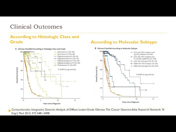
Clinical Outcomes
According to Histologic Class and Grade
According to Molecular Subtype
Comprehensive, Integrative
Clinical Outcomes
According to Histologic Class and Grade
According to Molecular Subtype
Comprehensive, Integrative
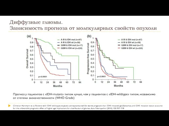
Диффузные глиомы.
Зависимость прогноза от молекулярных свойств опухоли
Прогноз у пациентов с
Диффузные глиомы.
Зависимость прогноза от молекулярных свойств опухоли
Прогноз у пациентов с
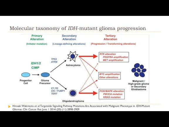
Molecular taxonomy of IDH-mutant glioma progression
Hiroaki Wakimoto et al. Targetable Signaling Pathway Mutations
Molecular taxonomy of IDH-mutant glioma progression
Hiroaki Wakimoto et al. Targetable Signaling Pathway Mutations
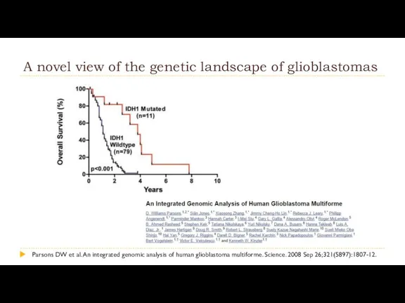
A novel view of the genetic landscape of glioblastomas
Parsons DW et
A novel view of the genetic landscape of glioblastomas
Parsons DW et
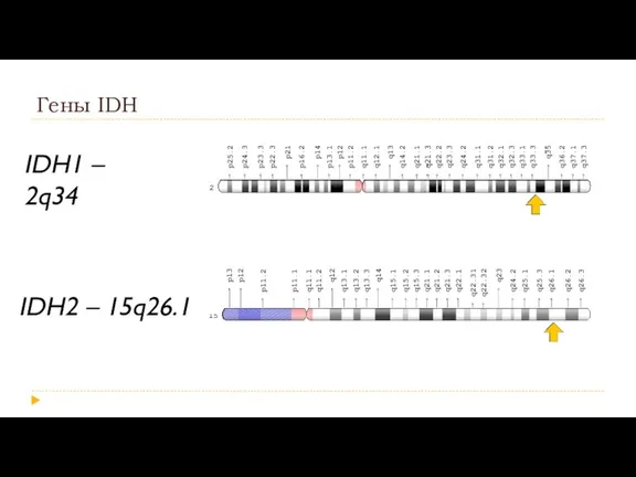
Гены IDH
IDH1 – 2q34
IDH2 – 15q26.1
Гены IDH
IDH1 – 2q34
IDH2 – 15q26.1
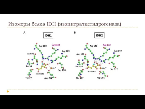
Изомеры белка IDH (изоцитратдегидрогеназа)
Изомеры белка IDH (изоцитратдегидрогеназа)
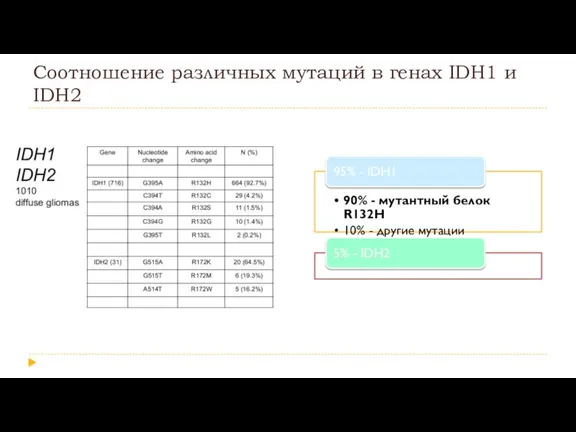
Соотношение различных мутаций в генах IDH1 и IDH2
Соотношение различных мутаций в генах IDH1 и IDH2
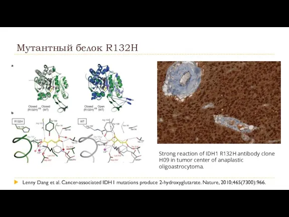
Мутантный белок R132H
Lenny Dang et al. Cancer-associated IDH1 mutations produce 2-hydroxyglutarate.
Мутантный белок R132H
Lenny Dang et al. Cancer-associated IDH1 mutations produce 2-hydroxyglutarate.
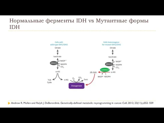
Нормальные ферменты IDH vs Мутантные формы IDH
Andrew R. Mullen and Ralph
Нормальные ферменты IDH vs Мутантные формы IDH
Andrew R. Mullen and Ralph
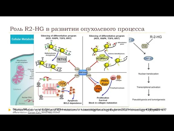
Роль R2-HG в развитии опухолевого процесса
Michael Heuser et al. Enigmas of
Роль R2-HG в развитии опухолевого процесса
Michael Heuser et al. Enigmas of
Diffuse astrocytic and oligodendroglial tumours
Diffuse astrocytoma, IDH-mutant
Gemistocytic astrocytoma, IDH-mutant
Diffuse astrocytic and oligodendroglial tumours
Diffuse astrocytoma, IDH-mutant
Gemistocytic astrocytoma, IDH-mutant
Diffuse astrocytoma WHO Grade II/
Anaplastic astrocytoma WHO Grade III
Diffuse astrocytoma WHO Grade II/
Anaplastic astrocytoma WHO Grade III
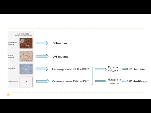
IDH-mutant
IDH-mutant
Секвенирование IDH1 и IDH2
Секвенирование IDH1 и IDH2
Мутация
найдена
IDH-mutant
Мутация не
найдена
IDH-wildtype
IDH-mutant
IDH-mutant
Секвенирование IDH1 и IDH2
Секвенирование IDH1 и IDH2
Мутация
найдена
IDH-mutant
Мутация не
найдена
IDH-wildtype
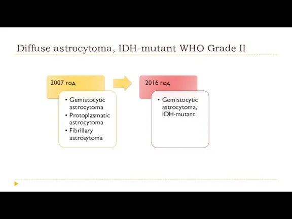
Diffuse astrocytoma, IDH-mutant WHO Grade II
Diffuse astrocytoma, IDH-mutant WHO Grade II
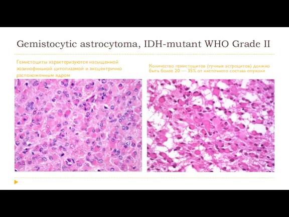
Gemistocytic astrocytoma, IDH-mutant WHO Grade II
Гемистоциты характеризуются насыщенной эозинофильной цитоплазмой и
Gemistocytic astrocytoma, IDH-mutant WHO Grade II
Гемистоциты характеризуются насыщенной эозинофильной цитоплазмой и
Diffuse astrocytic and oligodendroglial tumours
Diffuse astrocytoma, IDH-mutant
Gemistocytic astrocytoma, IDH-mutant
Diffuse astrocytic and oligodendroglial tumours
Diffuse astrocytoma, IDH-mutant
Gemistocytic astrocytoma, IDH-mutant
Сравнительная характеристика глиобластом
Сравнительная характеристика глиобластом
Glioblastoma WHO Grade IV
Glioblastoma WHO Grade IV
Glioblastoma, IDH-wildtype WHO Grade IV
Glioblastoma, IDH-wildtype WHO Grade IV
Epithelioid glioblastoma
В основном, возникает у детей и молодых людей.
В типичных
Epithelioid glioblastoma
В основном, возникает у детей и молодых людей.
В типичных
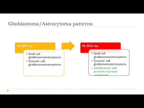
Glioblastoma/Astrocytoma patterns
Glioblastoma/Astrocytoma patterns
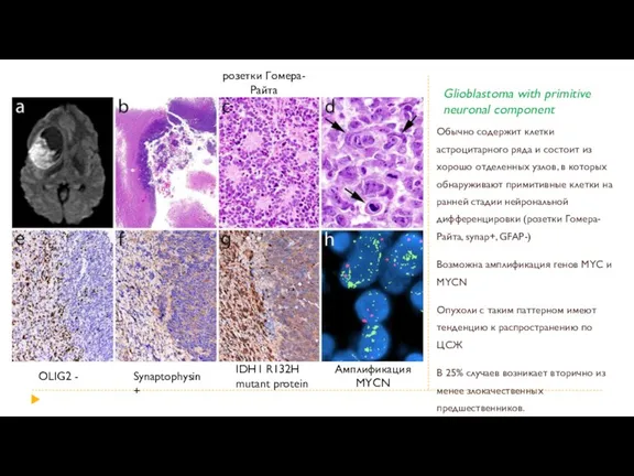
Glioblastoma with primitive neuronal component
Обычно содержит клетки астроцитарного ряда и состоит
Glioblastoma with primitive neuronal component
Обычно содержит клетки астроцитарного ряда и состоит

Diffuse astrocytic and oligodendroglial tumours
Diffuse astrocytoma, IDH-mutant
Gemistocytic astrocytoma, IDH-mutant
Diffuse astrocytic and oligodendroglial tumours
Diffuse astrocytoma, IDH-mutant
Gemistocytic astrocytoma, IDH-mutant
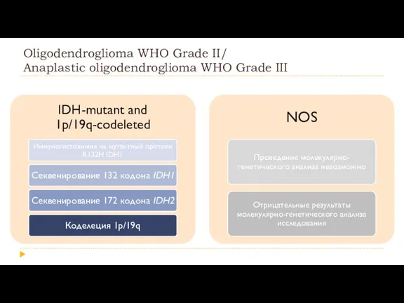
Oligodendroglioma WHO Grade II/
Anaplastic oligodendroglioma WHO Grade III
Oligodendroglioma WHO Grade II/
Anaplastic oligodendroglioma WHO Grade III
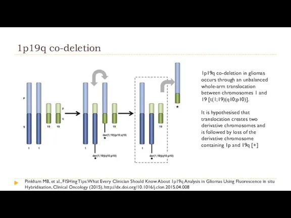
1p19q co-deletion
1p19q co-deletion in gliomas occurs through an unbalanced whole-arm translocation
1p19q co-deletion
1p19q co-deletion in gliomas occurs through an unbalanced whole-arm translocation
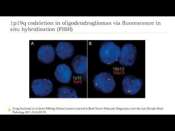
1p19q codeletion in oligodendrogliomas via fluorescence in situ hybridization (FISH)
Craig Horbinski
1p19q codeletion in oligodendrogliomas via fluorescence in situ hybridization (FISH)
Craig Horbinski

Oligodendroglioma WHO Grade II/
Anaplastic oligodendroglioma WHO Grade III
Oligodendroglioma WHO Grade II/
Anaplastic oligodendroglioma WHO Grade III
Diffuse astrocytic and oligodendroglial tumours
Diffuse astrocytoma, IDH-mutant
Gemistocytic astrocytoma, IDH-mutant
Diffuse astrocytic and oligodendroglial tumours
Diffuse astrocytoma, IDH-mutant
Gemistocytic astrocytoma, IDH-mutant
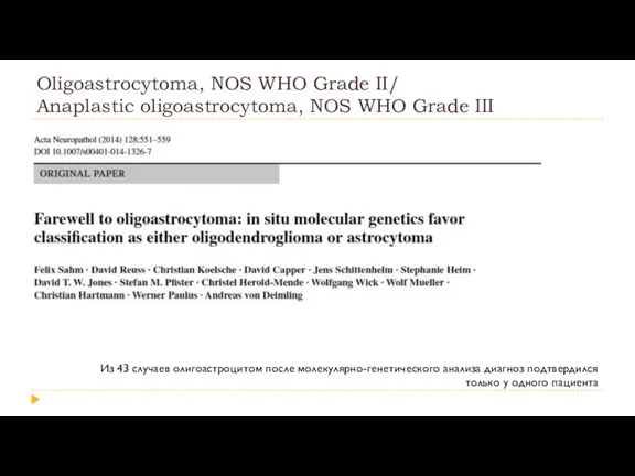
Oligoastrocytoma, NOS WHO Grade II/
Anaplastic oligoastrocytoma, NOS WHO Grade III
Из 43
Oligoastrocytoma, NOS WHO Grade II/
Anaplastic oligoastrocytoma, NOS WHO Grade III
Из 43
Diffuse astrocytic and oligodendroglial tumours
Diffuse astrocytoma, IDH-mutant
Gemistocytic astrocytoma, IDH-mutant
Diffuse astrocytic and oligodendroglial tumours
Diffuse astrocytoma, IDH-mutant
Gemistocytic astrocytoma, IDH-mutant
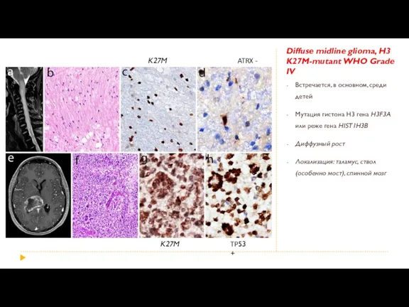
Diffuse midline glioma, H3 K27M-mutant WHO Grade IV
Встречается, в основном, среди
Diffuse midline glioma, H3 K27M-mutant WHO Grade IV
Встречается, в основном, среди
Глиоматоз головного мозга
Исключен из классификации ВОЗ 2016 года как отдельный вид
Глиоматоз головного мозга
Исключен из классификации ВОЗ 2016 года как отдельный вид
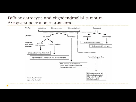
Diffuse astrocytic and oligodendroglial tumours
Алгоритм постановки диагноза.
Diffuse astrocytic and oligodendroglial tumours
Алгоритм постановки диагноза.
Other astrocytic tumours
Pilocytic astrocytoma
Pilomyxoid astrocytoma
Subependymal giant cell astrocytoma
Other astrocytic tumours
Pilocytic astrocytoma
Pilomyxoid astrocytoma
Subependymal giant cell astrocytoma
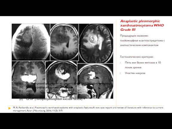
Anaplastic pleomorphic xanthoastrocytoma WHO Grade III
Предыдущее название: плейоморфная ксантоастроцитома с анапластическим
Anaplastic pleomorphic xanthoastrocytoma WHO Grade III
Предыдущее название: плейоморфная ксантоастроцитома с анапластическим
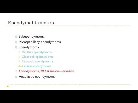
Ependymal tumours
Subependymoma
Myxopapillary ependymoma
Ependymoma
Papillary ependymoma
Clear cell ependymoma
Ependymal tumours
Subependymoma
Myxopapillary ependymoma
Ependymoma
Papillary ependymoma
Clear cell ependymoma
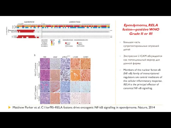
Ependymoma, RELA fusion—positive WHO Grade II or III
Большая часть супратенториальных опухолей
Ependymoma, RELA fusion—positive WHO Grade II or III
Большая часть супратенториальных опухолей
Neuronal and mixed neuronal-glial tumours
Dysembryoplastic neuroepithelial tumour
Gangliocytoma
Ganglioglioma
Anaplastic
Neuronal and mixed neuronal-glial tumours
Dysembryoplastic neuroepithelial tumour
Gangliocytoma
Ganglioglioma
Anaplastic
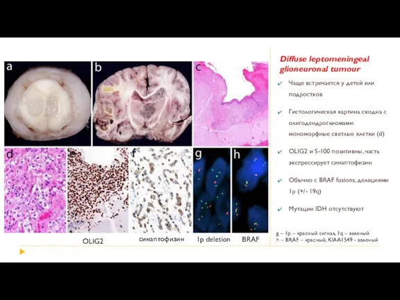
Diffuse leptomeningeal glioneuronal tumour
Чаще встречается у детей или подростков
Гистологическая картина
Diffuse leptomeningeal glioneuronal tumour
Чаще встречается у детей или подростков
Гистологическая картина
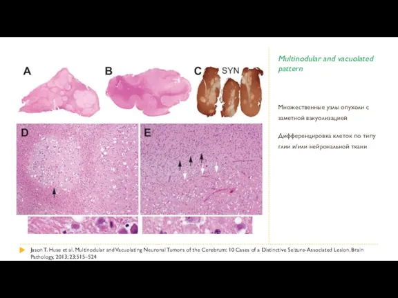
Multinodular and vacuolated pattern
Множественные узлы опухоли с заметной вакуолизацией
Дифференцировка клеток по
Multinodular and vacuolated pattern
Множественные узлы опухоли с заметной вакуолизацией
Дифференцировка клеток по
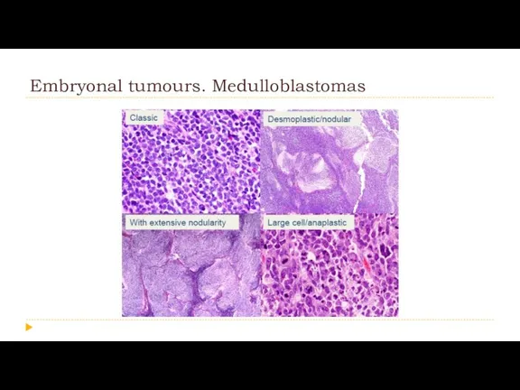
Embryonal tumours. Medulloblastomas
Embryonal tumours. Medulloblastomas
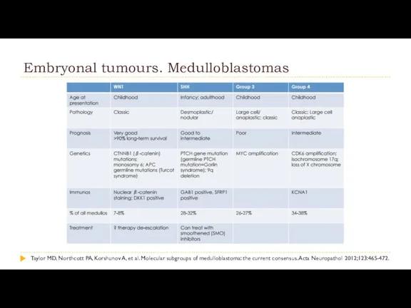
Embryonal tumours. Medulloblastomas
Taylor MD, Northcott PA, Korshunov A, et al. Molecular
Embryonal tumours. Medulloblastomas
Taylor MD, Northcott PA, Korshunov A, et al. Molecular
Embryonal tumours. Medulloblastomas
Embryonal tumours. Medulloblastomas
Корреляция между генетическим профилем, морфологической картиной и прогнозом для пациента.
Корреляция между генетическим профилем, морфологической картиной и прогнозом для пациента.
Other Embryonal tumours
Embryonal tumour with multilayered rosettes, C19MC-aItered
Embryonal tumour with
Other Embryonal tumours
Embryonal tumour with multilayered rosettes, C19MC-aItered
Embryonal tumour with
Embryonal tumour with multilayered rosettes
R Neelima et al. Embryonal tumor with
Embryonal tumour with multilayered rosettes
R Neelima et al. Embryonal tumor with

Embryonal tumour with multilayered rosettes
Embryonal tumour with multilayered rosettes
Other Embryonal tumours
Embryonal tumour with multilayered rosettes, C19MC-aItered
Embryonal tumour with
Other Embryonal tumours
Embryonal tumour with multilayered rosettes, C19MC-aItered
Embryonal tumour with
Atypical teratoid/rhabdoid tumour
CNS embryonal tumour with rhabdoid features
AK Taneja et
Atypical teratoid/rhabdoid tumour
CNS embryonal tumour with rhabdoid features
AK Taneja et
Atypical teratoid/rhabdoid tumour
CNS embryonal tumour with rhabdoid features
Atypical teratoid/rhabdoid tumour
CNS embryonal tumour with rhabdoid features

Tumours of the cranial and paraspinal nerves
Schwannoma
Cellular schwannoma
Plexiform
Tumours of the cranial and paraspinal nerves
Schwannoma
Cellular schwannoma
Plexiform
Melanotic schwannoma
Встречается редко, в литературе описано около 20 случаев.
Чаще всего
Melanotic schwannoma
Встречается редко, в литературе описано около 20 случаев.
Чаще всего
Tumours of the cranial and paraspinal nerves
Schwannoma
Cellular schwannoma
Plexiform
Tumours of the cranial and paraspinal nerves
Schwannoma
Cellular schwannoma
Plexiform
Hybrid nerve sheath tumours
Представляет собой не один вид, а скорее целую
Hybrid nerve sheath tumours
Представляет собой не один вид, а скорее целую

Tumours of the cranial and paraspinal nerves
Schwannoma
Cellular schwannoma
Plexiform
Tumours of the cranial and paraspinal nerves
Schwannoma
Cellular schwannoma
Plexiform
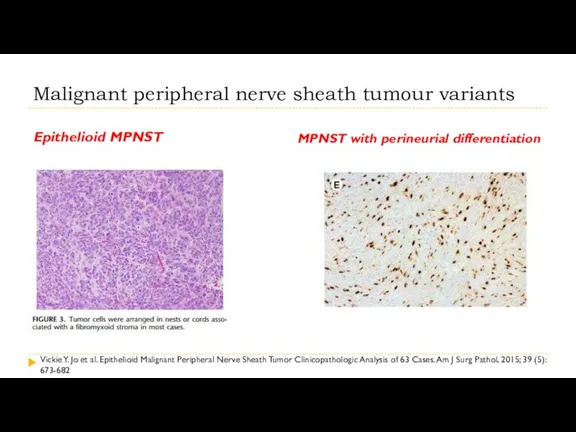
Malignant peripheral nerve sheath tumour variants
Epithelioid MPNST
MPNST with perineurial differentiation
Vickie
Malignant peripheral nerve sheath tumour variants
Epithelioid MPNST
MPNST with perineurial differentiation
Vickie
Meningiomas
Meningioma
Meningothelial meningioma
Fibrous meningioma
Transitional meningioma
Psammomatous meningioma
Angiomatous meningioma
Meningiomas
Meningioma
Meningothelial meningioma
Fibrous meningioma
Transitional meningioma
Psammomatous meningioma
Angiomatous meningioma
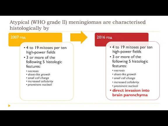
Atypical (WHO grade II) meningiomas are characterised histologically by
Atypical (WHO grade II) meningiomas are characterised histologically by
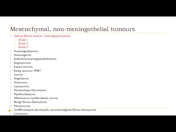
Mesenchymal, non-meningothelial tumours
Solitary fibrous turnour / haemangiopericytoma
Grade 1
Grade
Mesenchymal, non-meningothelial tumours
Solitary fibrous turnour / haemangiopericytoma
Grade 1
Grade
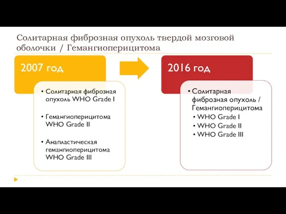
Солитарная фиброзная опухоль твердой мозговой оболочки / Гемангиоперицитома
Солитарная фиброзная опухоль твердой мозговой оболочки / Гемангиоперицитома
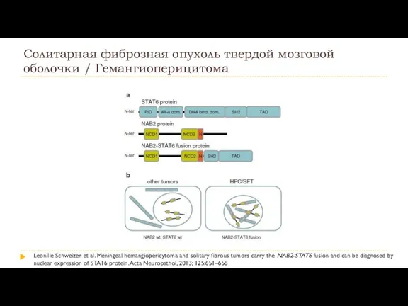
Солитарная фиброзная опухоль твердой мозговой оболочки / Гемангиоперицитома
Leonille Schweizer et al. Meningeal
Солитарная фиброзная опухоль твердой мозговой оболочки / Гемангиоперицитома
Leonille Schweizer et al. Meningeal
Солитарная фиброзная опухоль твердой мозговой оболочки / Гемангиоперицитома
NAB2
STAT6
HPC
SFT
HPC
SFT
Leonille Schweizer et
Солитарная фиброзная опухоль твердой мозговой оболочки / Гемангиоперицитома
NAB2
STAT6
HPC
SFT
HPC
SFT
Leonille Schweizer et
Ключевые моменты
1. Мы перешли в эру постановки диагноза с помощью молекулярной
Ключевые моменты
1. Мы перешли в эру постановки диагноза с помощью молекулярной

 Патология эмоциональной сферы. Расстройства влечений. Патология двигательной и волевой сферы
 Выделительная система
 Ауыз қуыс кілегей қабық ауруларына тағайындалатын дәрілік терпияның салыстырмалы сипаттамасы
 Дисциркуляторная энцефалопатия и болезнь мелких сосудов
 Жүкті әйелдердегі ауруханадан тыс пневмония
 Анафилаксикалық шок кезіндегі жедел көмек
 Рак тела матки
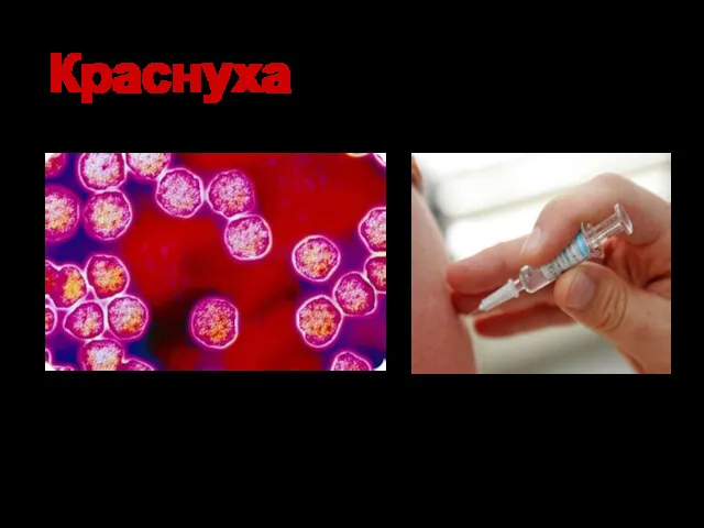 Краснуха - острая вирусная антропонозная инфекция
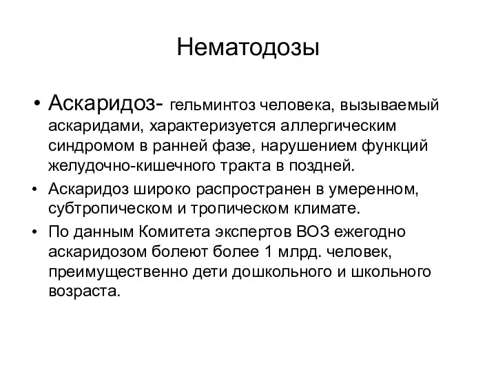 Нематодозы. Аскаридоз
 Аномалии количества зубов
 Операции на органах шеи
 Методы диагностики гортани
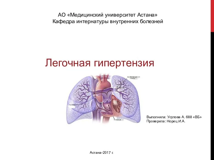 Легочная гипертензия
 Сестринский уход за пациентами с хронической сердечной недостаточностью
 Лечение открытых переломов
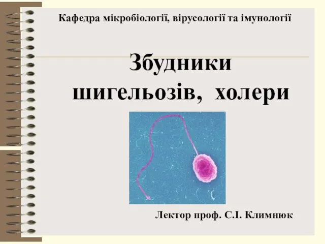 Збудники шигельозів, холери
 Роль гормонов в регуляции роста, развития и гомеостаза
 Тыныс алу ағзаларының аурулары
 Тері физиологиясы
 Бустеры Expert Boost. Уход за кожей
 Таным туралы ілім. Медициналық, ғылыми танымның ерекшелігі
 Placental abruption
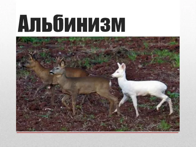 Альбинизм. Типы альбинизма
 Современные аспекты сердечно-легочной и церебральной реанимации в стоматологической практике
 Факторы риска при инфакте миокарда в развитии низкой физической активности найти информацию научных статей
 Балалардағы бұлшық ет жүйесінің анатомо-физиологиялық ерекшеліктері
 Профилактическая медицина
 Аккредитация медицинских работников январь 2023