- Клеточное строение костного мозга

Содержание
- 2. Все клетки, которые можно встретить в костном мозге в норме и при патологии можно разделить на
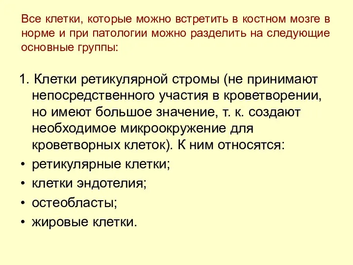
- 3. Ретикулярные клетки Reticular cells, polychromatic NRBCs, normal marrow Two reticular cells (center), 4 lymphocytes, 5 polychromatic
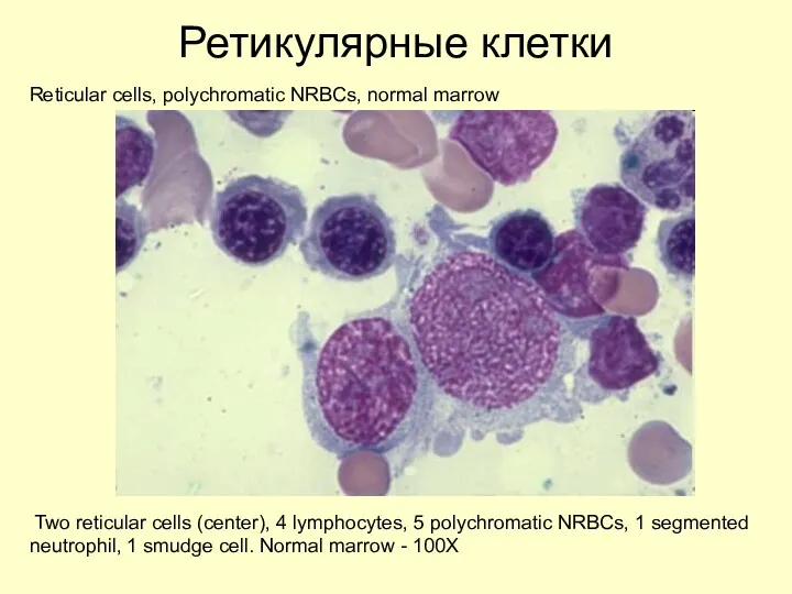
- 4. Ретикулярные клетки Reticular cell, normal marrow One reticular cell. Normal marrow - 100X
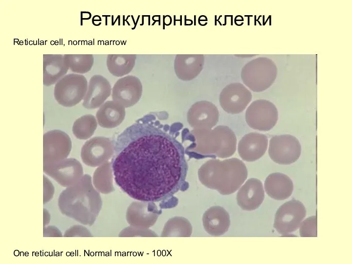
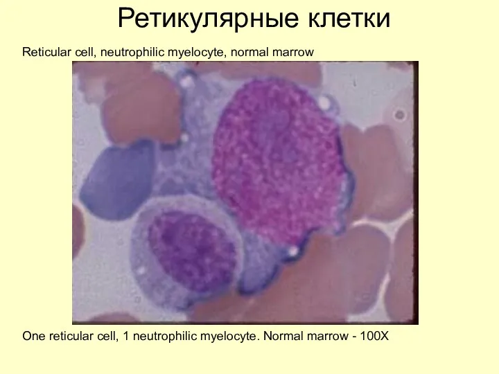
- 5. Ретикулярные клетки Reticular cell, neutrophilic myelocyte, normal marrow One reticular cell, 1 neutrophilic myelocyte. Normal marrow
- 6. Клетки эндотелия Endothelial cells. Strand of endothelial cells. Normal marrow -20X
- 7. Клетки эндотелия Endothelial cells Higher magnification of strand of endothelial cells. Normal marrow - 100X
- 8. Остеобласты Osteoblasts Clump of osteoblasts. Normal marrow particle preparation -50X Osteoblast are large in size, often
- 9. Остеобласты Osteoblasts, Normal marrow Same clump of osteoblasts. Normal marrow particle preparation -100X
- 10. Остеобласты Рlasma cell, osteoblasts One small and 1 large plasma cell in top left frame. 2
- 11. Жировые клетки Fat cells. Aplastic anemia marrow Multiple fat cells. Aplastic anemia marrow -20X
- 12. Жировые клетки Fat cells, Aplastic anemia marrow Two very large fat cells. Aplastic anemia marrow -50X
- 13. 2. Миелокариоциты - клетки кроветворной ткани костного мозга с их производными - зрелыми клетками крови. К
- 14. Недифференцированные бласты
- 15. Миелобласт Myeloblast, late neutrophil, normal marrow One myeloblast, 1 late neutrophil. Normal marrow - 100X
- 16. Миелобласт Myeloblast, neutrophilic metamyelocyte, normal marrow One myeloblast, 1 neutrophilic metamyelocyte. Normal marrow - 100X
- 17. Миелобласт Myeloblast, late NRBC, normal marrow One myeloblast / monoblast, 1 late NRBC. Normal marrow -
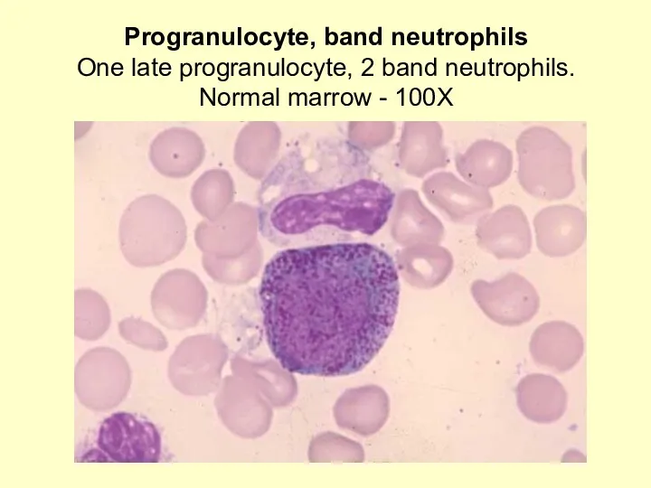
- 18. Progranulocyte, band neutrophils One late progranulocyte, 2 band neutrophils. Normal marrow - 100X
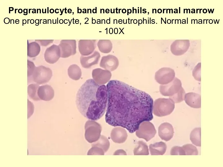
- 19. Progranulocyte, band neutrophils, normal marrow One progranulocyte, 2 band neutrophils. Normal marrow - 100X
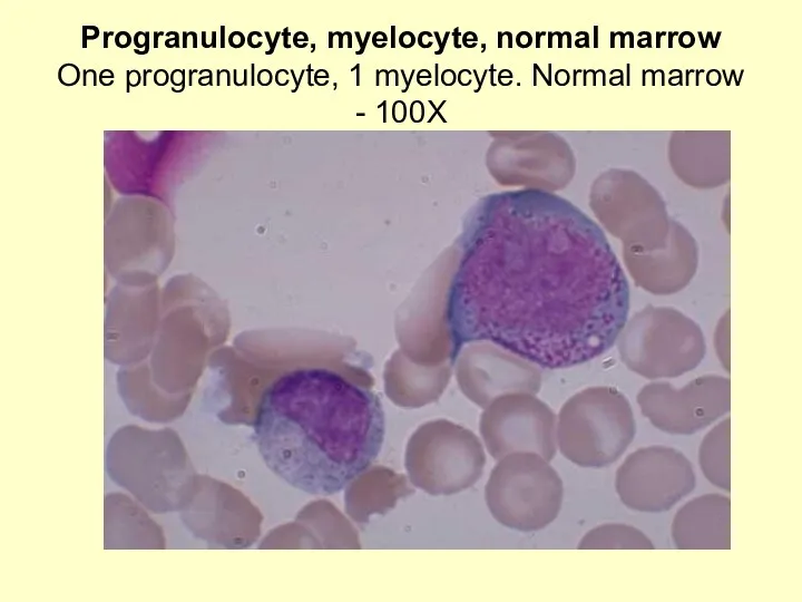
- 20. Progranulocyte, myelocyte, normal marrow One progranulocyte, 1 myelocyte. Normal marrow - 100X
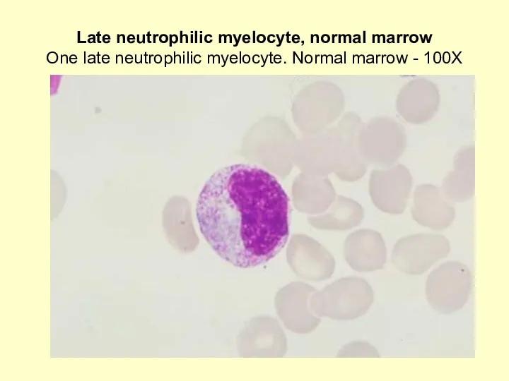
- 21. Late neutrophilic myelocyte, normal marrow One late neutrophilic myelocyte. Normal marrow - 100X
- 22. Neutrophilic myelocyte, normal marrow One neutrophilic myelocyte. Normal marrow - 100X
- 23. Myelocytes, abnormal platelets, AML blood Two myelocytes, one with and the other without primary or coarse
- 24. Myelocyte, disrupted band neutrophil One early myelocyte with many azure granules, 1 disrupted band neutrophil. Normal
- 25. Eosinophilic myelocyte, Normal marrow Left frame: 1 normal eosinophilic myelocyte. Center frame: 1 eosinophilic myelocyte with
- 26. Eosinophilic myelocyte, neutrophilic myelocyte One eosinophilic myelocyte (lower left), 1 neutrophilic myelocyte, 1 segmented neutrophil, 1
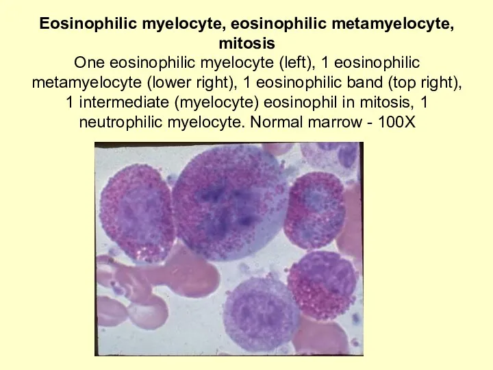
- 27. Eosinophilic myelocyte, eosinophilic metamyelocyte, mitosis One eosinophilic myelocyte (left), 1 eosinophilic metamyelocyte (lower right), 1 eosinophilic
- 28. Eosinophilic myelocyte, neutrophilic myelocytes One eosinophilic myelocyte (left), 2 neutrophilic myelocytes. Normal marrow - 100X
- 29. Metamyelocyte, normal marrow One metamyelocyte - late. Normal marrow - 100X
- 30. Metamyelocyte, smudge cell, normal marrow One metamyelocyte, 1 smudge cell. Normal marrow - 100X
- 31. Metamyelocyte, smudge cell, normal marrow One metamyelocyte - early, 1 smudge cell. Normal marrow - 100X
- 32. Metamyelocyte, neutrophilic myelocyte, normal marrow One metamyelocyte - early, 1 neutrophilic myelocyte. Normal marrow - 100X
- 33. Eosinophilic myelocyte, eosinophilic metamyelocyte, mitosis One eosinophilic myelocyte (left), 1 eosinophilic metamyelocyte (lower right), 1 eosinophilic
- 34. Band neutrophils, monocyte Three band neutrophils, 1 monocyte (top left center) Note differences in chromatin and
- 35. Band neutrophils Three band neutrophils - 100X
- 36. Band eosinophil, mature neutrophil, Normal blood One band eosinophil, 1 mature neutrophil. Normal blood - 100X
- 37. Mature Neutrophils, Platelet Satellitism One huge clump of nine Mature Neutrophils, each of which is surrounded
- 38. Young megakaryocyte, neutrophilic myelocytes, normal marrow Young megakaryocyte showing early granulation adjacent to lobulated nuclei. 1
- 39. Segmented neutrophils, band neutrophils, normal blood Segmented neutrophils and band neutrophils composite; 2 band neutrophils (top
- 40. Eosinophil, Normal blood One 3-lobed eosinophil, 1 2-lobed eosinophil, 2 mature neutrophils; composite. Normal blood -
- 41. Basophils, mature neutrophils, CML blood Three mature basophils, 2 mature neutrophils. Chronic myeloid leukemia blood -
- 42. Basophil One mature basophil. Normal blood - 100X
- 43. Монобласт
- 44. Промоноцит
- 45. Young monocyte, normal blood One young monocyte with immature chromatin and nucleoli. Normal blood - 100X
- 46. Immature Monocytes, cytoplasmic fragmentation, M-5 Two Immature Monocytes, one of which shows cytoplasmic fragmentation. These fragments
- 47. Young monocytes, normal blood Two young monocytes, 1 with many vacuoles. Normal blood - 100X
- 48. Macrophage, mature megakaryocyte, normal marrow A macrophage with an oval shaped nucleus and a highly vacuolated
- 49. Osteoclast One osteoclast with 12 nuclei. Normal marrow -100X
- 50. Very small lymphocyte, monocyte, normal blood One very small lymphocyte, 1 monocyte. Normal blood - 50X
- 51. Lymphocytes, normal blood Two lymphocytes (1 with granules); Normal blood - 100X
- 52. Large reactive lymphocyte, normal blood One very large lymphocyte (reactive); Normal blood - 100X
- 53. Monocyte, large lymphocyte, normal blood One monocyte (left), 1 large lymphocyte. Normal blood - 100X
- 54. КЛЕТКИ ЛЕЙКОЛИЗА
- 55. Plasmacytoid lymphoid, infectious mononucleosis blood One small mature lymphocyte, 1 Plasmacytoid Lymphoid with a contorted nucleus.
- 56. Plasmacytoid lymphocyte, severe arthritis with osteoporosis Left frame: 1 Plasmacytoid Lymphocyte with a double nucleus, 1
- 57. Monocyte, Plasmacytoid Lymphocyte, drug reaction buffy coat One monocyte, 1 Plasmacytoid Lymphocyte and 1 eosinophil. Drug
- 58. Plasma cell, alcoholic, liver disease One monocyte at left edge and one Plasma Cell at right
- 59. Plasma Cell One large Plasma Cell, oval in shape, with an eccentrically located nucleus, a very
- 60. ЭРИТРОБЛАСТ
- 61. Plasma cells, proerythroblast, NRBC Two Plasma Cells with low nuclear/cytoplasmic ratio, deep basophilic blue cytoplasm, distinct
- 62. Plasma cell, proerythroblast One Plasma Cell and 1 Proerythroblast. Early erythroblasts have a deep blue cytoplasm,
- 63. Mature lymphocyte, proerythroblast, basophilic erythroblast
- 64. Late NRBC, Mature Lymphocyte, AML One Late NRBC at upper left and one Mature Lymphocyte at
- 65. Blasts, polychromatic nrbcs, acute megakaryocytic leukemia Two Blasts of medium size with fine nuclear chromatin, prominent
- 66. Binucleated Plasma Cell, orthochromatic NRBC One Binucleated Plasma Cell. Compare its nuclear chromatin, which is typical
- 67. РЕТИКУЛОЦИТЫ
- 68. Эритроцит
- 69. Megakaryoblast , fold in nucleus, normal marrow One megakaryoblast with an apparent fold in the nucleus.
- 70. Promegakaryocyte with nuclear separation, normal marrow One promegakaryocyte with nuclear separation and some nuclear lobulation. Normal
- 71. Abnormal megakaryocyte, Wright's-Giemsa stain, AML marrow Mature Abnormal Megakaryocyte with two apparent nuclei; one is very
- 72. Megakaryocyte and bare nuclei, megakaryocytic leukemia Top left frame: Small mature megakaryocyte with lobulated nuclei and
- 74. Скачать презентацию
Все клетки, которые можно встретить в костном мозге в норме и
Все клетки, которые можно встретить в костном мозге в норме и
Ретикулярные клетки
Reticular cells, polychromatic NRBCs, normal marrow
Two reticular cells (center), 4
Ретикулярные клетки
Reticular cells, polychromatic NRBCs, normal marrow
Two reticular cells (center), 4
Ретикулярные клетки
Reticular cell, normal marrow
One reticular cell. Normal marrow - 100X
Ретикулярные клетки
Reticular cell, normal marrow
One reticular cell. Normal marrow - 100X
Ретикулярные клетки
Reticular cell, neutrophilic myelocyte, normal marrow
One reticular cell, 1 neutrophilic
Ретикулярные клетки
Reticular cell, neutrophilic myelocyte, normal marrow
One reticular cell, 1 neutrophilic
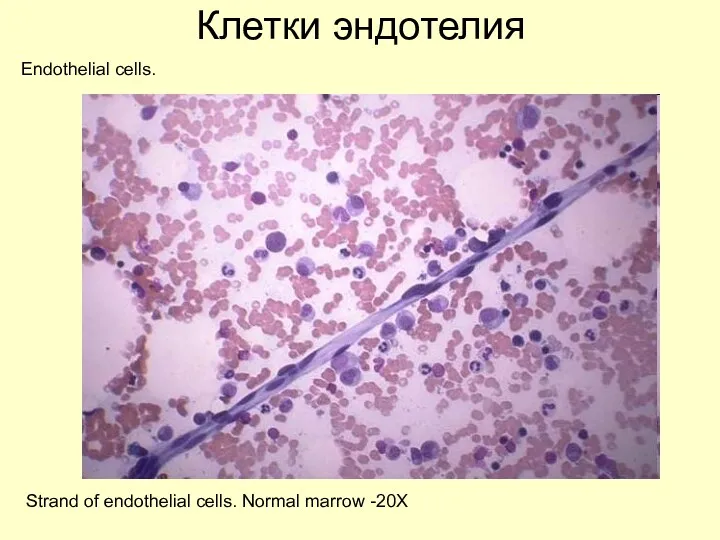
Клетки эндотелия
Endothelial cells.
Strand of endothelial cells. Normal marrow -20X
Клетки эндотелия
Endothelial cells.
Strand of endothelial cells. Normal marrow -20X
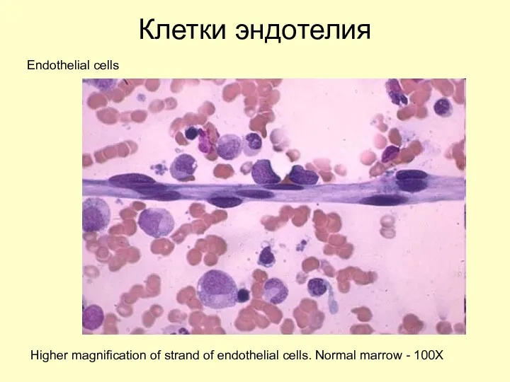
Клетки эндотелия
Endothelial cells
Higher magnification of strand of endothelial cells. Normal marrow
Клетки эндотелия
Endothelial cells
Higher magnification of strand of endothelial cells. Normal marrow
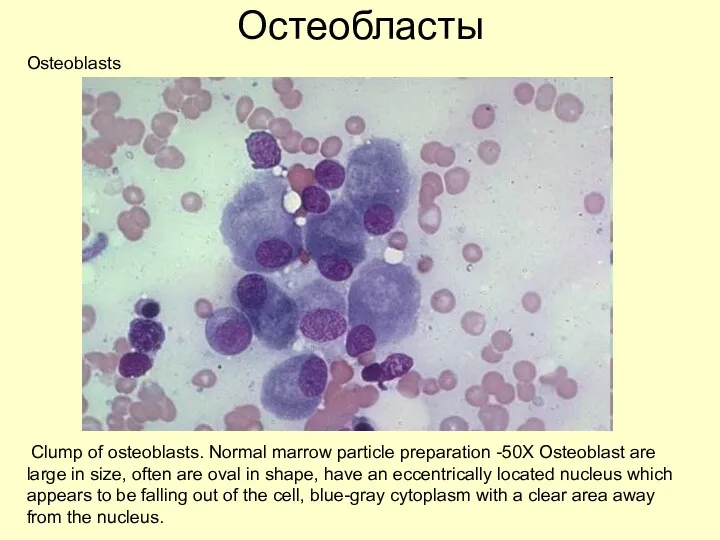
Остеобласты
Osteoblasts
Clump of osteoblasts. Normal marrow particle preparation -50X Osteoblast are large
Остеобласты
Osteoblasts
Clump of osteoblasts. Normal marrow particle preparation -50X Osteoblast are large
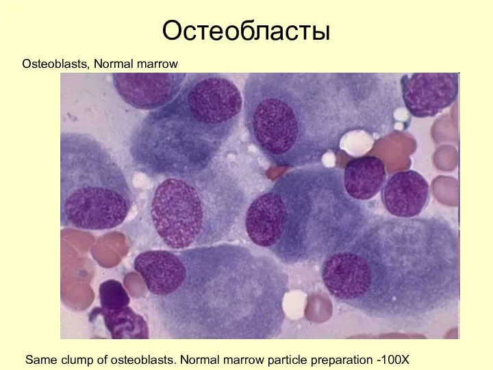
Остеобласты
Osteoblasts, Normal marrow
Same clump of osteoblasts. Normal marrow particle preparation -100X
Остеобласты
Osteoblasts, Normal marrow
Same clump of osteoblasts. Normal marrow particle preparation -100X
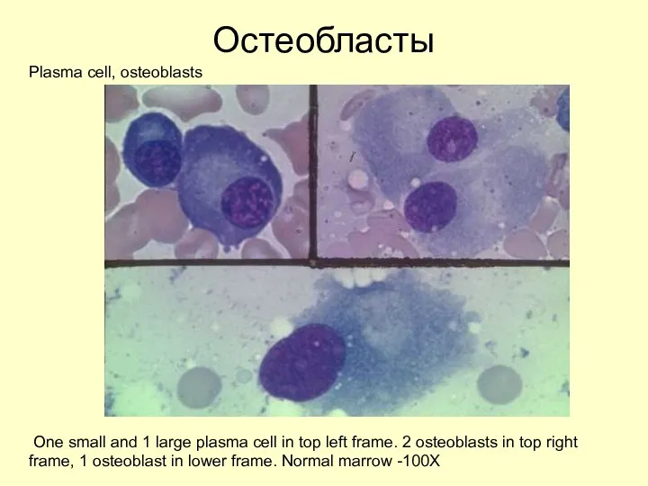
Остеобласты
Рlasma cell, osteoblasts
One small and 1 large plasma cell in top
Остеобласты
Рlasma cell, osteoblasts
One small and 1 large plasma cell in top
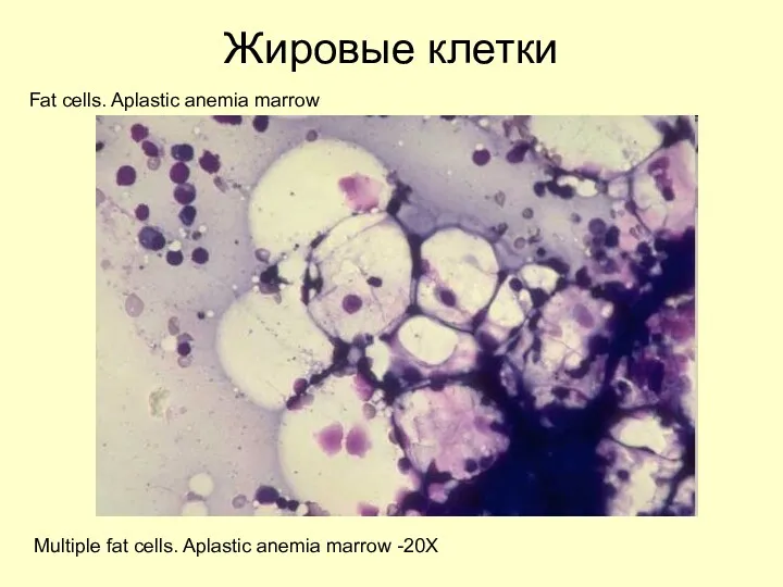
Жировые клетки
Fat cells. Aplastic anemia marrow
Multiple fat cells. Aplastic anemia marrow
Жировые клетки
Fat cells. Aplastic anemia marrow
Multiple fat cells. Aplastic anemia marrow
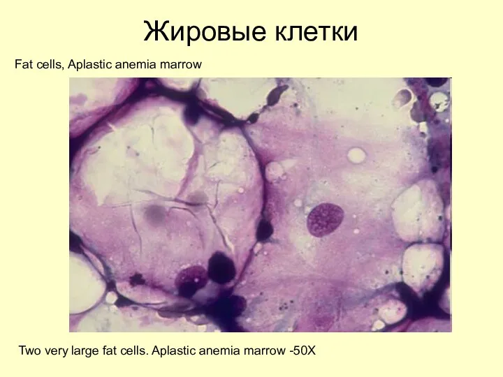
Жировые клетки
Fat cells, Aplastic anemia marrow
Two very large fat cells. Aplastic
Жировые клетки
Fat cells, Aplastic anemia marrow
Two very large fat cells. Aplastic
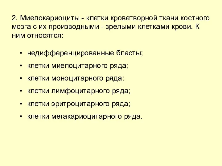
2. Миелокариоциты - клетки кроветворной ткани костного мозга с их производными
2. Миелокариоциты - клетки кроветворной ткани костного мозга с их производными
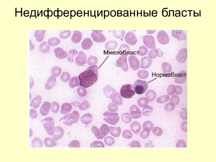
Недифференцированные бласты
Недифференцированные бласты
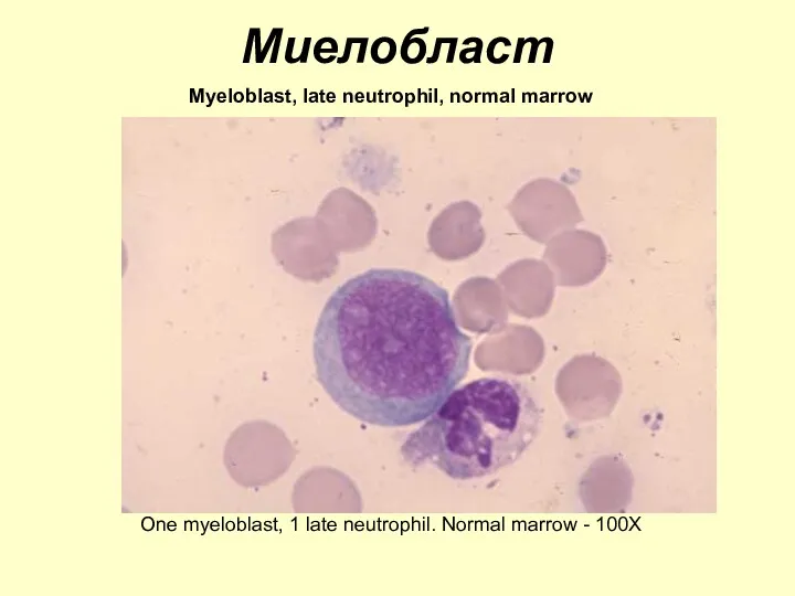
Миелобласт
Myeloblast, late neutrophil, normal marrow
One myeloblast, 1 late neutrophil. Normal marrow
Миелобласт
Myeloblast, late neutrophil, normal marrow
One myeloblast, 1 late neutrophil. Normal marrow
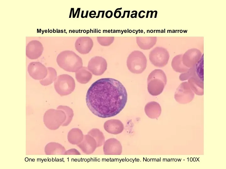
Миелобласт
Myeloblast, neutrophilic metamyelocyte, normal marrow
One myeloblast, 1 neutrophilic metamyelocyte. Normal marrow
Миелобласт
Myeloblast, neutrophilic metamyelocyte, normal marrow
One myeloblast, 1 neutrophilic metamyelocyte. Normal marrow
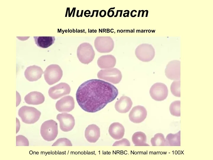
Миелобласт
Myeloblast, late NRBC, normal marrow
One myeloblast / monoblast, 1 late NRBC.
Миелобласт
Myeloblast, late NRBC, normal marrow
One myeloblast / monoblast, 1 late NRBC.
Progranulocyte, band neutrophils
One late progranulocyte, 2 band neutrophils. Normal marrow -
Progranulocyte, band neutrophils One late progranulocyte, 2 band neutrophils. Normal marrow -
Progranulocyte, band neutrophils, normal marrow
One progranulocyte, 2 band neutrophils. Normal marrow
Progranulocyte, band neutrophils, normal marrow One progranulocyte, 2 band neutrophils. Normal marrow
Progranulocyte, myelocyte, normal marrow
One progranulocyte, 1 myelocyte. Normal marrow - 100X
Progranulocyte, myelocyte, normal marrow
One progranulocyte, 1 myelocyte. Normal marrow - 100X
Late neutrophilic myelocyte, normal marrow
One late neutrophilic myelocyte. Normal marrow -
Late neutrophilic myelocyte, normal marrow One late neutrophilic myelocyte. Normal marrow -
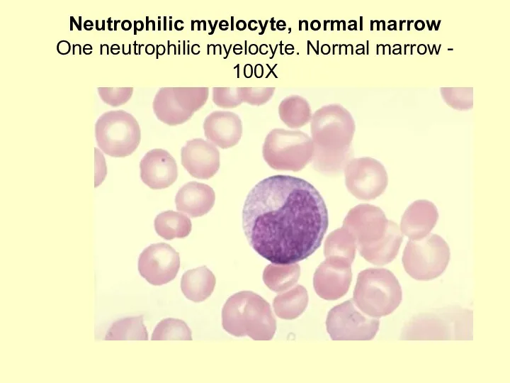
Neutrophilic myelocyte, normal marrow
One neutrophilic myelocyte. Normal marrow - 100X
Neutrophilic myelocyte, normal marrow
One neutrophilic myelocyte. Normal marrow - 100X
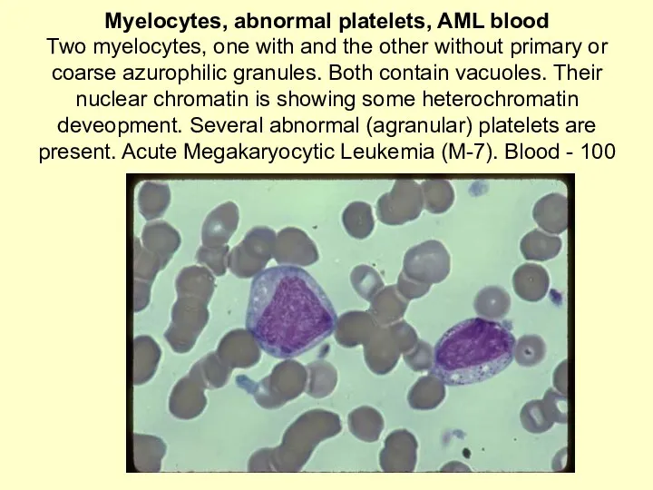
Myelocytes, abnormal platelets, AML blood
Two myelocytes, one with and the other
Myelocytes, abnormal platelets, AML blood Two myelocytes, one with and the other
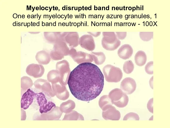
Myelocyte, disrupted band neutrophil
One early myelocyte with many azure granules, 1
Myelocyte, disrupted band neutrophil One early myelocyte with many azure granules, 1
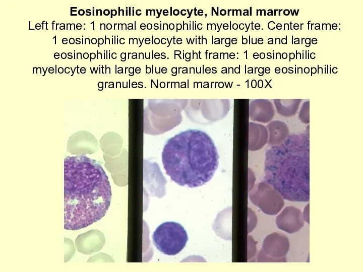
Eosinophilic myelocyte, Normal marrow
Left frame: 1 normal eosinophilic myelocyte. Center frame:
Eosinophilic myelocyte, Normal marrow Left frame: 1 normal eosinophilic myelocyte. Center frame:
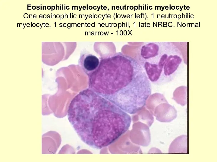
Eosinophilic myelocyte, neutrophilic myelocyte
One eosinophilic myelocyte (lower left), 1 neutrophilic myelocyte,
Eosinophilic myelocyte, neutrophilic myelocyte One eosinophilic myelocyte (lower left), 1 neutrophilic myelocyte,
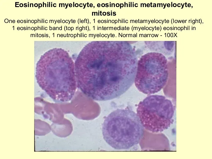
Eosinophilic myelocyte, eosinophilic metamyelocyte, mitosis
One eosinophilic myelocyte (left), 1 eosinophilic metamyelocyte
Eosinophilic myelocyte, eosinophilic metamyelocyte, mitosis One eosinophilic myelocyte (left), 1 eosinophilic metamyelocyte
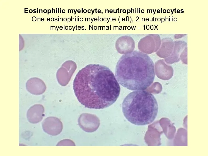
Eosinophilic myelocyte, neutrophilic myelocytes
One eosinophilic myelocyte (left), 2 neutrophilic myelocytes. Normal
Eosinophilic myelocyte, neutrophilic myelocytes One eosinophilic myelocyte (left), 2 neutrophilic myelocytes. Normal
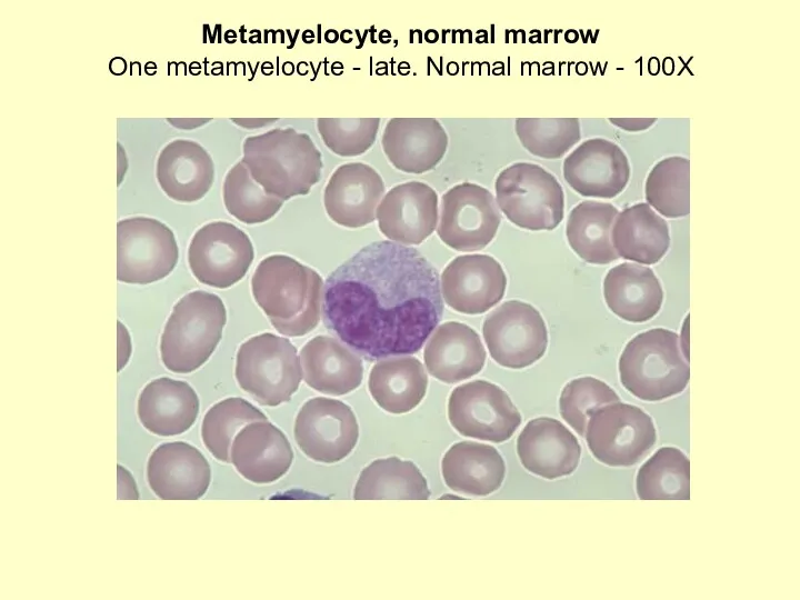
Metamyelocyte, normal marrow
One metamyelocyte - late. Normal marrow - 100X
Metamyelocyte, normal marrow
One metamyelocyte - late. Normal marrow - 100X
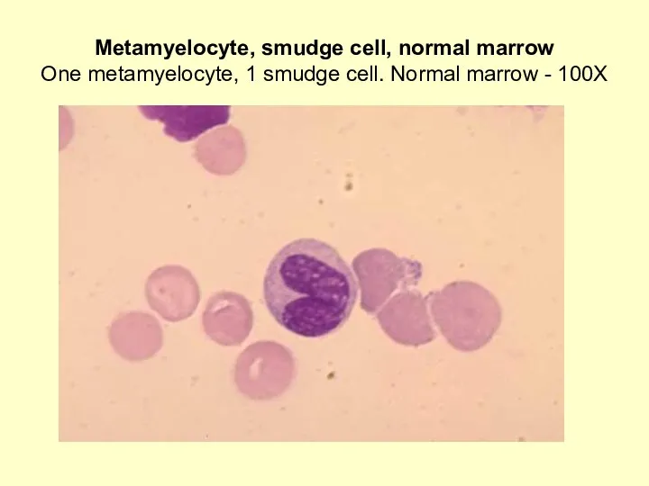
Metamyelocyte, smudge cell, normal marrow
One metamyelocyte, 1 smudge cell. Normal marrow
Metamyelocyte, smudge cell, normal marrow One metamyelocyte, 1 smudge cell. Normal marrow
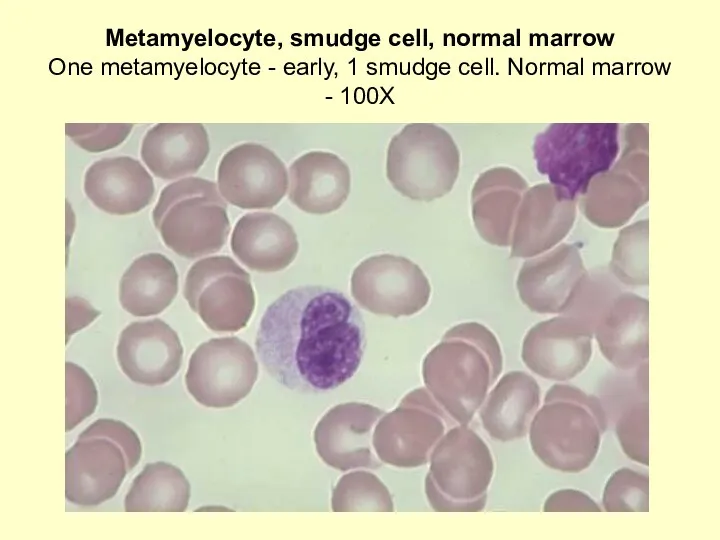
Metamyelocyte, smudge cell, normal marrow
One metamyelocyte - early, 1 smudge cell.
Metamyelocyte, smudge cell, normal marrow One metamyelocyte - early, 1 smudge cell.
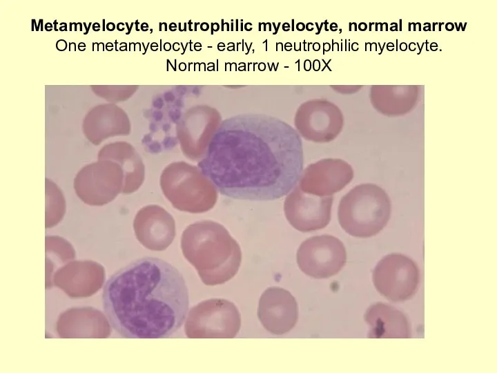
Metamyelocyte, neutrophilic myelocyte, normal marrow
One metamyelocyte - early, 1 neutrophilic myelocyte.
Metamyelocyte, neutrophilic myelocyte, normal marrow One metamyelocyte - early, 1 neutrophilic myelocyte.
Eosinophilic myelocyte, eosinophilic metamyelocyte, mitosis
One eosinophilic myelocyte (left), 1 eosinophilic metamyelocyte
Eosinophilic myelocyte, eosinophilic metamyelocyte, mitosis One eosinophilic myelocyte (left), 1 eosinophilic metamyelocyte
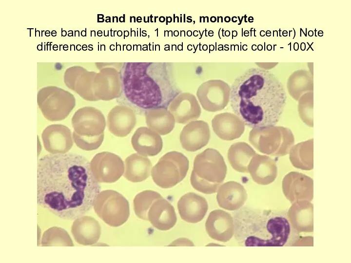
Band neutrophils, monocyte
Three band neutrophils, 1 monocyte (top left center) Note
Band neutrophils, monocyte Three band neutrophils, 1 monocyte (top left center) Note
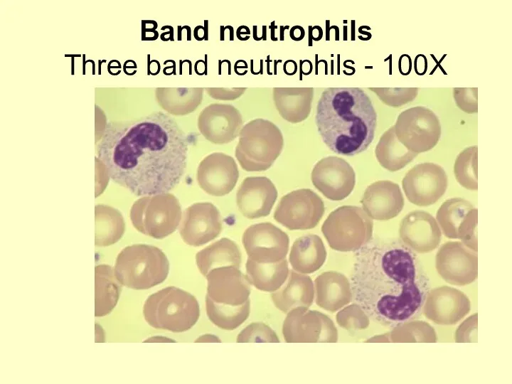
Band neutrophils
Three band neutrophils - 100X
Band neutrophils
Three band neutrophils - 100X
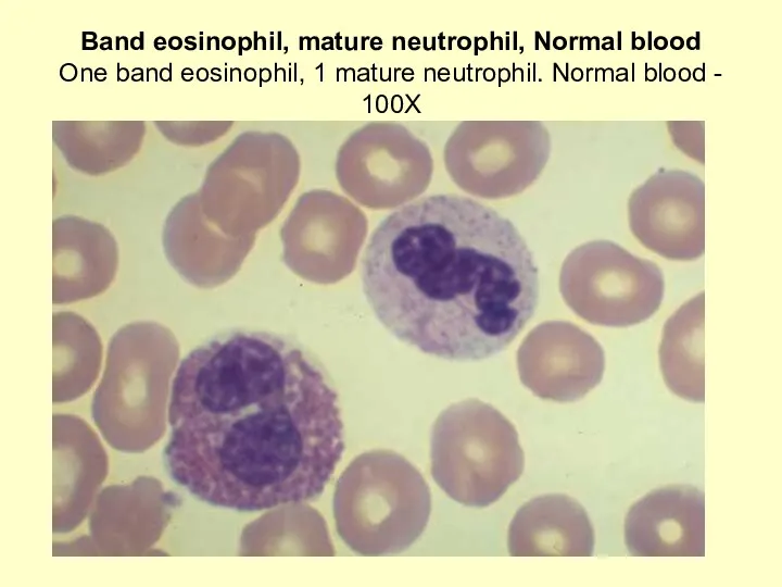
Band eosinophil, mature neutrophil, Normal blood
One band eosinophil, 1 mature neutrophil.
Band eosinophil, mature neutrophil, Normal blood One band eosinophil, 1 mature neutrophil.
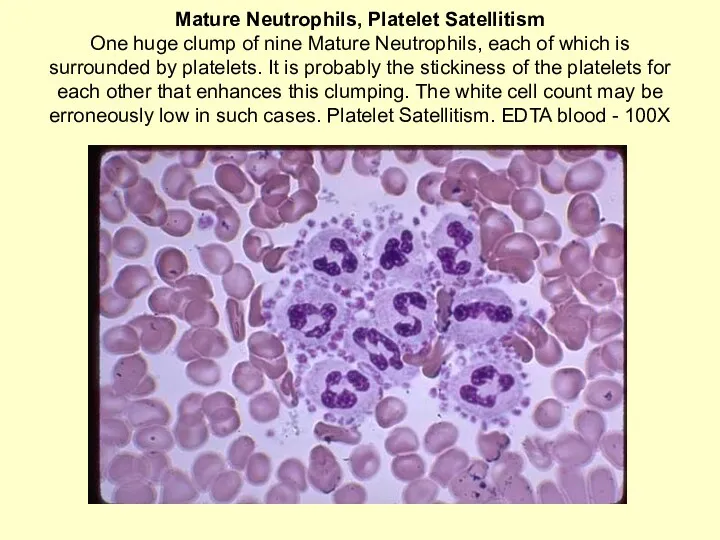
Mature Neutrophils, Platelet Satellitism
One huge clump of nine Mature Neutrophils, each
Mature Neutrophils, Platelet Satellitism One huge clump of nine Mature Neutrophils, each
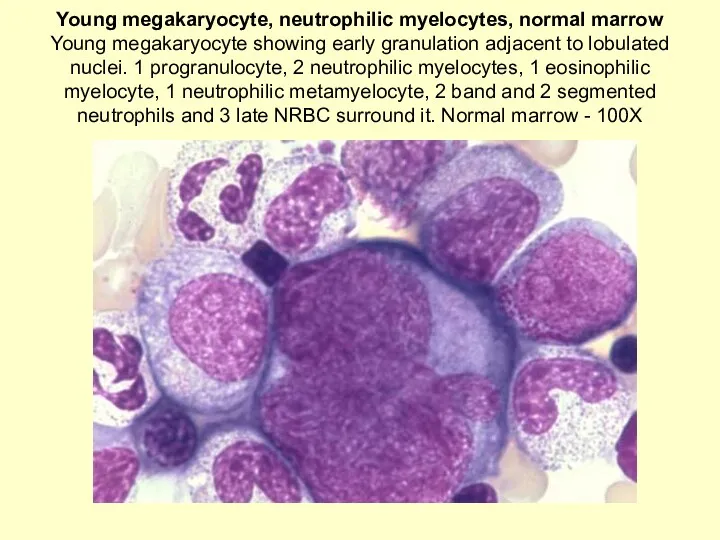
Young megakaryocyte, neutrophilic myelocytes, normal marrow
Young megakaryocyte showing early granulation adjacent
Young megakaryocyte, neutrophilic myelocytes, normal marrow Young megakaryocyte showing early granulation adjacent
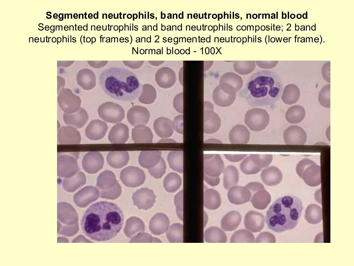
Segmented neutrophils, band neutrophils, normal blood
Segmented neutrophils and band neutrophils composite;
Segmented neutrophils, band neutrophils, normal blood Segmented neutrophils and band neutrophils composite;
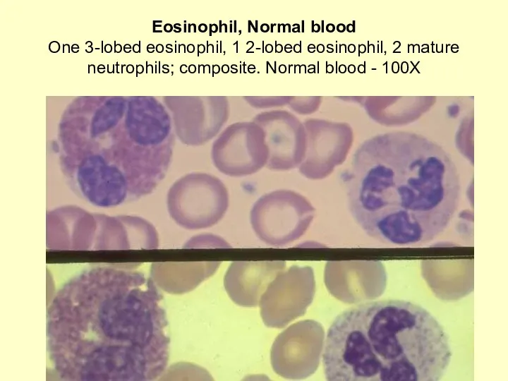
Eosinophil, Normal blood
One 3-lobed eosinophil, 1 2-lobed eosinophil, 2 mature neutrophils;
Eosinophil, Normal blood One 3-lobed eosinophil, 1 2-lobed eosinophil, 2 mature neutrophils;
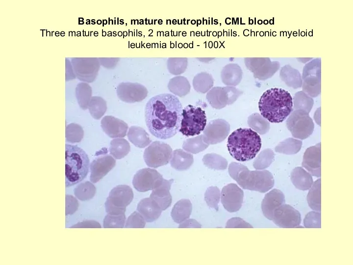
Basophils, mature neutrophils, CML blood
Three mature basophils, 2 mature neutrophils. Chronic
Basophils, mature neutrophils, CML blood Three mature basophils, 2 mature neutrophils. Chronic
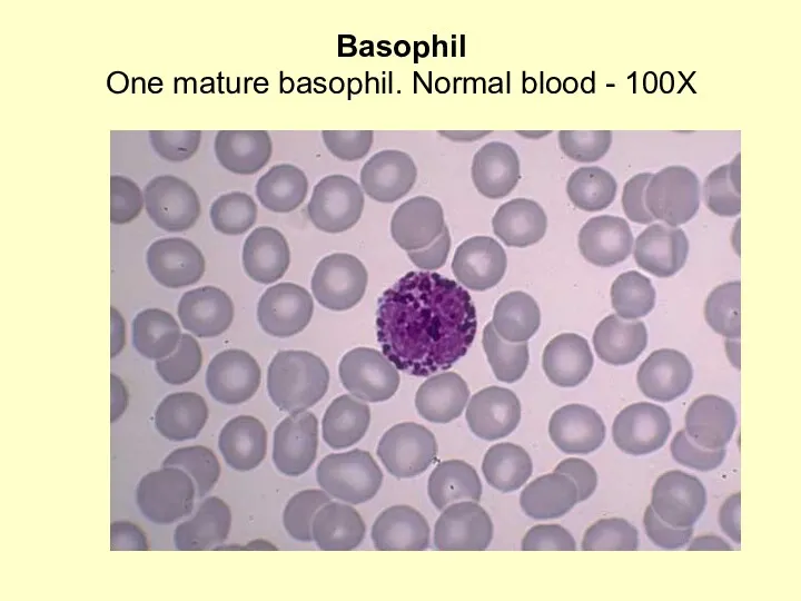
Basophil
One mature basophil. Normal blood - 100X
Basophil
One mature basophil. Normal blood - 100X
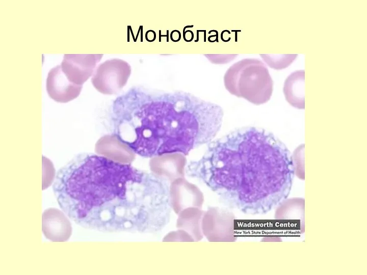
Монобласт
Монобласт
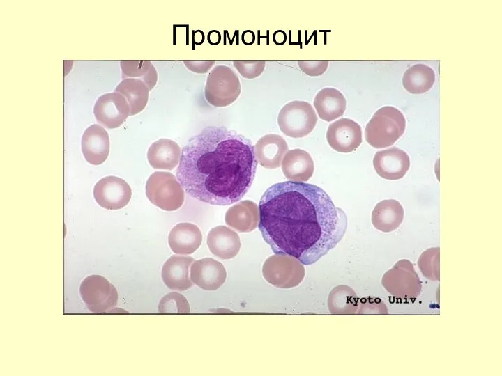
Промоноцит
Промоноцит
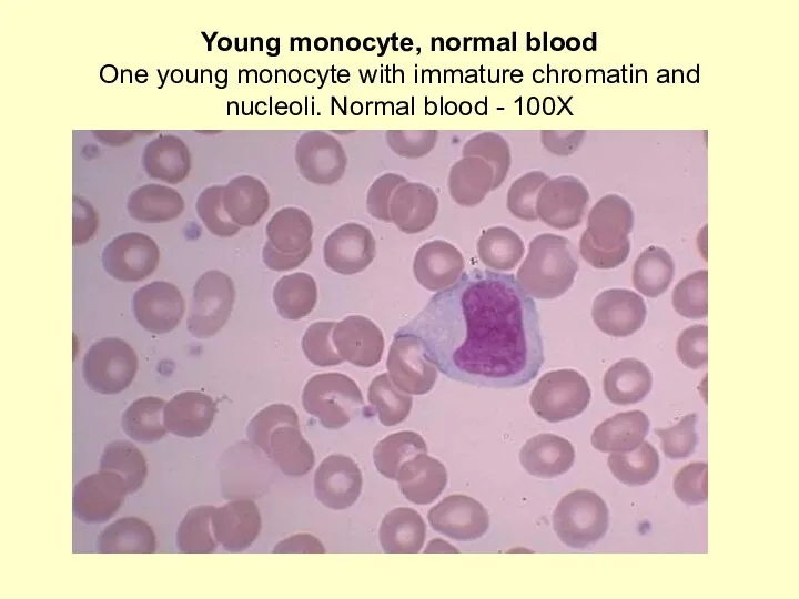
Young monocyte, normal blood
One young monocyte with immature chromatin and nucleoli.
Young monocyte, normal blood One young monocyte with immature chromatin and nucleoli.
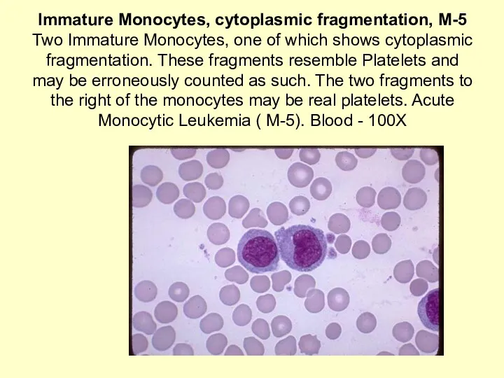
Immature Monocytes, cytoplasmic fragmentation, M-5
Two Immature Monocytes, one of which shows
Immature Monocytes, cytoplasmic fragmentation, M-5 Two Immature Monocytes, one of which shows
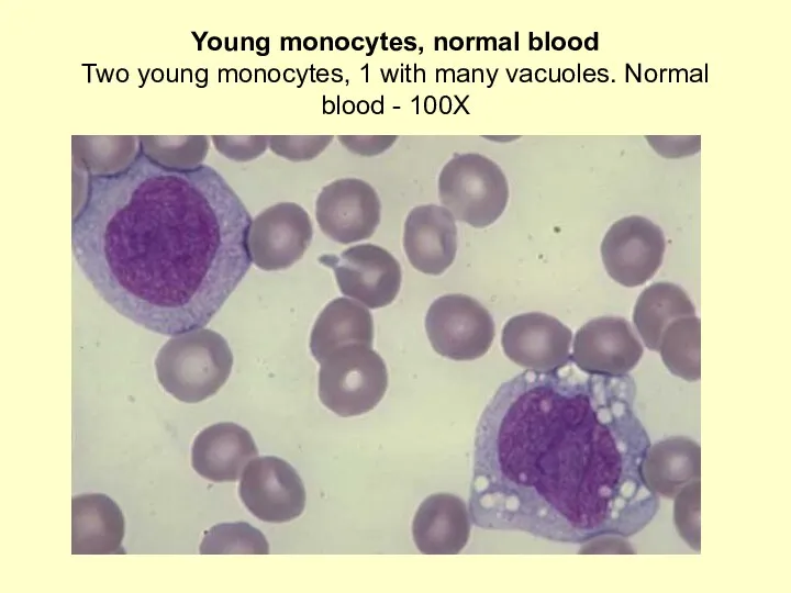
Young monocytes, normal blood
Two young monocytes, 1 with many vacuoles. Normal
Young monocytes, normal blood Two young monocytes, 1 with many vacuoles. Normal

Macrophage, mature megakaryocyte, normal marrow
A macrophage with an oval shaped nucleus
Macrophage, mature megakaryocyte, normal marrow A macrophage with an oval shaped nucleus
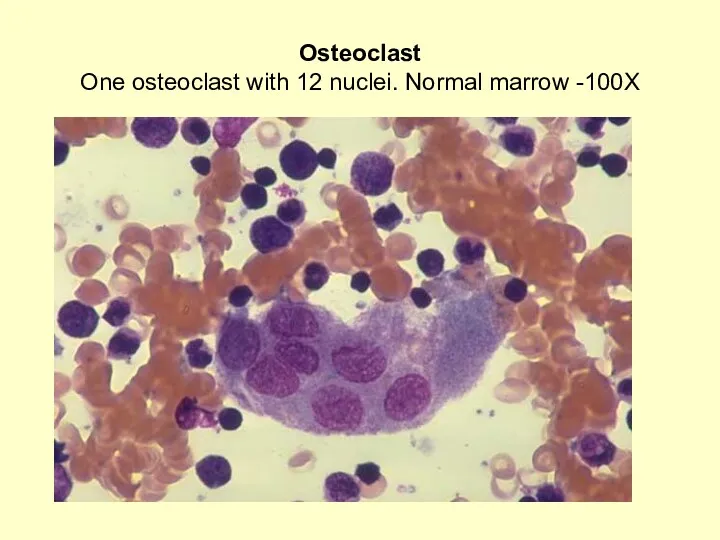
Osteoclast
One osteoclast with 12 nuclei. Normal marrow -100X
Osteoclast
One osteoclast with 12 nuclei. Normal marrow -100X
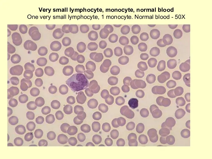
Very small lymphocyte, monocyte, normal blood
One very small lymphocyte, 1 monocyte.
Very small lymphocyte, monocyte, normal blood One very small lymphocyte, 1 monocyte.
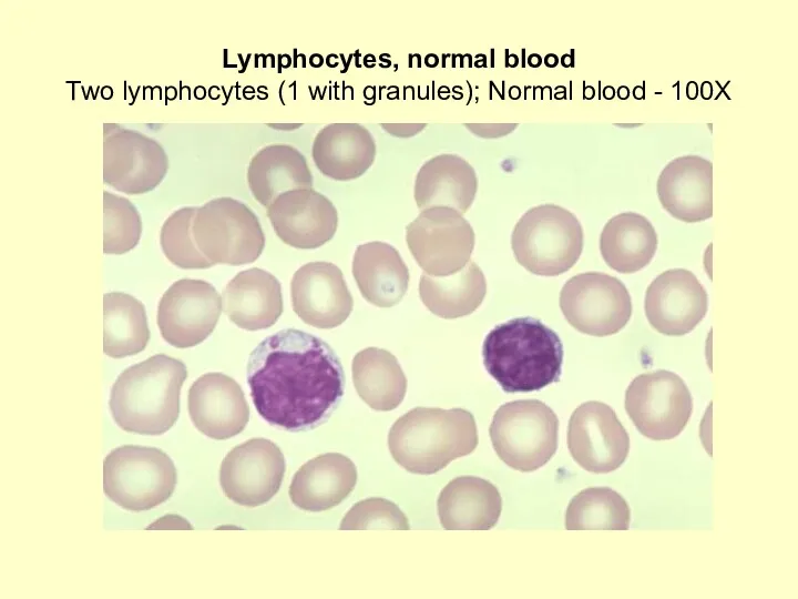
Lymphocytes, normal blood
Two lymphocytes (1 with granules); Normal blood - 100X
Lymphocytes, normal blood
Two lymphocytes (1 with granules); Normal blood - 100X
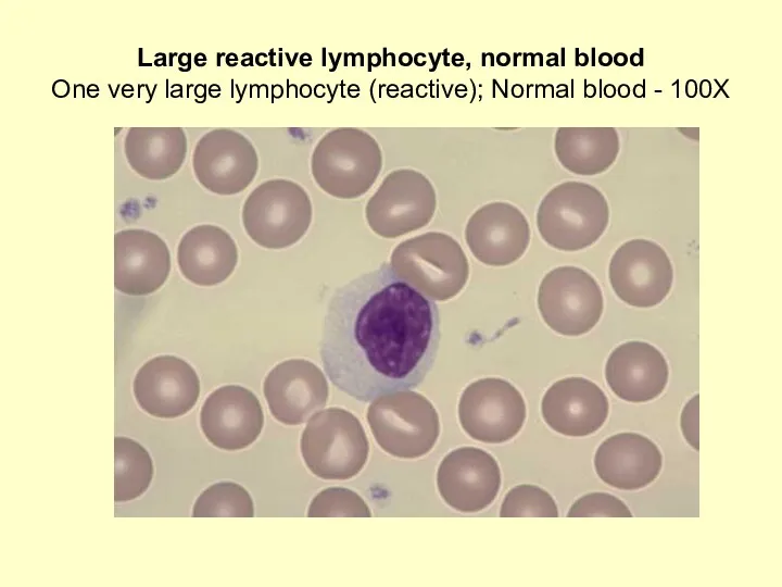
Large reactive lymphocyte, normal blood
One very large lymphocyte (reactive); Normal blood
Large reactive lymphocyte, normal blood One very large lymphocyte (reactive); Normal blood
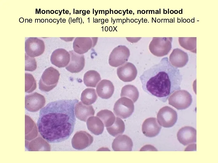
Monocyte, large lymphocyte, normal blood
One monocyte (left), 1 large lymphocyte. Normal
Monocyte, large lymphocyte, normal blood One monocyte (left), 1 large lymphocyte. Normal
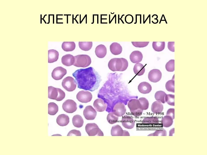
КЛЕТКИ ЛЕЙКОЛИЗА
КЛЕТКИ ЛЕЙКОЛИЗА
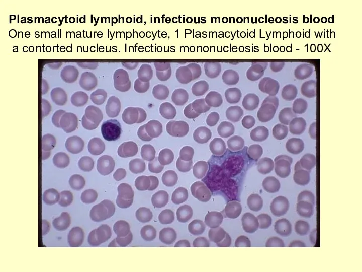
Plasmacytoid lymphoid, infectious mononucleosis blood
One small mature lymphocyte, 1 Plasmacytoid Lymphoid
Plasmacytoid lymphoid, infectious mononucleosis blood One small mature lymphocyte, 1 Plasmacytoid Lymphoid
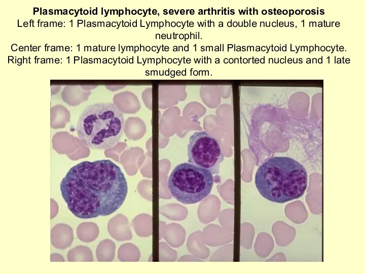
Plasmacytoid lymphocyte, severe arthritis with osteoporosis
Left frame: 1 Plasmacytoid Lymphocyte with
Plasmacytoid lymphocyte, severe arthritis with osteoporosis Left frame: 1 Plasmacytoid Lymphocyte with
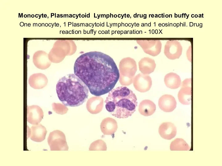
Monocyte, Plasmacytoid Lymphocyte, drug reaction buffy coat
One monocyte, 1 Plasmacytoid Lymphocyte
Monocyte, Plasmacytoid Lymphocyte, drug reaction buffy coat One monocyte, 1 Plasmacytoid Lymphocyte
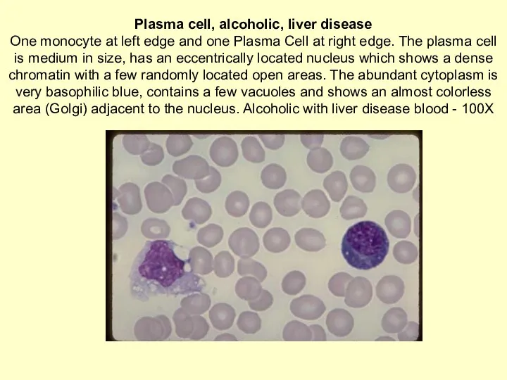
Plasma cell, alcoholic, liver disease
One monocyte at left edge and one
Plasma cell, alcoholic, liver disease One monocyte at left edge and one
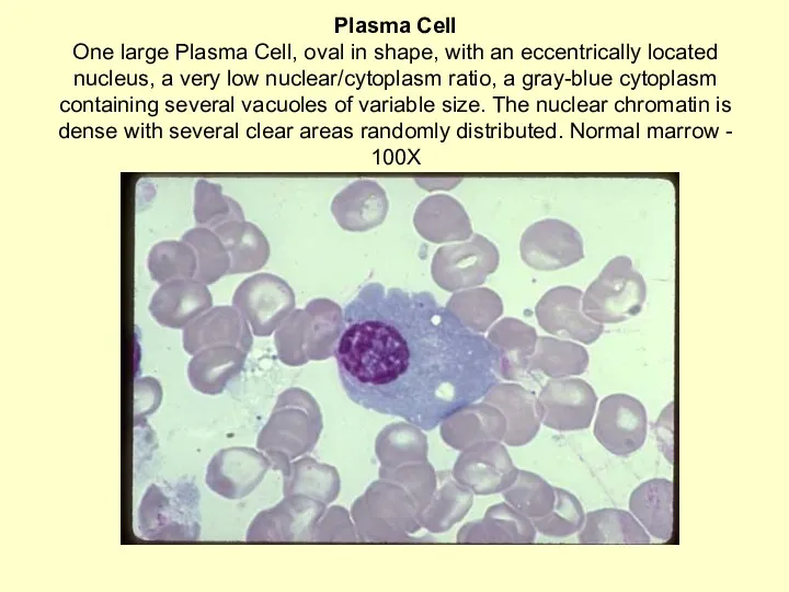
Plasma Cell
One large Plasma Cell, oval in shape, with an eccentrically
Plasma Cell One large Plasma Cell, oval in shape, with an eccentrically
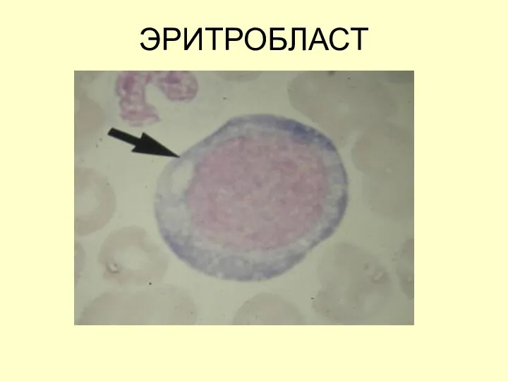
ЭРИТРОБЛАСТ
ЭРИТРОБЛАСТ
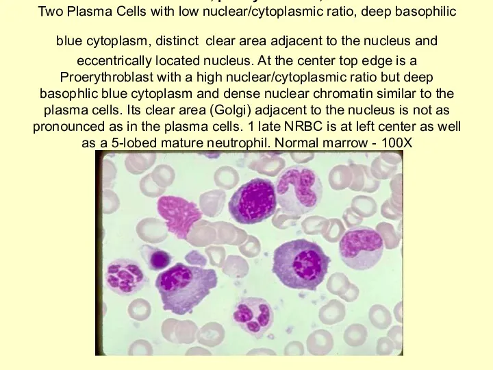
Plasma cells, proerythroblast, NRBC
Two Plasma Cells with low nuclear/cytoplasmic ratio, deep
Plasma cells, proerythroblast, NRBC Two Plasma Cells with low nuclear/cytoplasmic ratio, deep
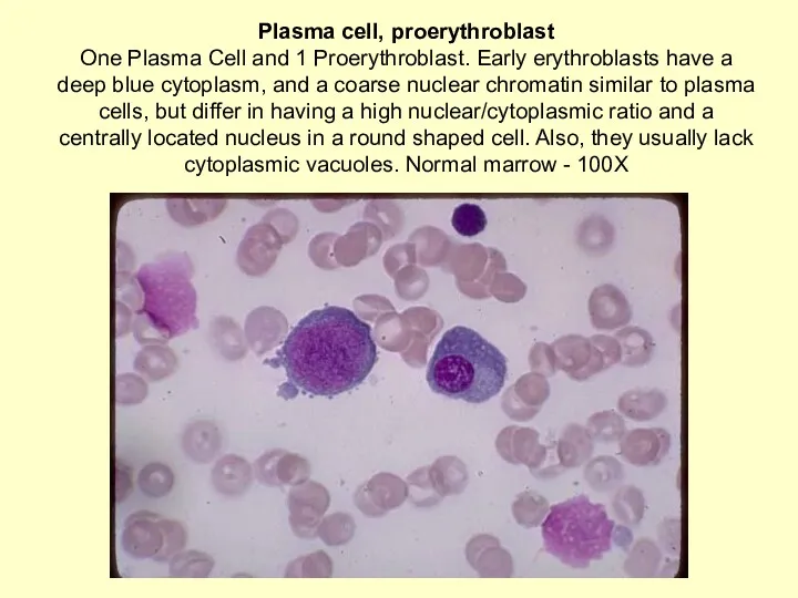
Plasma cell, proerythroblast
One Plasma Cell and 1 Proerythroblast. Early erythroblasts have
Plasma cell, proerythroblast One Plasma Cell and 1 Proerythroblast. Early erythroblasts have
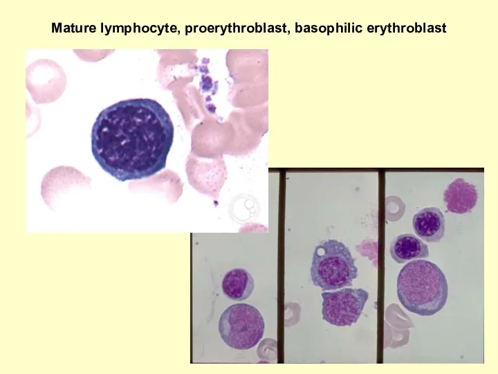
Mature lymphocyte, proerythroblast, basophilic erythroblast
Mature lymphocyte, proerythroblast, basophilic erythroblast
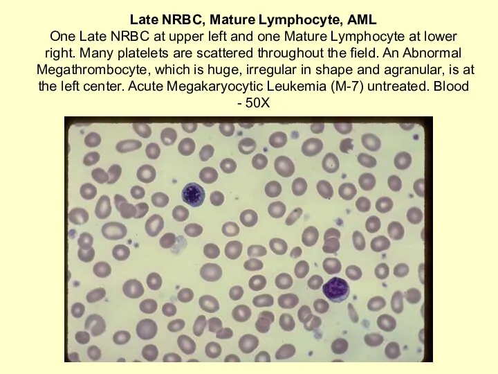
Late NRBC, Mature Lymphocyte, AML
One Late NRBC at upper left and
Late NRBC, Mature Lymphocyte, AML One Late NRBC at upper left and
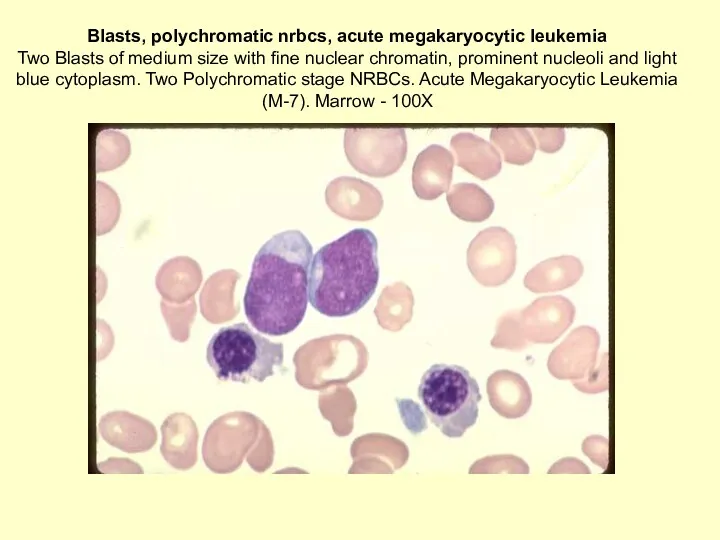
Blasts, polychromatic nrbcs, acute megakaryocytic leukemia
Two Blasts of medium size with
Blasts, polychromatic nrbcs, acute megakaryocytic leukemia
Two Blasts of medium size with
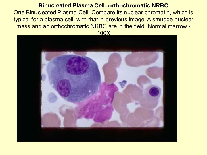
Binucleated Plasma Cell, orthochromatic NRBC
One Binucleated Plasma Cell. Compare its nuclear
Binucleated Plasma Cell, orthochromatic NRBC One Binucleated Plasma Cell. Compare its nuclear

РЕТИКУЛОЦИТЫ
РЕТИКУЛОЦИТЫ
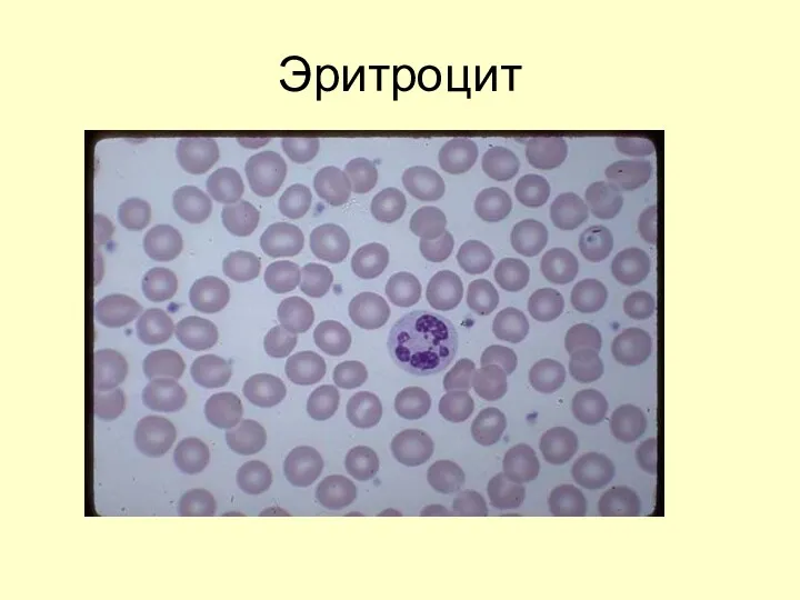
Эритроцит
Эритроцит
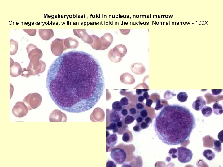
Megakaryoblast , fold in nucleus, normal marrow
One megakaryoblast with an apparent
Megakaryoblast , fold in nucleus, normal marrow
One megakaryoblast with an apparent
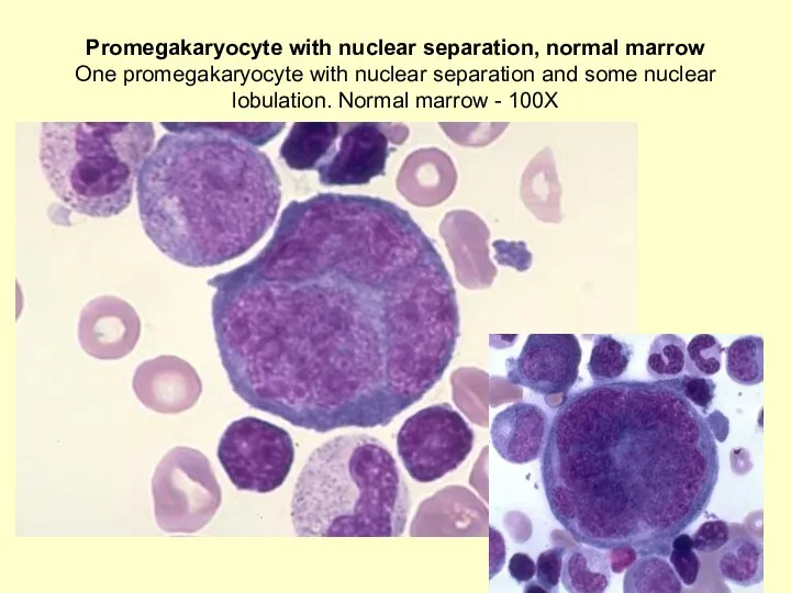
Promegakaryocyte with nuclear separation, normal marrow
One promegakaryocyte with nuclear separation and
Promegakaryocyte with nuclear separation, normal marrow One promegakaryocyte with nuclear separation and
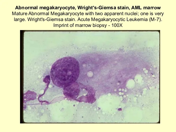
Abnormal megakaryocyte, Wright's-Giemsa stain, AML marrow
Mature Abnormal Megakaryocyte with two apparent
Abnormal megakaryocyte, Wright's-Giemsa stain, AML marrow Mature Abnormal Megakaryocyte with two apparent
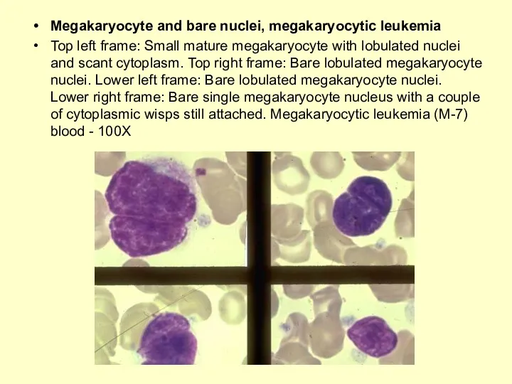
Megakaryocyte and bare nuclei, megakaryocytic leukemia
Top left frame: Small mature megakaryocyte
Megakaryocyte and bare nuclei, megakaryocytic leukemia
Top left frame: Small mature megakaryocyte
 Диуретики (мочегонные средства )
 Тамақтан улану кезіндегі алғашқы жәрдем
 Неотложные состояния в хирургии
 Группа антибиотиков макролиды
 Гранулематоз Вегенера
 Tuberculosis. Mycobacterium tuberculosis
 Неотложная помощь при тепловом ударе
 Туа пайда болған жоғарғы ішек өтімсіздігі
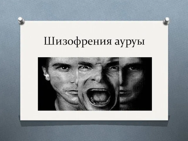 Шизофрения ауруы
 Закрытая травма сердца
 Поствакцинальные реакции и осложнения
 Энтеровирусная инфекция
 Адаптация. Срочная и долговременная адаптация
 Неврология. Функциональная анатомия спинного мозга. Нервная система и её функции
 Көк ірің таяқшасы
 Острая кишечная непроходимость
 Биомедицинская этика как форма профессиальной защиты личности врача
 Холера: клиника, лечение, дифференциальный диагноз
 Денсаулық сақтауды жоспарлау және қаржыландыру
 Гигиенические требования к условиям обучения в общеобразовательных учреждениях
 Гемоглобин. Соединения и виды гемоглобина. Практическая значимость их определения
 Болезни оперированного желудка
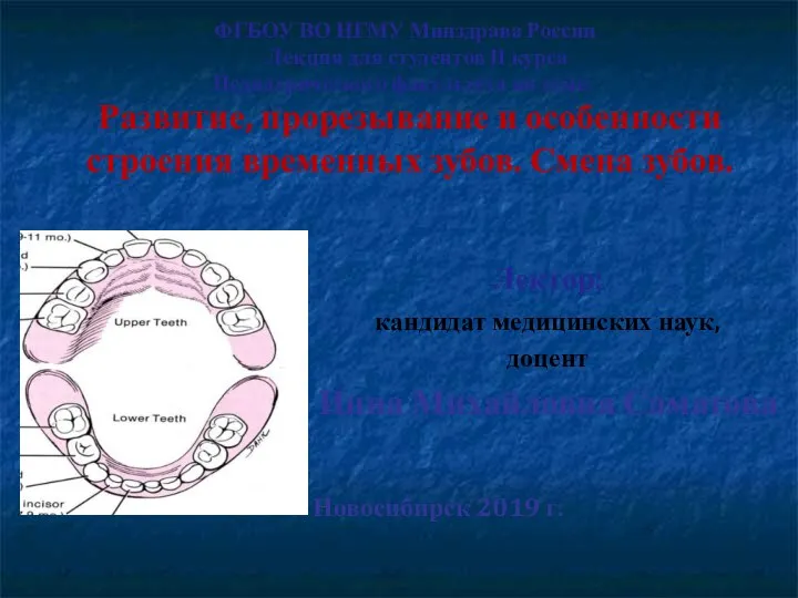 Развитие, прорезывание и особенности строения временных зубов. Смена зубов
 Травматология и ортопедия повреждений и заболеваний плечевого сустава
 Гендік мутациялар
 Даму барысындағы ақаулар
 Гипертермический синдром у детей
 Эндоцервикоз шейки матки