- Leishmaniasis. Department of Infectious Diseases Leishmaniasis

Содержание
- 2. Leishmaniasis Leishmaniasis is a zoonosis. Transmitted among mammalian hosts by female sand flies.
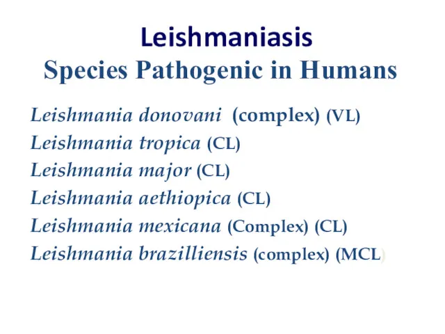
- 3. Leishmaniasis Leishmania donovani (complex) (VL) Leishmania tropica (CL) Leishmania major (CL) Leishmania aethiopica (CL) Leishmania mexicana
- 4. Three important Species Leishmania donovani (VL ) VISCERAL LEISHMANIASIS : involving endothelial tissue liver, spleen, and
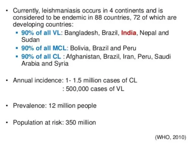
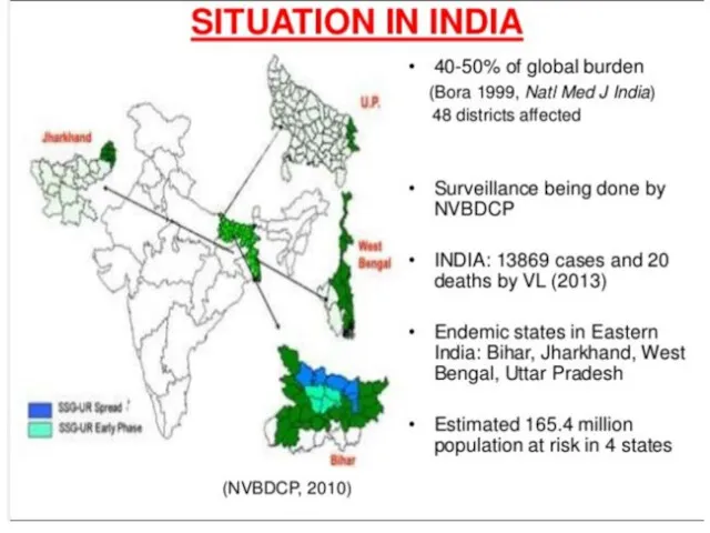
- 5. Geographical distribution of leishmaniasis
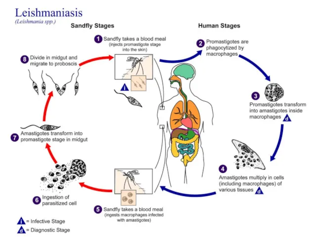
- 8. Life Cycle of leishmaniasis Promastigote Amasitgote Transformation
- 9. Promastigote stage flagella Promastigote stage inside the Sandfly Sand fly : Vectors Intermediate host, transmitted disease
- 10. Promastigotes in rosettes in a culture of an orient sore on N.N.N. medium (Giemsa stain).
- 11. Ovoid small intracellular parasites in a bone marrow aspirate. The typical rod shaped kinetoplast is seen
- 12. Life cycle
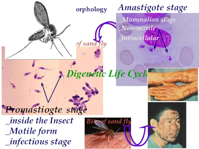
- 14. Bite of sand fly Bite of sand fly Leishmania Morphology Digenetic Life Cycle Promastiogte stage _inside
- 15. Transmission of Leishmaniasis _ by sand flies. _ artificial transmission of leishmania via the sharing of
- 16. Cutaneous Leishmaniasis Cutaneous forms of the disease normally produce skin ulcers on the exposed parts of
- 17. A cutaneous leishmaniasis lesion on the arm. The skin sores will heal by themselves, but this
- 18. Cutaneous Leishmaniasis
- 19. The Baghdad boil Baghdad-boil, 2004 Several hundred US soldiers in Iraq.
- 20. Leishmania tropica Causes ulceration of the skin called Cutaneous Leshmaniasis Dry or urban C.L. Dry sore
- 21. Mucocutaneous Leishmaniasis Mucocutaneous leishmaniasis (Espundia) Leishmania braziliensis & L . maxicana
- 22. mucocutaneous forms of leishmaniasis , lesions can lead to partial or total destruction of the mucosa
- 23. Visceral Leishmaniasis Visceral disease (Kala-azar)
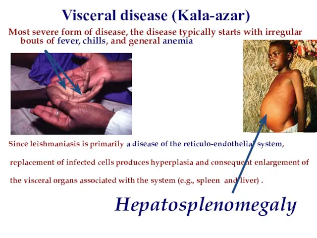
- 24. Visceral disease (Kala-azar) Most severe form of disease, the disease typically starts with irregular bouts of
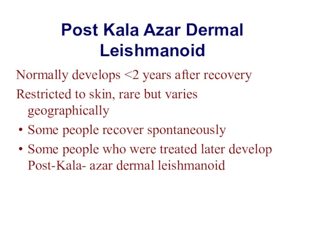
- 25. Post Kala Azar Dermal Leishmanoid Normally develops Restricted to skin, rare but varies geographically Some people
- 26. Hepatosplenomegaly Post Kala Azar Dermal Leishmanoid
- 27. Dogs can act as reservoirs of Leishmania parasites. They also exhibit symptoms of infection.
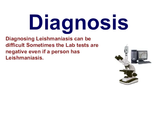
- 28. Diagnosis Diagnosing Leishmaniasis can be difficult Sometimes the Lab tests are negative even if a person
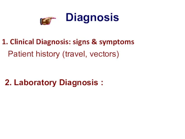
- 29. Diagnosis 1. Clinical Diagnosis: signs & symptoms 2. Laboratory Diagnosis : Patient history (travel, vectors)
- 30. Cutaneous leishmaniasis : Tissue sample (scraping, aspirate or punch biopsy) for smear and culture Visceral leishmaniasis
- 31. Animal inoculation Inoculate serum of infected person in lab. animals.
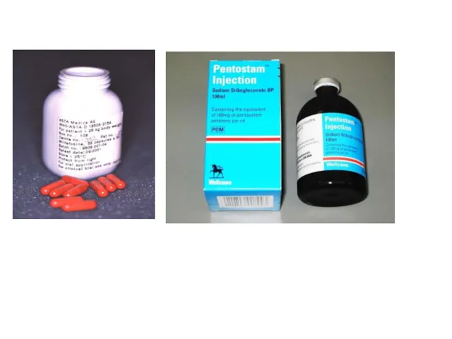
- 32. Cutaneous and mucocutaneous treatment Antimony components : Meglumine antimoniate (Glucantime) and Sodium stibogluconate (Pentostam) are drugs
- 33. Visceral leishmaniasis treatment Pentostam or Glucantime 20 mg /kg/d IV or IM for 28d Amphotricin B:
- 34. Visceral leishmaniasis treatment (con.) Miltefosine (Impavido) (2.5 mg/kg /d p.o. for 28 d) It was developed
- 37. Скачать презентацию

Leishmaniasis
Leishmaniasis is a zoonosis.
Transmitted among mammalian hosts by female sand
Leishmaniasis
Leishmaniasis is a zoonosis.
Transmitted among mammalian hosts by female sand
Leishmaniasis
Leishmania donovani (complex) (VL)
Leishmania tropica (CL)
Leishmania major (CL)
Leishmania aethiopica (CL)
Leishmania
Leishmaniasis
Leishmania donovani (complex) (VL)
Leishmania tropica (CL)
Leishmania major (CL)
Leishmania aethiopica (CL)
Leishmania
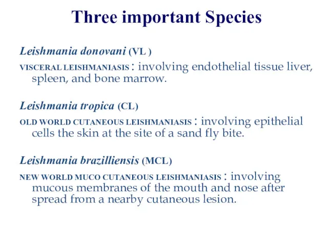
Three important Species
Leishmania donovani (VL )
VISCERAL LEISHMANIASIS : involving
Three important Species
Leishmania donovani (VL )
VISCERAL LEISHMANIASIS : involving
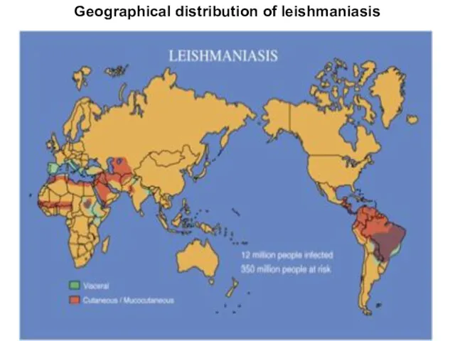
Geographical distribution of leishmaniasis
Geographical distribution of leishmaniasis
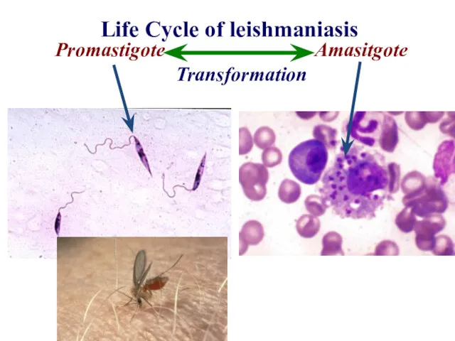
Life Cycle of leishmaniasis
Promastigote Amasitgote
Transformation
Life Cycle of leishmaniasis
Promastigote Amasitgote
Transformation
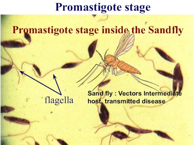
Promastigote stage
flagella
Promastigote stage inside the Sandfly
Sand fly : Vectors Intermediate
Promastigote stage
flagella
Promastigote stage inside the Sandfly
Sand fly : Vectors Intermediate
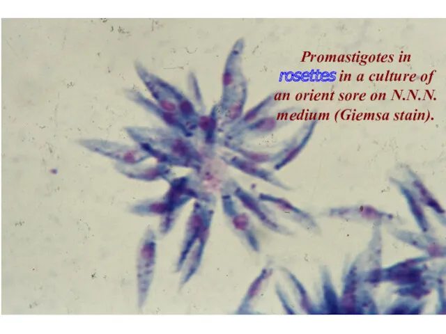
Promastigotes in rosettes in a culture of an orient sore on
Promastigotes in rosettes in a culture of an orient sore on
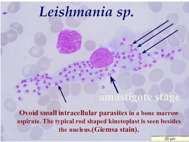
Ovoid small intracellular parasites in a bone marrow aspirate. The typical
Ovoid small intracellular parasites in a bone marrow aspirate. The typical
Life cycle
Life cycle
Bite of sand fly
Bite of sand fly
Leishmania Morphology
Digenetic Life Cycle
Promastiogte stage
_inside
Bite of sand fly
Bite of sand fly
Leishmania Morphology
Digenetic Life Cycle
Promastiogte stage _inside
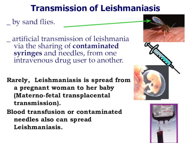
Transmission of Leishmaniasis
_ by sand flies.
_ artificial transmission of leishmania
Transmission of Leishmaniasis
_ by sand flies.
_ artificial transmission of leishmania
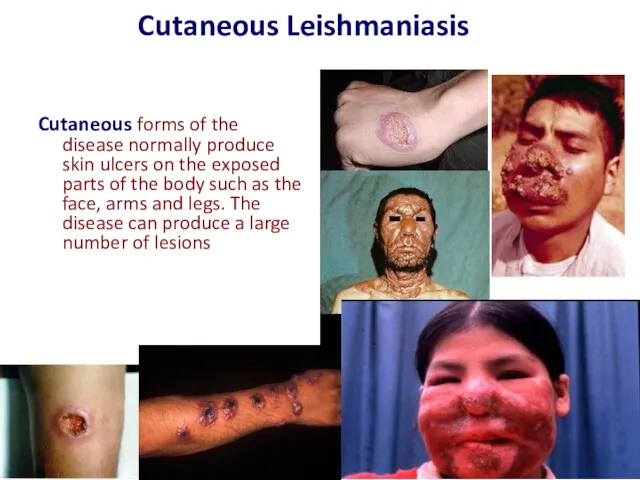
Cutaneous Leishmaniasis
Cutaneous forms of the disease normally produce skin ulcers on
Cutaneous Leishmaniasis
Cutaneous forms of the disease normally produce skin ulcers on
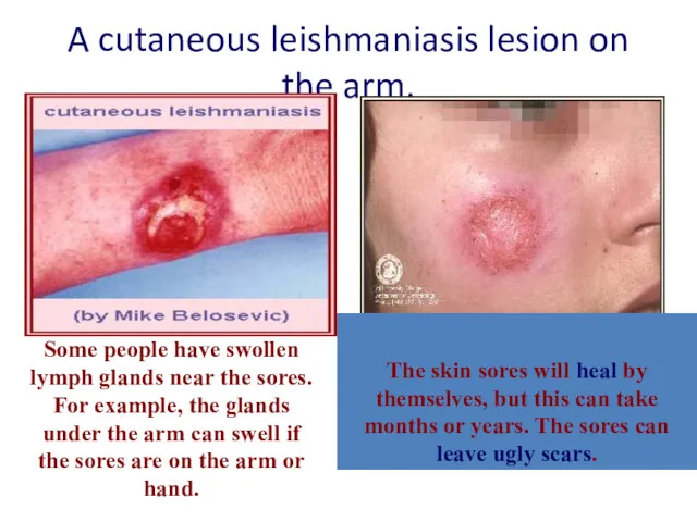
A cutaneous leishmaniasis lesion on the arm.
The skin sores will
A cutaneous leishmaniasis lesion on the arm.
The skin sores will
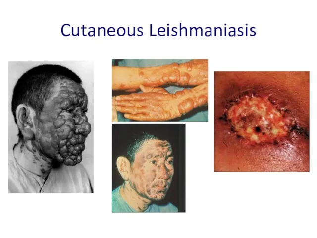
Cutaneous Leishmaniasis
Cutaneous Leishmaniasis
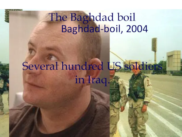
The Baghdad boil
Baghdad-boil, 2004
Several hundred US soldiers in Iraq.
The Baghdad boil
Baghdad-boil, 2004
Several hundred US soldiers in Iraq.
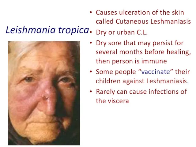
Leishmania tropica
Causes ulceration of the skin called Cutaneous Leshmaniasis
Dry or urban
Leishmania tropica
Causes ulceration of the skin called Cutaneous Leshmaniasis
Dry or urban
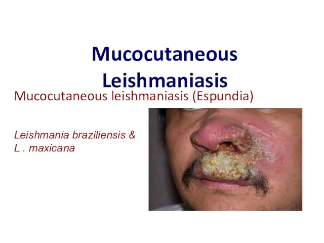
Mucocutaneous Leishmaniasis
Mucocutaneous leishmaniasis (Espundia)
Leishmania braziliensis & L . maxicana
Mucocutaneous Leishmaniasis
Mucocutaneous leishmaniasis (Espundia)
Leishmania braziliensis & L . maxicana
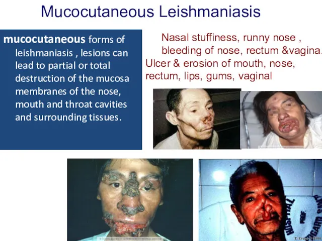
mucocutaneous forms of leishmaniasis , lesions can lead to partial or
mucocutaneous forms of leishmaniasis , lesions can lead to partial or
Visceral Leishmaniasis
Visceral disease (Kala-azar)
Visceral Leishmaniasis
Visceral disease (Kala-azar)
Visceral disease (Kala-azar)
Most severe form of disease, the disease typically starts
Visceral disease (Kala-azar)
Most severe form of disease, the disease typically starts
Post Kala Azar Dermal Leishmanoid
Normally develops <2 years after recovery
Restricted to
Post Kala Azar Dermal Leishmanoid
Normally develops <2 years after recovery
Restricted to
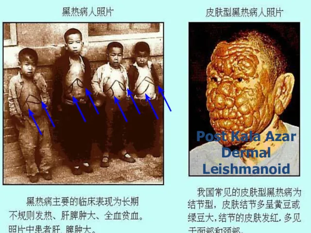
Hepatosplenomegaly
Post Kala Azar Dermal Leishmanoid
Hepatosplenomegaly
Post Kala Azar Dermal Leishmanoid
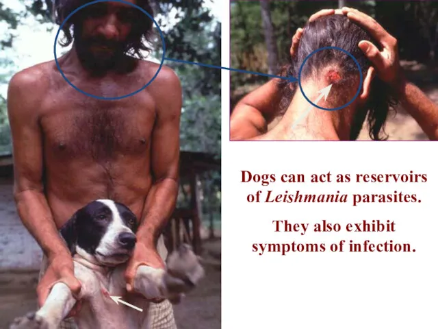
Dogs can act as reservoirs of Leishmania parasites.
They also exhibit
Dogs can act as reservoirs of Leishmania parasites.
They also exhibit
Diagnosis
Diagnosing Leishmaniasis can be difficult Sometimes the Lab tests are negative
Diagnosis
Diagnosing Leishmaniasis can be difficult Sometimes the Lab tests are negative
Diagnosis
1. Clinical Diagnosis: signs & symptoms
2. Laboratory Diagnosis :
Patient history (travel,
Diagnosis
1. Clinical Diagnosis: signs & symptoms
2. Laboratory Diagnosis :
Patient history (travel,
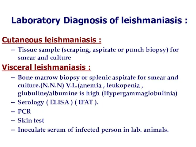
Cutaneous leishmaniasis :
Tissue sample (scraping, aspirate or punch biopsy) for smear
Cutaneous leishmaniasis :
Tissue sample (scraping, aspirate or punch biopsy) for smear
Animal inoculation
Inoculate serum of infected person in lab. animals.
Animal inoculation
Inoculate serum of infected person in lab. animals.
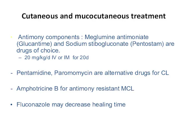
Cutaneous and mucocutaneous treatment
Antimony components : Meglumine antimoniate (Glucantime) and
Cutaneous and mucocutaneous treatment
Antimony components : Meglumine antimoniate (Glucantime) and
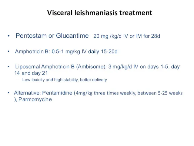
Visceral leishmaniasis treatment
Pentostam or Glucantime 20 mg /kg/d IV
Visceral leishmaniasis treatment
Pentostam or Glucantime 20 mg /kg/d IV
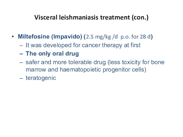
Visceral leishmaniasis treatment (con.)
Miltefosine (Impavido) (2.5 mg/kg /d p.o. for 28
Visceral leishmaniasis treatment (con.)
Miltefosine (Impavido) (2.5 mg/kg /d p.o. for 28
 Первая доврачебная помощь при ДТП
 Семейная гиперхолестеринемия
 Требования к организации питания пациентов в буфетных. Обязанности старшей медицинской сестры
 Консервативное лечение атеросклероза
 Қант диабеті кезіндегі пациентті және туыстарын оқыту
 Уход за онкологическими больными
 Вакцинация детей. Календарь прививок
 Омытқа жотасының қызметі және маңызы
 Нематодозы. Аскаридоз. Трихоцефалез
 Заболевания детей раннего возраста. Заболевания слизистой полости рта (стоматиты, молочница)
 Аутоиммунный гепатит и беременность
 Нарушения липидного обмена
 Кровотечения в последовом и раннем послеродовом периоде
 Меридиан почек VIII
 Ауыз қуысы және оның ағзаларының дамуы (онтогенез). Ақаулары
 Сестринская помощь при пиелонефритах
 Мочевыделительная система
 Клиническая анатомия головы и шеи
 Принципы купирования острого инфаркта миокарда
 Психические расстройства при сосудистых заболеваниях головного мозга
 Методы радионуклидной диагностики органов и систем человека
 Болезни печени
 Балалардағы безгек
 Врожденный вывих бедра
 Синдром Прадера- Вилли
 Устройство стоматологического кабинета
 Тері биохимиясы
 Теоретическое пособие по массажу