- Лимфопролиферативные заболевания

Содержание
- 2. Patients with hematological malignancies in Belarus (adults) (2007).
- 3. Limphoproliferative diseases
- 4. B-cell lymphopoiesis
- 5. B-cell malignan-cies
- 6. T-cell differen-tiation stages
- 7. Lymphopoiesis in lymph nodes.
- 8. B-cell malignancies
- 9. Morphology of leukocytes
- 10. Acute leukemia. Originated from bone marrow (>25% blasts). Usually monoclonal disease. Lineage committed morphology (FAB classif.)
- 11. Acute leukemia (WHO classification, 2008). Mixed phenotype acute leukemia (T or B- myeloid, NK-cell…) B lymphoblastic
- 12. Cytogenetic and genetic features of ALL.
- 13. Chronic lymphocytic leukemia (WHO classification, 2008). Mature B-cell neoplasms Chronic lymphocytic leukemia/small lymphocytic lymphoma, B-cell prolymphocytic
- 14. Chronic lymphocytic leukemia (WHO classification, 2008). Mature T-cell and NK-cell neoplasms: T-cell prolymphocytic leukemia, T-cell large
- 15. Adverse prognostic factors of CLL Diffuse infiltration of bone marrow by lymphocytes; Advanced age; Male gender;
- 16. Typical B cell phenotype in CLL
- 17. Strategy for CLL therapy. First line of therapy: Fludarabine, Cyclophosphamine, Rituximabe (FCR). Chemotherapy, MABs such as
- 18. Types of lymphomas.
- 19. Hodgkin Lymphoma et al. (WHO, 2008). Hodgkin lymphoma: - classical Hodgkin lymphoma, - Lymphocyte-rich classical Hodgkin
- 20. Histological diagnosis of HD. The Reed–Sternberg cells are identified as large often bi-nucleated cells with prominent
- 21. The adverse prognostic factors for HD Age ≥ 45 years Stage IV disease Hemoglobin Lymphocyte count
- 22. Stages and Therapy of HD Therapy strategy: radiation therapy +/- chemotherapy. Prognosis: The 5-year survival rate
- 23. Non-Hodgkin lymphoma Causes The many different forms of lymphoma likely have different causes. These possible causes
- 26. Cytogenetic analysis for B-cell malignancies t(11;14) is mainly found in mantle cell lymphoma, but also in
- 27. Diagnosis of DLBCL by MicroArray technique: Germinal center B cell DLBCL vs activated (post-germinal center) B
- 28. Burkitt’s lymphoma (rare type of NHL) (endemic= EBV positive)
- 29. Immunophenotypic diagnosis of Burkitt’s lymphoma The cells of BL typically express monotypic surface IgM, CD19, CD20,
- 30. T (8,14) in Burkitt’s lymphoma
- 31. Path from Normal plasma cells through Monoclonal Gammopathy of Undetermined Significance to Multiple Myeloma.
- 32. Plasma cell malignancies
- 33. Morphology of malignant plasma cells in blood (H&E staining)
- 34. Immunophenotyping of Plasma Cells
- 36. Multiple Myeloma diagnosis and therapy. Diagnosis: Roentgen + BM biopsy+.. Therapy: chemotherapy, BMT. Survival: 5-8 years.
- 37. Serum paraprotein detection
- 38. M-protein and diseases. More than 50% of patients with serum M protein have an initial clinical
- 39. Waldenstrom macroglobulinemia: pathogenesis Immunophenotype of BM cells in WM Ig light chain - Positive CD19 -
- 40. Diagnosis and Therapy of WM.
- 41. Light chain Disease (Bence-Jones proteins). A Bence Jones protein is a monoclonal globulion protein or immunoglobulin
- 42. (Bence-Jones protein in serum/urine (up) and serum (down))
- 43. HEAVY CHAIN DISEASE Heavy chain disease is a form of paraproteinemia with a proliferation of cells
- 45. Скачать презентацию
 Ауаның физикалық қасиетінің, химиялық және биологиялық құрамының гигиеналық маңызы. Климат және ауа
 Вакцинация және адамның құқықтары
 Побочные действия антибиотиков
 Клиника интеллектуальных нарушений при деменции
 Физиологические особенности проявления двигательных качеств (часть 2)
 Анатомияны тірі адамда окып білу әдістері: антропометрия. Дене бітімінің түрлері (типтері)
 Введение в патологическую физиологию. Причины и условия возникновения болезни
 Современные лабораторные маркеры аутоиммунных заболеваний (СКВ, РА, склеродермия, дерматомиозит)
 Судово-медична експертиза ушкоджень гостримим предметами
 Компоненты здорового образа жизни и пути их формирования. Методы, формы и средства гигиенического воспитания
 Аномалии конституции (диатезы) у детей
 Патофизиология клетки
 Чипсы: польза или вред
 Жансыздандыру. Жергілікті анестетиктер
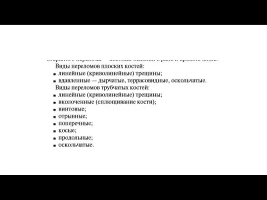 Виды переломов плоских костей
 Қызылорда медицина жоғары колледжінде жасөспірімдер арасында семіздіктің ақпараттандырылу деңгейі қандай
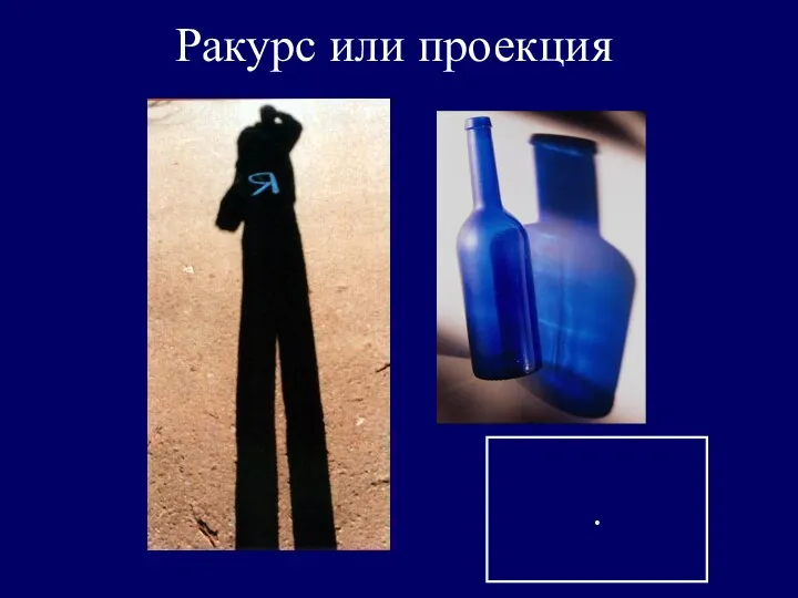 Ракурс или проекция
 Воздействие сотовой связи на человека
 Дитячий церебральний параліч
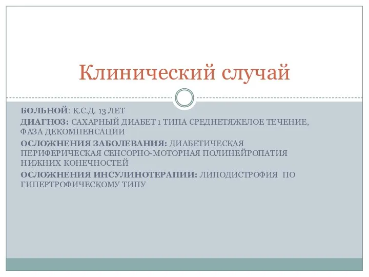 Клинический случай. Сахарный диабет 1 типа среднетяжелое течение, фаза декомпенсации
 Диагностика во фтизиатрии
 Преэклампсия. Актуальность
 Лекарственные растения
 Экзема
 Сыпной тиф и другие риккетсиозы
 Роль региональной ассоциации в развитии сестринского дела в Республике Коми
 Апластикалық анемиялар
 The lungs