- Medical protozology

Содержание
- 2. Protozoa (singular, protozoan), from the Greek ‘protos’ and ‘zoon’ meaning “first animal”, are members of eukaryotic
- 3. Occurrence of protozoa Protozoa are found in all moist habitats. They are common in sea, in
- 4. Morphology of protozoa Protozoa are predominantly microscopic, ranging in size from 2 to more than 100μm.
- 5. Importance of protozoa Protozoa serve as an important link in the food chain and ecological balance
- 6. Transmission In most parasitic protozoa, the developmental stages are often transmitted from one host to another
- 7. Pathogenesis Protozoan organisms are virtually always acquired from an exogenous source, and as such, they have
- 8. Classification of Protozoa Protozoa of medical importance are classified based on their morphology and locomotive system
- 9. 1. Parasitic amoeba
- 10. Amoebiasis. Entamoeba histolytica
- 12. Symptoms: Abdominal pain, Mild diarrhea, bloody diarrhea, Perforation and tissue death. This last complication may cause
- 13. Invasion of the intestinal lining causes amoebic bloody diarrhea or amoebic colitis. If the parasite reaches
- 14. Transmission: It is usually transmitted by fecal-oral route, but it can also be transmitted indirectly through
- 15. Diagnosis: Microscopy of feces Serological tests. Serology becomes positive about 2 weeks after invasion. Treatment: Entamoeba
- 16. Prevention: Wash hands with soap and hot running water Clean bathrooms and toilets often Avoid sharing
- 17. 2. Parasitic ciliates
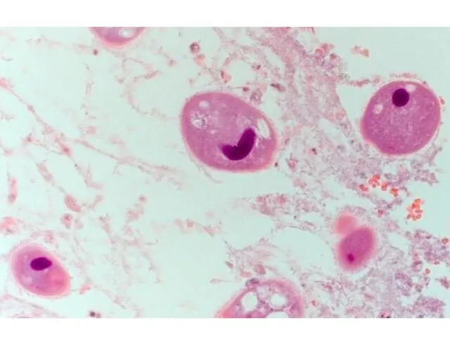
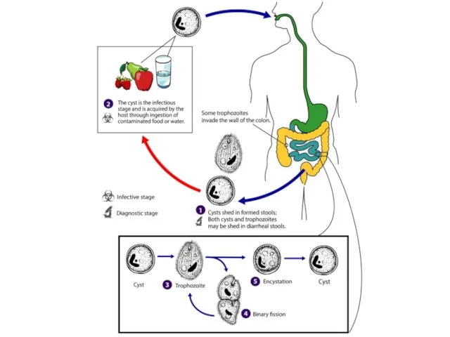
- 18. Balantidiasis. Balantidium coli
- 21. Symptoms: Can be local due to involvement of the intestinal mucosa, systemic in nature and include
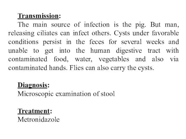
- 22. Transmission: The main source of infection is the pig. But man, releasing ciliates can infect others.
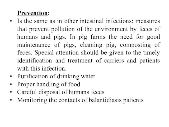
- 23. Prevention: Is the same as in other intestinal infections: measures that prevent pollution of the environment
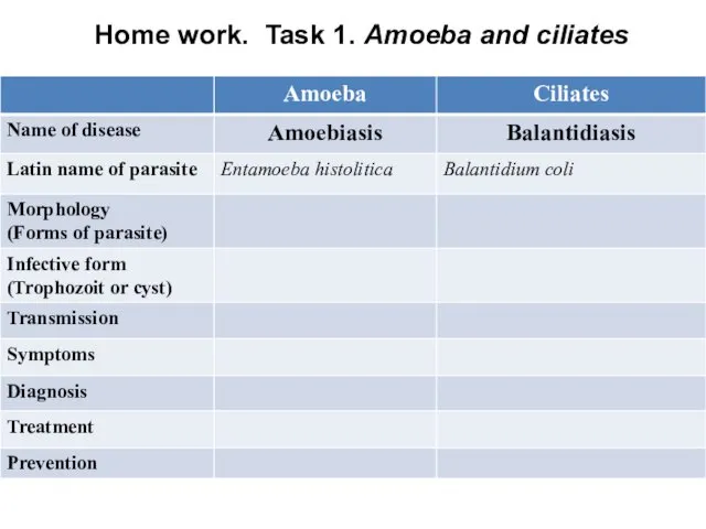
- 24. Home work. Task 1. Amoeba and ciliates
- 25. 3. Parasitic Flagellates: 3.1. Intestinal and vaginal Flagellates
- 26. Giardiasis or lambliosis Giardia lamblia
- 27. Transmission: The source of infection is people. Transmission is by ingestion of the infective cyst. Infection
- 29. Symptoms: The main habitat of giardia in human body are duodenum and initial part of colon.
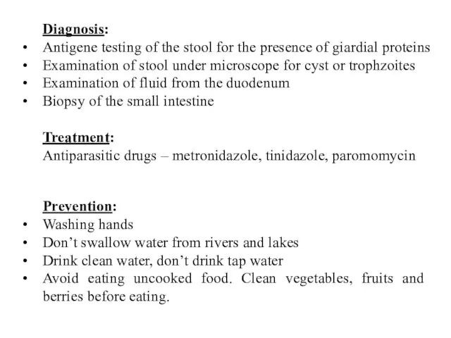
- 30. Diagnosis: Antigene testing of the stool for the presence of giardial proteins Examination of stool under
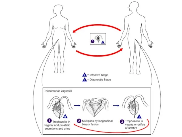
- 31. Trichomoniasis. Trichomonas vaginalis
- 32. Is a sexually transmitted infection. It caused by Trichomonas vaginalis. It is very common. This tiny
- 34. Symptoms: Vaginal discharge, which may be white, gray, yellow or green and usually has unpleasant smell.
- 35. Diagnosis: Physical test and laboratory test: cell culture, DNA examing samples of vaginal fluids. Treatment: Metronidazole
- 36. Home work. Task 2. Amoeba, Ciliates, intestinal and vaginal flagellates
- 37. 3. Parasitic Flagellates: 3.2. Hemoflagellates
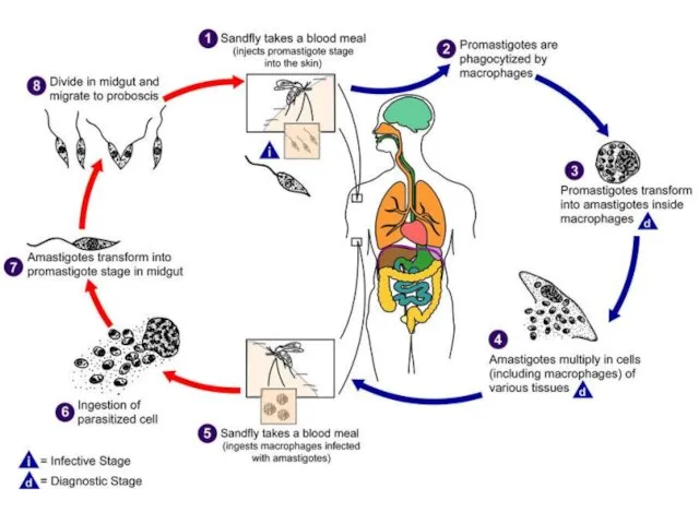
- 38. Leishmaniasis. Is a disease caused by protozoan parasites of the genus Leishmania and spread by the
- 39. Morphology
- 41. Pathogenesis In visceral leishmaniasis, the organs of the reticuloendothelial system (liver, spleen and bone marrow) are
- 42. Clinical features Symptoms begin with intermittent fever, weakness, and diarrhea; chills and sweating that may resemble
- 44. Diagnosis: Seeing the parasites under the microscope. Visceral disease can be diagnosed by blood tests. Examination
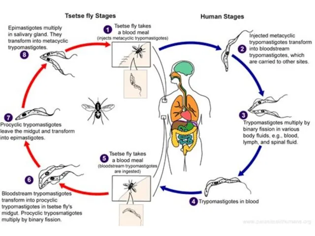
- 45. Trypanasomiasis This disease caused by Tripanosoma species. In humans this include African and South-American types. Trypanosoma
- 46. Morphology Typical trypanosome structure is an elongated spindle-shaped body that more or less tapers at both
- 47. African type of Trypanosomiasis. It caused by Trypanosoma brucei. Common name: sleeping sickness. Definitive host: man,
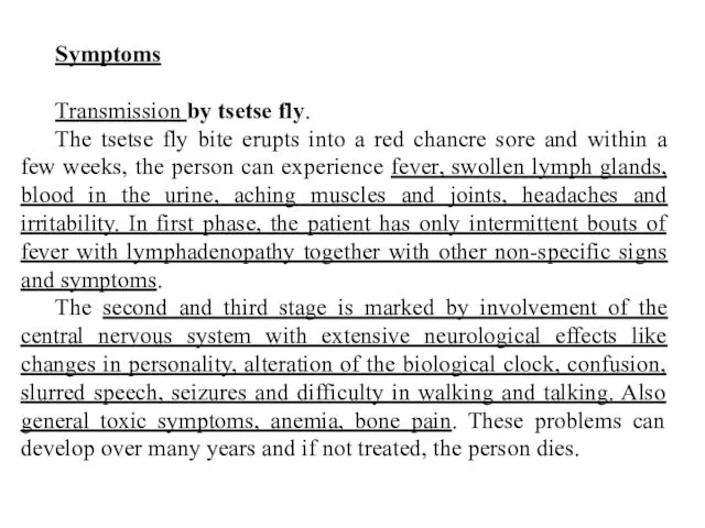
- 48. Symptoms Transmission by tsetse fly. The tsetse fly bite erupts into a red chancre sore and
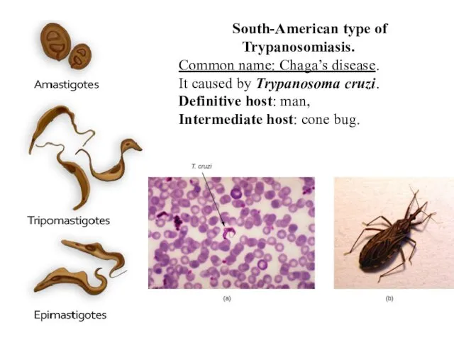
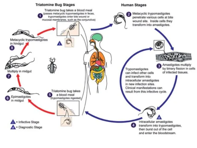
- 50. South-American type of Trypanosomiasis. Common name: Chaga’s disease. It caused by Trypanosoma cruzi. Definitive host: man,
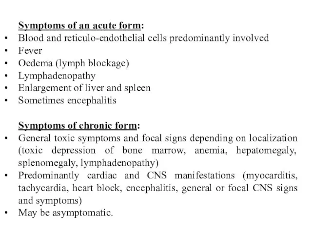
- 52. Symptoms of an acute form: Blood and reticulo-endothelial cells predominantly involved Fever Oedema (lymph blockage) Lymphadenopathy
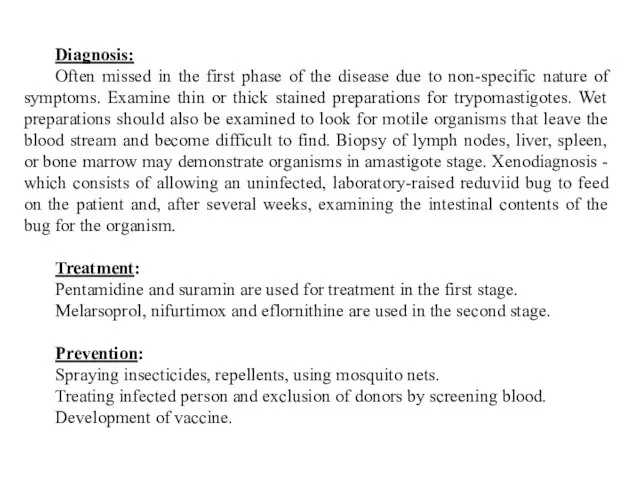
- 53. Diagnosis: Often missed in the first phase of the disease due to non-specific nature of symptoms.
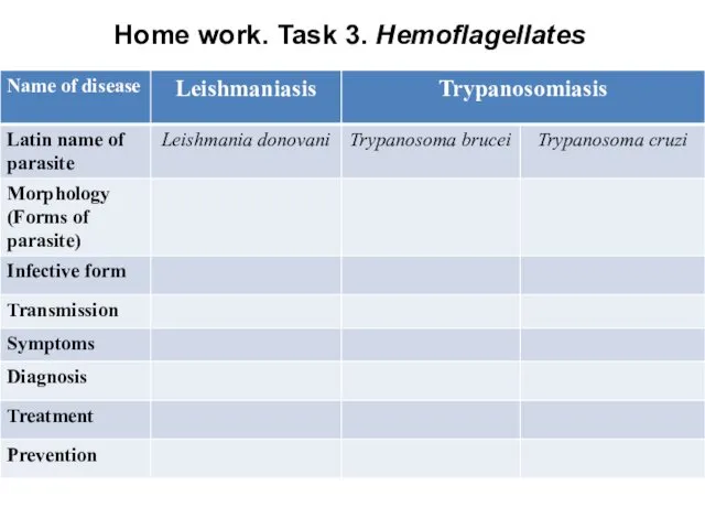
- 54. Home work. Task 3. Hemoflagellates
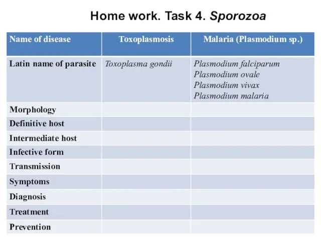
- 55. 4. Sporozoa
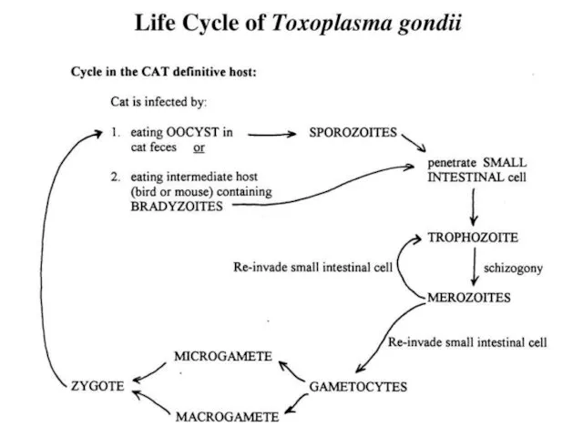
- 56. Toxoplasmosis. Toxoplasma gondii
- 58. Transmission: The definitive host is the domestic cat and other felines. Humans and other mammals are
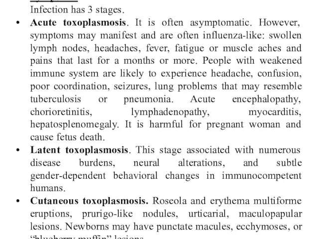
- 60. Symptoms: Infection has 3 stages. Acute toxoplasmosis. It is often asymptomatic. However, symptoms may manifest and
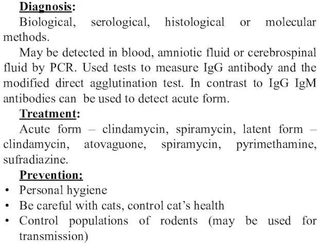
- 61. Diagnosis: Biological, serological, histological or molecular methods. May be detected in blood, amniotic fluid or cerebrospinal
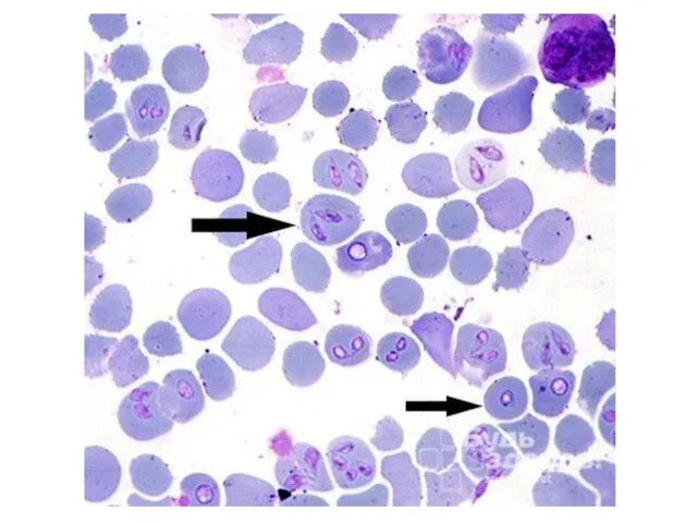
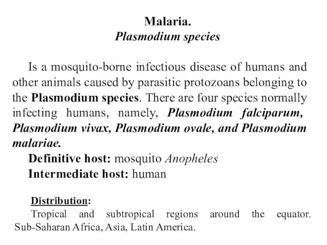
- 62. Malaria. Plasmodium species Is a mosquito-borne infectious disease of humans and other animals caused by parasitic
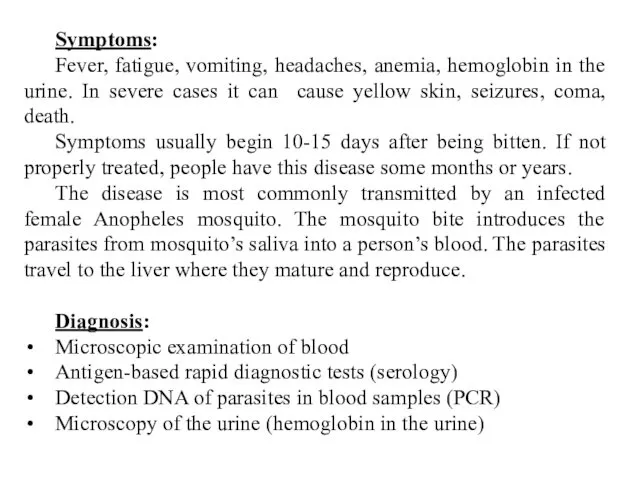
- 64. Symptoms: Fever, fatigue, vomiting, headaches, anemia, hemoglobin in the urine. In severe cases it can cause
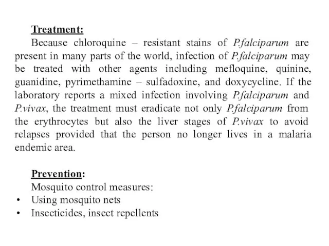
- 65. Treatment: Because chloroquine – resistant stains of P.falciparum are present in many parts of the world,
- 66. Home work. Task 4. Sporozoa
- 68. Скачать презентацию
Protozoa (singular, protozoan), from the Greek ‘protos’ and ‘zoon’ meaning “first
Protozoa (singular, protozoan), from the Greek ‘protos’ and ‘zoon’ meaning “first
Occurrence of protozoa
Protozoa are found in all moist habitats. They are
Occurrence of protozoa
Protozoa are found in all moist habitats. They are
Morphology of protozoa
Protozoa are predominantly microscopic, ranging in size from 2
Morphology of protozoa
Protozoa are predominantly microscopic, ranging in size from 2
Importance of protozoa
Protozoa serve as an important link in the food
Importance of protozoa
Protozoa serve as an important link in the food
Transmission
In most parasitic protozoa, the developmental stages are often transmitted from
Transmission
In most parasitic protozoa, the developmental stages are often transmitted from
Pathogenesis
Protozoan organisms are virtually always acquired from an exogenous source, and
Pathogenesis
Protozoan organisms are virtually always acquired from an exogenous source, and
Classification of Protozoa
Protozoa of medical importance are classified based on their
Classification of Protozoa
Protozoa of medical importance are classified based on their
1. Parasitic amoeba
1. Parasitic amoeba
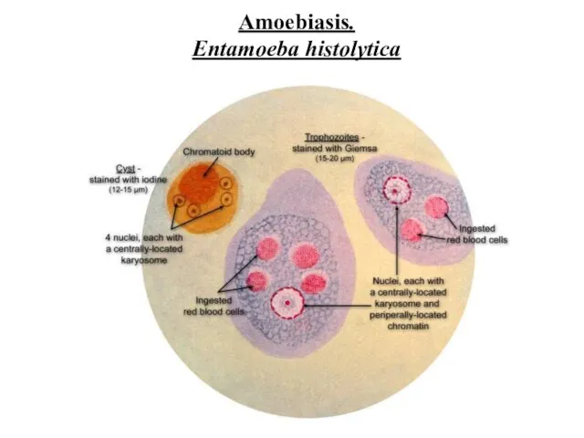
Amoebiasis.
Entamoeba histolytica
Amoebiasis.
Entamoeba histolytica
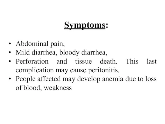
Symptoms:
Abdominal pain,
Mild diarrhea, bloody diarrhea,
Perforation and tissue death. This last
Symptoms:
Abdominal pain,
Mild diarrhea, bloody diarrhea,
Perforation and tissue death. This last
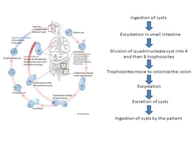
Invasion of the intestinal lining causes amoebic bloody diarrhea or amoebic
Invasion of the intestinal lining causes amoebic bloody diarrhea or amoebic
Transmission:
It is usually transmitted by fecal-oral route, but it can also
Transmission:
It is usually transmitted by fecal-oral route, but it can also
Diagnosis:
Microscopy of feces
Serological tests. Serology becomes positive about 2 weeks after
Diagnosis:
Microscopy of feces
Serological tests. Serology becomes positive about 2 weeks after
Prevention:
Wash hands with soap and hot running water
Clean bathrooms and toilets
Prevention:
Wash hands with soap and hot running water
Clean bathrooms and toilets
2. Parasitic ciliates
2. Parasitic ciliates
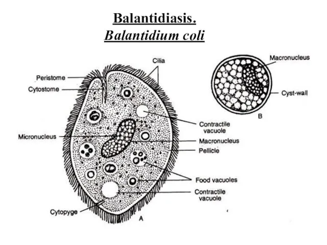
Balantidiasis.
Balantidium coli
Balantidiasis.
Balantidium coli
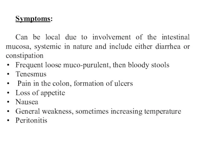
Symptoms:
Can be local due to involvement of the intestinal mucosa, systemic
Symptoms:
Can be local due to involvement of the intestinal mucosa, systemic
Transmission:
The main source of infection is the pig. But man, releasing
Transmission:
The main source of infection is the pig. But man, releasing
Prevention:
Is the same as in other intestinal infections: measures that prevent
Prevention:
Is the same as in other intestinal infections: measures that prevent
Home work. Task 1. Amoeba and ciliates
Home work. Task 1. Amoeba and ciliates
3. Parasitic Flagellates:
3.1. Intestinal and vaginal Flagellates
3. Parasitic Flagellates:
3.1. Intestinal and vaginal Flagellates
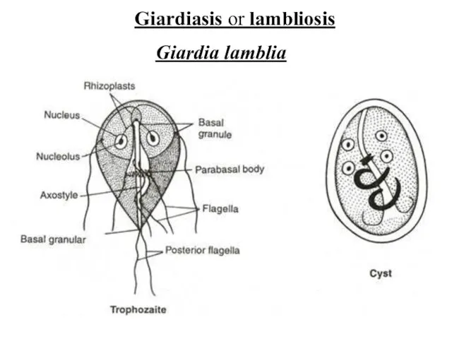
Giardiasis or lambliosis
Giardia lamblia
Giardiasis or lambliosis
Giardia lamblia
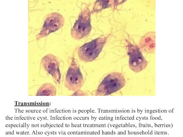
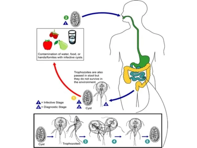
Transmission:
The source of infection is people. Transmission is by ingestion of
Transmission:
The source of infection is people. Transmission is by ingestion of
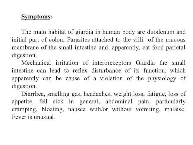
Symptoms:
The main habitat of giardia in human body are duodenum and
Symptoms:
The main habitat of giardia in human body are duodenum and
Diagnosis:
Antigene testing of the stool for the presence of giardial proteins
Examination
Diagnosis:
Antigene testing of the stool for the presence of giardial proteins
Examination
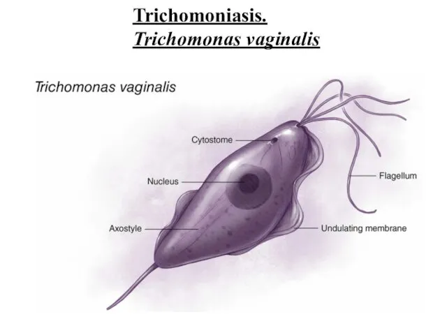
Trichomoniasis.
Trichomonas vaginalis
Trichomoniasis.
Trichomonas vaginalis
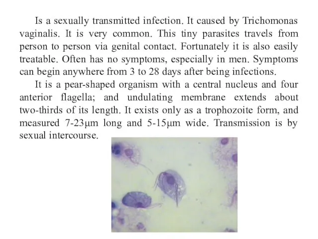
Is a sexually transmitted infection. It caused by Trichomonas vaginalis. It
Is a sexually transmitted infection. It caused by Trichomonas vaginalis. It
Symptoms:
Vaginal discharge, which may be white, gray, yellow or green and
Symptoms:
Vaginal discharge, which may be white, gray, yellow or green and
Diagnosis:
Physical test and laboratory test: cell culture, DNA examing samples of
Diagnosis:
Physical test and laboratory test: cell culture, DNA examing samples of
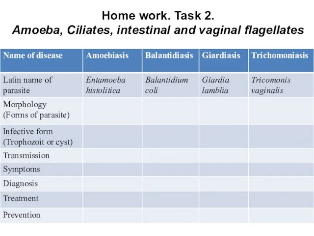
Home work. Task 2.
Amoeba, Ciliates, intestinal and vaginal flagellates
Home work. Task 2.
Amoeba, Ciliates, intestinal and vaginal flagellates
3. Parasitic Flagellates:
3.2. Hemoflagellates
3. Parasitic Flagellates:
3.2. Hemoflagellates
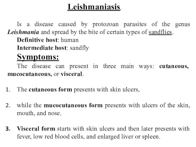
Leishmaniasis.
Is a disease caused by protozoan parasites of the genus Leishmania
Leishmaniasis.
Is a disease caused by protozoan parasites of the genus Leishmania

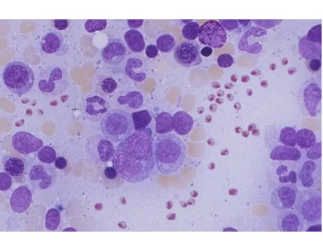
Morphology
Morphology
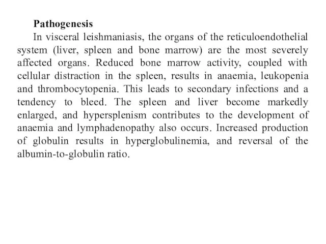
Pathogenesis
In visceral leishmaniasis, the organs of the reticuloendothelial system (liver, spleen
Pathogenesis
In visceral leishmaniasis, the organs of the reticuloendothelial system (liver, spleen
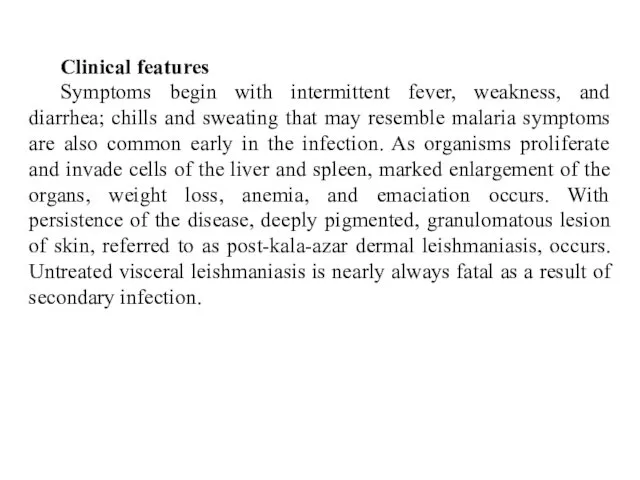
Clinical features
Symptoms begin with intermittent fever, weakness, and diarrhea; chills and
Clinical features
Symptoms begin with intermittent fever, weakness, and diarrhea; chills and
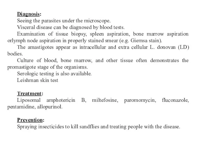
Diagnosis:
Seeing the parasites under the microscope.
Visceral disease can be
Diagnosis:
Seeing the parasites under the microscope.
Visceral disease can be

Trypanasomiasis
This disease caused by Tripanosoma species. In humans this include African
Trypanasomiasis
This disease caused by Tripanosoma species. In humans this include African
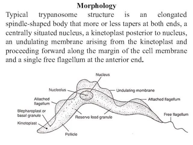
Morphology
Typical trypanosome structure is an elongated spindle-shaped body that more or
Morphology
Typical trypanosome structure is an elongated spindle-shaped body that more or
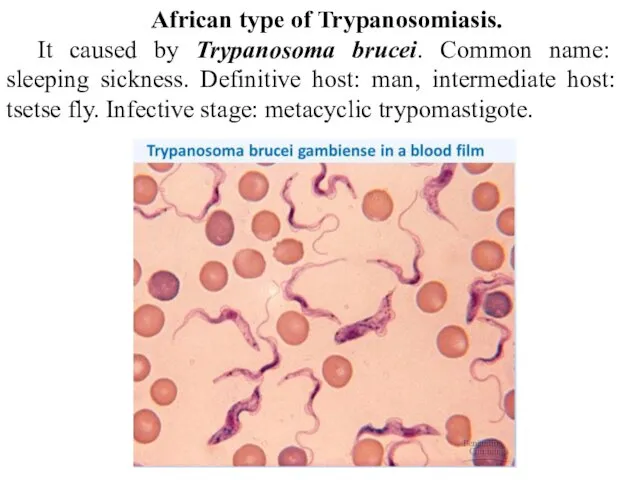
African type of Trypanosomiasis.
It caused by Trypanosoma brucei. Common name: sleeping
African type of Trypanosomiasis.
It caused by Trypanosoma brucei. Common name: sleeping
Symptoms
Transmission by tsetse fly.
The tsetse fly bite erupts into a
Symptoms
Transmission by tsetse fly.
The tsetse fly bite erupts into a
South-American type of Trypanosomiasis.
Common name: Chaga’s disease.
It caused by Trypanosoma
South-American type of Trypanosomiasis.
Common name: Chaga’s disease.
It caused by Trypanosoma
Symptoms of an acute form:
Blood and reticulo-endothelial cells predominantly involved
Fever
Oedema (lymph
Symptoms of an acute form:
Blood and reticulo-endothelial cells predominantly involved
Fever
Oedema (lymph
Diagnosis:
Often missed in the first phase of the disease due
Diagnosis:
Often missed in the first phase of the disease due
Home work. Task 3. Hemoflagellates
Home work. Task 3. Hemoflagellates
4. Sporozoa
4. Sporozoa
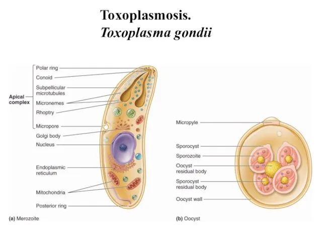
Toxoplasmosis.
Toxoplasma gondii
Toxoplasmosis.
Toxoplasma gondii
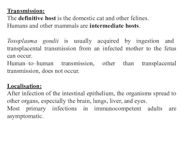
Transmission:
The definitive host is the domestic cat and other felines.
Humans
Transmission:
The definitive host is the domestic cat and other felines.
Humans
Symptoms:
Infection has 3 stages.
Acute toxoplasmosis. It is often asymptomatic. However, symptoms
Symptoms:
Infection has 3 stages.
Acute toxoplasmosis. It is often asymptomatic. However, symptoms
Diagnosis:
Biological, serological, histological or molecular methods.
May be detected in blood, amniotic
Diagnosis:
Biological, serological, histological or molecular methods.
May be detected in blood, amniotic
Malaria.
Plasmodium species
Is a mosquito-borne infectious disease of humans and other animals
Malaria.
Plasmodium species
Is a mosquito-borne infectious disease of humans and other animals

Symptoms:
Fever, fatigue, vomiting, headaches, anemia, hemoglobin in the urine. In severe
Symptoms:
Fever, fatigue, vomiting, headaches, anemia, hemoglobin in the urine. In severe
Treatment:
Because chloroquine – resistant stains of P.falciparum are present in many
Treatment:
Because chloroquine – resistant stains of P.falciparum are present in many
Home work. Task 4. Sporozoa
Home work. Task 4. Sporozoa
 Правильное питание – ваш путь к счастью и долголетию
 Ауаның ылғалдылығы, анықтау тәсілдері. Аэроионотерапия
 Орталық жүйке жүйесінің ноцецептивтік жүйесі. Неврологиядағы ауырсыну синдромы
 Воспалительные заболевания позвоночника. Хирургическое лечение
 Дифференциальная диагностика инфильтративных образований в легких. Лекция №10
 Түбір өзектерді аспаптармен өңдеу әдістер
 Клиническая фармакология бронхолитических препаратов
 Общая фармакология. Фармакокинетика
 Государственный санитарно-эпидемиологический надзор в Республике Казахстан
 Лечение заболеваний нервной системы человека с помощью иппотерапии
 Патофизиология водно-солевого обмена
 Диагностика инфаркта миокарда
 Медицинская реабилитация
 Государственная система управления здравоохранением. Современные формы управления в системе здравоохранения
 Медицина және денсаулық сақтау теориялары
 Моногенді аурулар
 Неотложная помощь при экстремальных состояниях у детей
 Медицина в зарубежных странах
 ВИЧ-инфекция и СПИД: без мифов и иллюзий
 Жедел аппендициттің асқынған түрінде хирургиялық тактика
 Анализ мокроты и плеврального выпота
 Военно-полевая хирургия
 Ишемическая болезнь сердца
 Инструкция по проведению генеральной уборки. Требования к уборочному инвентарю
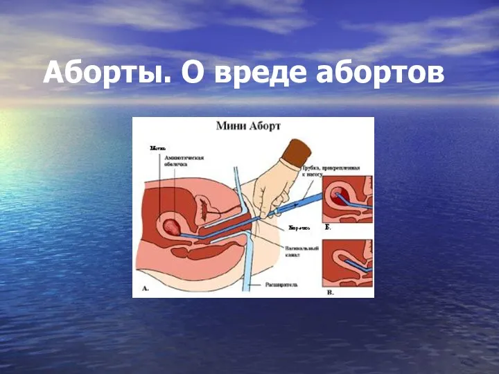 Аборты. О вреде абортов
 Первая медицинская помощь при остановке сердца
 Аппараты физиотерапевтические ЭСМА 12.20 Комби, ЭСМА 12.16 Универсал. Инструкция пользователя
 Современные принципы сердечно-легочной реанимации и профилактики внезапной сердечной смерти