- Medical protozoology sarcodina and flagellata

Содержание
- 2. General definitions Protozoa consist of a vast set of single-cell microorganisms that belong to protozoa phylum.
- 3. PHYLUM: PROTOZOA CLASS: Amoebae (Rhizopoda) SARCODINA CLASS: Ciliatae INFUZORIA CLASS: Telosporidea SPOROZOA CLASS: Zoomastigophora FLAGELLATA Entamoeba
- 4. PHYLUM: PROTOZOA protozoa inhabiting the gastrointestinal tract: In oral cavity In the small intestine In the
- 5. CLASS Amoebae (Rhizopoda) SARCODINA
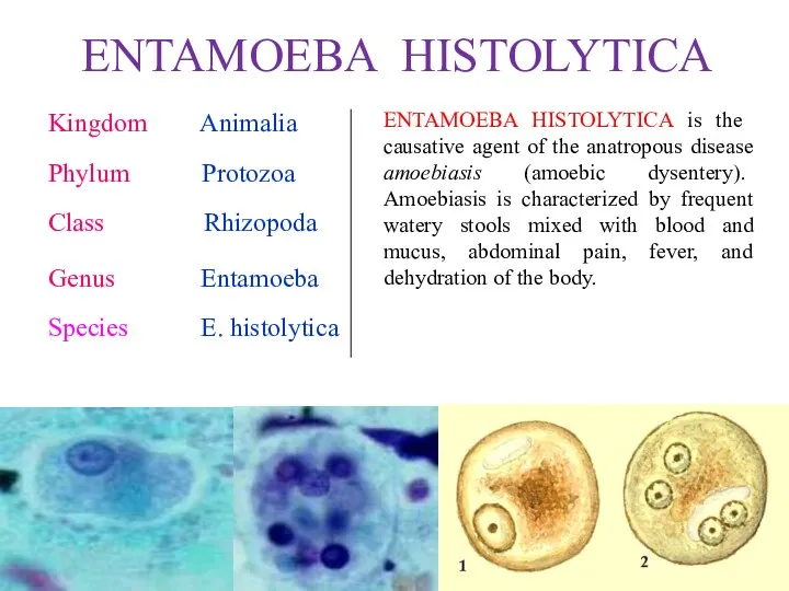
- 6. ENTAMOEBA HISTOLYTICA Kingdom Animalia Phylum Protozoa Class Rhizopoda Genus Entamoeba Species E. histolytica ENTAMOEBA HISTOLYTICA is
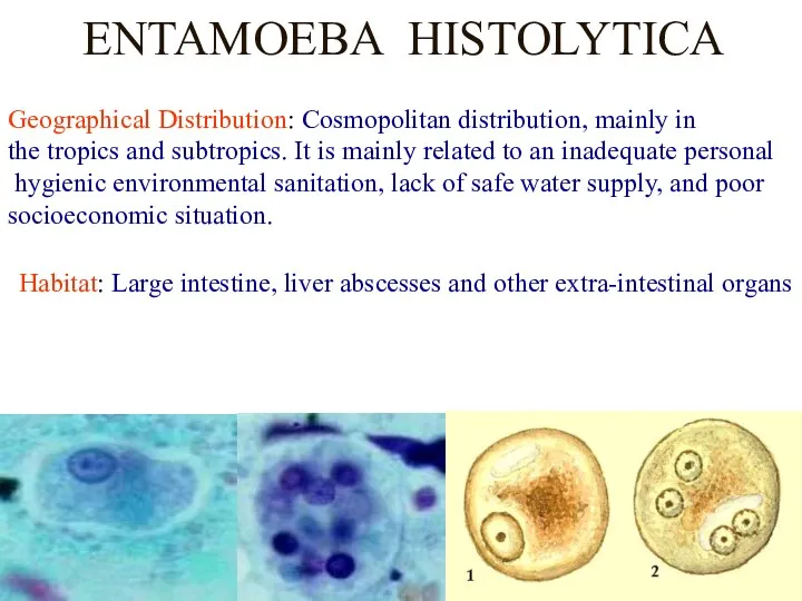
- 7. ENTAMOEBA HISTOLYTICA Geographical Distribution: Cosmopolitan distribution, mainly in the tropics and subtropics. It is mainly related
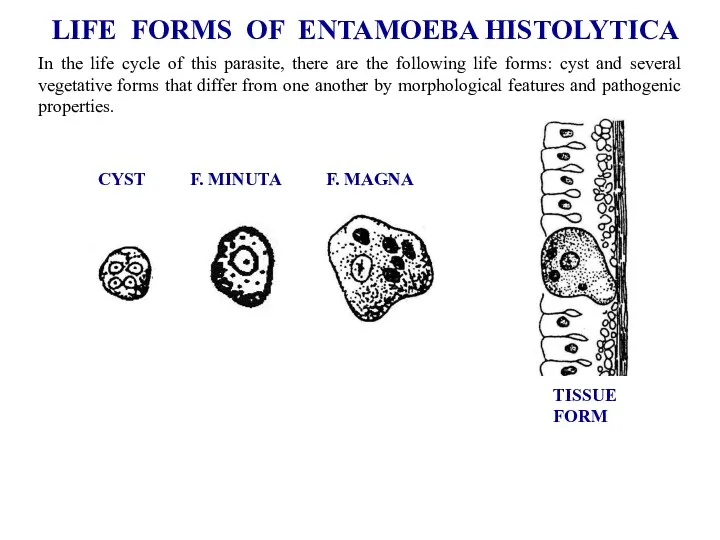
- 8. LIFE FORMS OF ENTAMOEBA HISTOLYTICA CYST F. MINUTA F. MAGNA TISSUE FORM In the life cycle
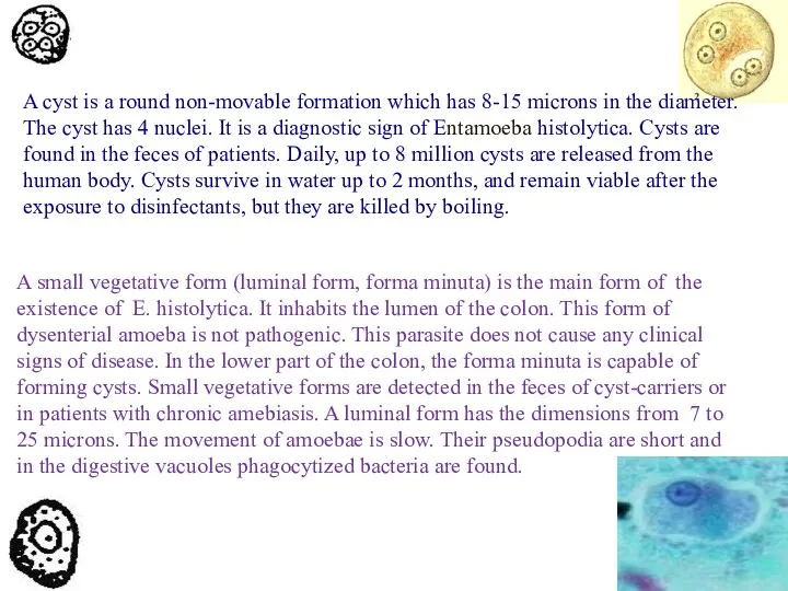
- 9. A cyst is a round non-movable formation which has 8-15 microns in the diameter. The cyst
- 10. A large vegetative form (forma magna) is a large cell which has the size from 20
- 11. The tissue form is also a pathogenic stage of dysenteric amoeba. Its size is 20-25 microns.
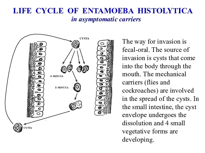
- 12. LIFE CYCLE OF ENTAMOEBA HISTOLYTICA in asymptomatic carriers The way for invasion is fecal-oral. The source
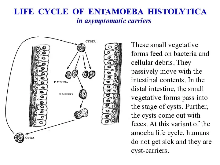
- 13. LIFE CYCLE OF ENTAMOEBA HISTOLYTICA in asymptomatic carriers These small vegetative forms feed on bacteria and
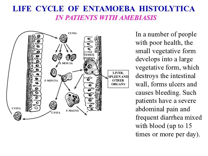
- 14. LIFE CYCLE OF ENTAMOEBA HISTOLYTICA IN PATIENTS WITH AMEBIASIS In a number of people with poor
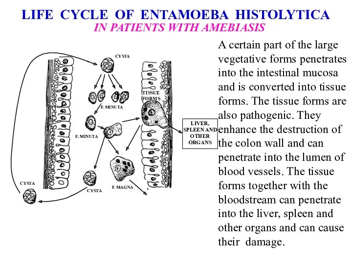
- 15. LIFE CYCLE OF ENTAMOEBA HISTOLYTICA IN PATIENTS WITH AMEBIASIS A certain part of the large vegetative
- 16. Patients with amoebic dysentery must be hospitalized. In the absence of proper treatment, such patients have
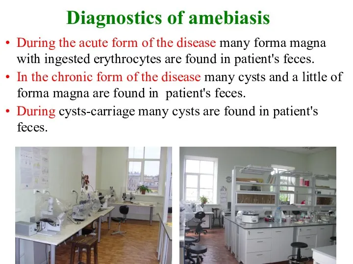
- 17. Diagnostics of amebiasis During the acute form of the disease many forma magna with ingested erythrocytes
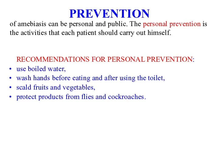
- 18. PREVENTION RECOMMENDATIONS FOR PERSONAL PREVENTION: use boiled water, wash hands before eating and after using the
- 19. RECOMMENDATIONS FOR PUBLIC PREVENTION: closing of access to local water-sources, import of fresh water, identification and
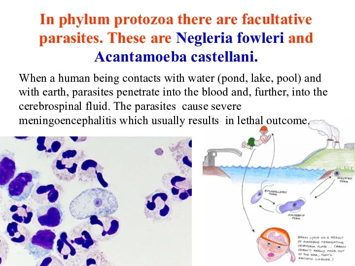
- 20. In phylum protozoa there are facultative parasites. These are Negleria fowleri and Acantamoeba castellani. When a
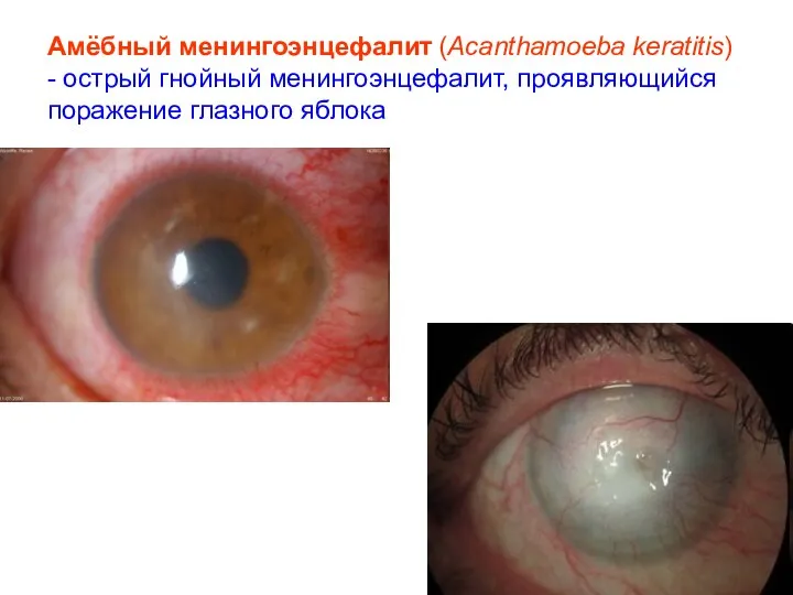
- 21. Амёбный менингоэнцефалит (Acanthamoeba keratitis) - острый гнойный менингоэнцефалит, проявляющийся поражение глазного яблока
- 22. CLASS ZOOMASTIGOPHORA (FLAGELLATA)
- 23. All members of the flagellata class can be divided into two groups: parasites which have one
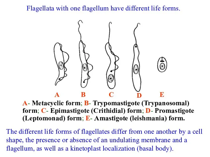
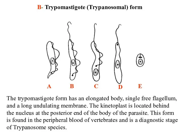
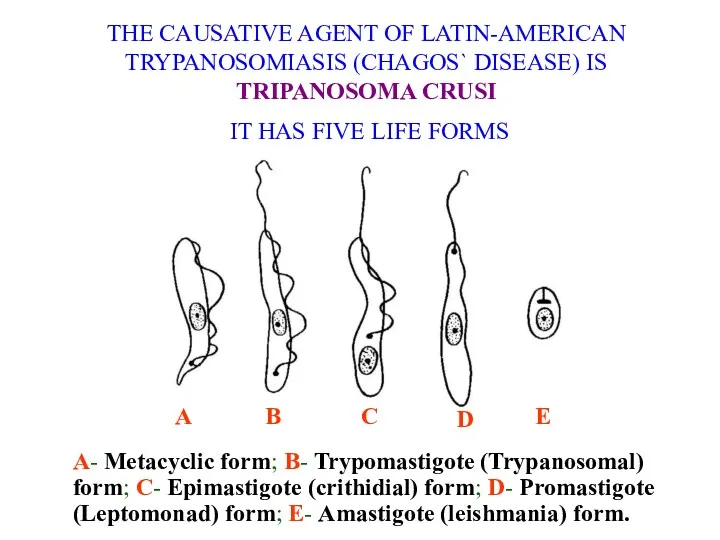
- 24. Flagellata with one flagellum have different life forms. А- Metacyclic form; B- Trypomastigote (Trypanosomal) form; C-
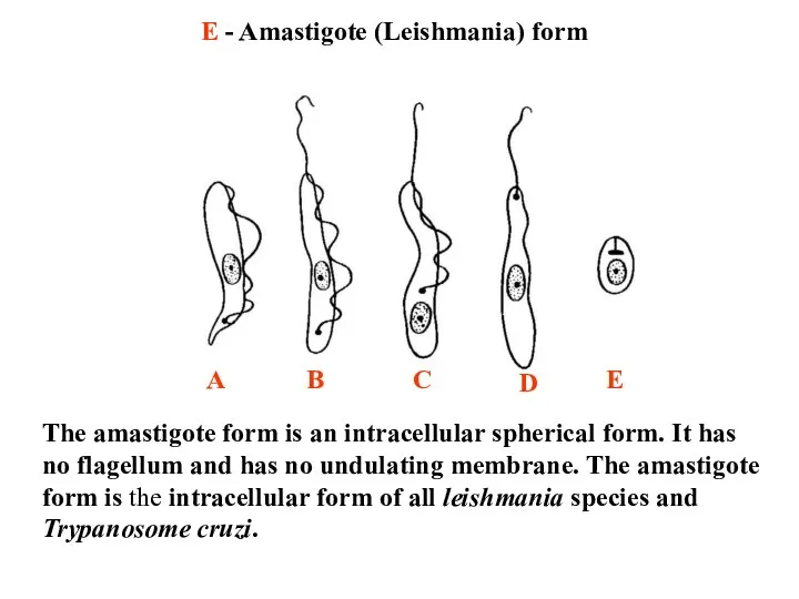
- 25. E - Amastigote (Leishmania) form The amastigote form is an intracellular spherical form. It has no
- 26. D - Promastigote (Leptomonad) form The promastigote form has an elongated body and a free flagellum
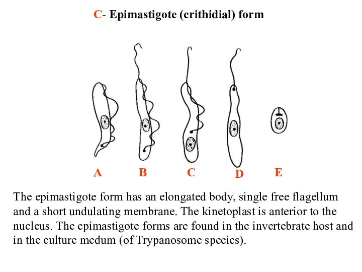
- 27. C- Epimastigote (crithidial) form The epimastigote form has an elongated body, single free flagellum and a
- 28. B- Trypomastigote (Trypanosomal) form The trypomastigote form has an elongated body, single free flagellum, and a
- 29. А- Metacyclic form The metacyclic form is morphologically similar to trypomastigote stage but it has no
- 30. The causative agents of leishmaniasis
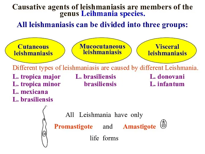
- 31. Causative agents of leishmaniasis are members of the genus Leihmania species. All leishmaniasis can be divided
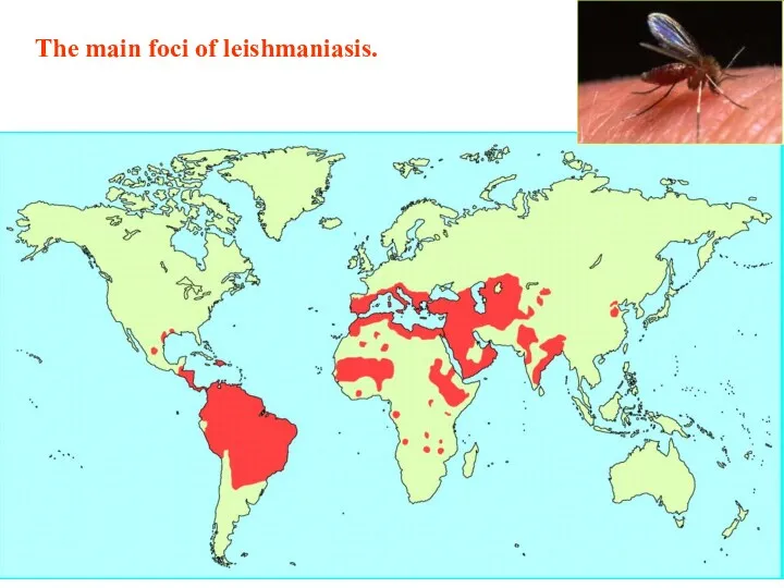
- 32. The main foci of leishmaniasis.
- 33. The life cycle of the causative agent of cutaneous leishmaniasis on the example of L. tropica
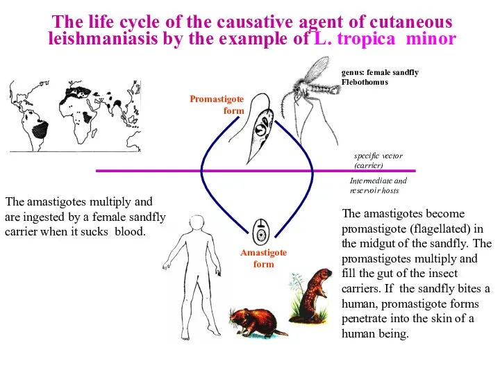
- 34. The life cycle of the causative agent of cutaneous leishmaniasis by the example of L. tropica
- 35. At the place of a bite, there develops dry painless ulcer, 25-70 mm in diameter, usually
- 36. THE CUTANEOUS LEISHMANIASIS
- 37. The life cycle of the causative agent of visceral leishmaniasis on the example of L. donovani
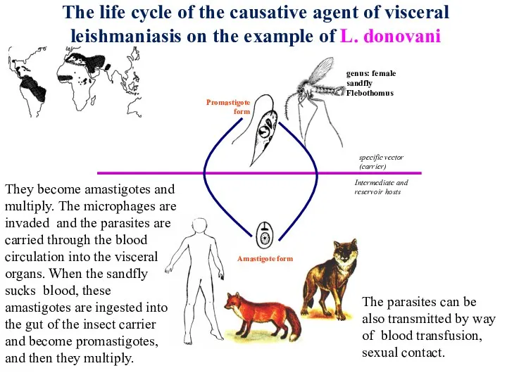
- 38. The life cycle of the causative agent of visceral leishmaniasis on the example of L. donovani
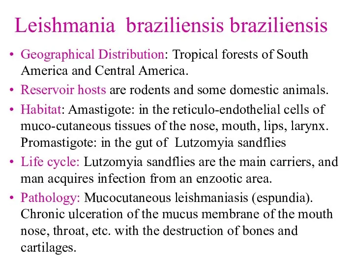
- 39. Leishmania braziliensis braziliensis Geographical Distribution: Tropical forests of South America and Central America. Reservoir hosts are
- 40. Laboratory Diagnosis of Leishmania species: Сutaneous and mucocutaneous leishmaniasis: 1. Amastigotes are revealed in stained smears
- 41. PREVENTION RECOMMENDATIONS FOR PUBLIC PREVENTION : Treatment of infected individuals, Destruction of specific carriers, Destruction of
- 42. The causative agents of African sleeping sickness (African trypanosomiasis)
- 43. Causative agents of African sleeping sickness are members of the species Tripanosoma brucei. There are two
- 44. The causative agents of African sleeping sickness have three forms of life: Trypomastigote (Trypanosomalform) Epimastigote (crithidial)
- 45. The African sleeping sickness is a natural-focal disease. It is typical of the East, West, and
- 46. The life cycle of Tripanosoma brucei gambiense Glossina palpalis Metacyclic form Epimastigote (crithidial) form The gut
- 47. The life cycle of Tripanosoma brucei rhodesiense Glossina morsitans Metacyclic form Epimastigote (crithidial) form gut The
- 48. Patients with African sleeping sickness They are not physically active. They stay for a long time
- 49. African sleeping sickness
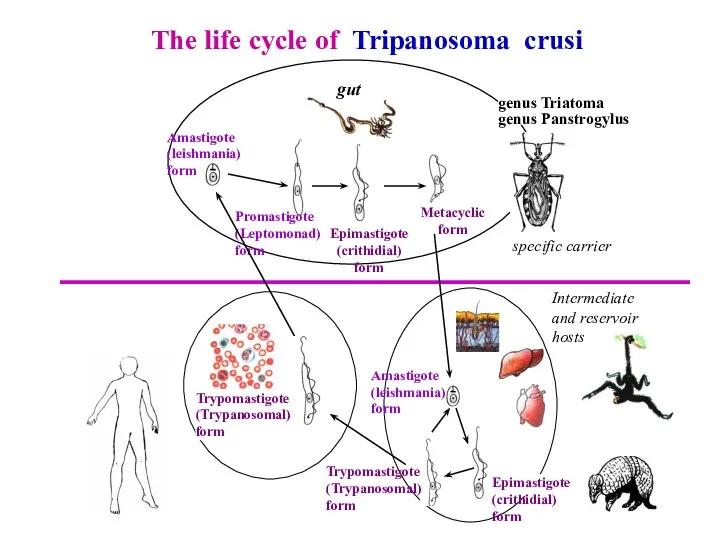
- 50. THE CAUSATIVE AGENT OF LATIN-AMERICAN TRYPANOSOMIASIS (CHAGOS` DISEASE) IS TRIPANOSOMA CRUSI IT HAS FIVE LIFE FORMS
- 51. The life cycle of Tripanosoma crusi genus Triatoma genus Panstrogylus Metacyclic form Epimastigote (crithidial) form gut
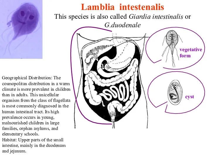
- 52. Lamblia intestenalis This species is also called Giardia intestinalis or G.duodenale Geographical Distribution: The cosmopolitan distribution
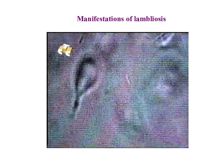
- 53. Manifestations of lambliosis
- 54. Trichomonas vaginalis A sick person A healthy person Sexual contacts gynecological instruments Using common hygiene products,
- 55. TRICHOMONAS HOMINIS
- 57. Скачать презентацию
General definitions
Protozoa consist of a vast set of single-cell microorganisms that
General definitions
Protozoa consist of a vast set of single-cell microorganisms that
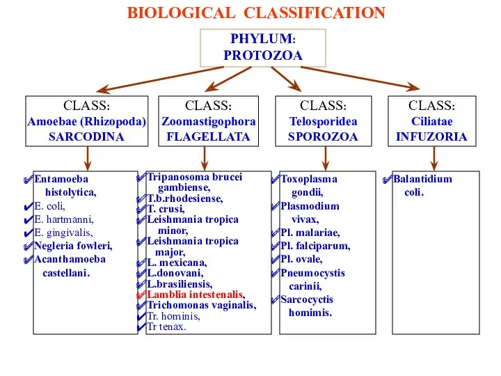
PHYLUM: PROTOZOA
CLASS:
Amoebae (Rhizopoda)
SARCODINA
CLASS:
Ciliatae
INFUZORIA
CLASS:
Telosporidea
SPOROZOA
CLASS:
Zoomastigophora
FLAGELLATA
Entamoeba
histolytica,
E. coli,
E. hartmanni,
E.
PHYLUM: PROTOZOA
CLASS:
Amoebae (Rhizopoda)
SARCODINA
CLASS:
Ciliatae
INFUZORIA
CLASS:
Telosporidea
SPOROZOA
CLASS:
Zoomastigophora
FLAGELLATA
Entamoeba
histolytica,
E. coli,
E. hartmanni,
E.
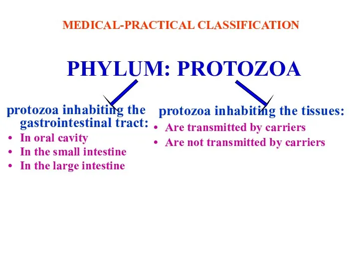
PHYLUM: PROTOZOA
protozoa inhabiting the gastrointestinal tract:
In oral cavity
In the small intestine
PHYLUM: PROTOZOA
protozoa inhabiting the gastrointestinal tract:
In oral cavity
In the small intestine
CLASS
Amoebae (Rhizopoda)
SARCODINA
CLASS
Amoebae (Rhizopoda)
SARCODINA
ENTAMOEBA HISTOLYTICA
Kingdom Animalia
Phylum Protozoa
Class Rhizopoda
Genus Entamoeba
Species E. histolytica
ENTAMOEBA
ENTAMOEBA HISTOLYTICA
Kingdom Animalia
Phylum Protozoa
Class Rhizopoda
Genus Entamoeba
Species E. histolytica
ENTAMOEBA
ENTAMOEBA HISTOLYTICA
Geographical Distribution: Cosmopolitan distribution, mainly in
the tropics and subtropics.
ENTAMOEBA HISTOLYTICA
Geographical Distribution: Cosmopolitan distribution, mainly in
the tropics and subtropics.
LIFE FORMS OF ENTAMOEBA HISTOLYTICA
CYST
F. MINUTA
F. MAGNA
TISSUE FORM
In the life cycle
LIFE FORMS OF ENTAMOEBA HISTOLYTICA
CYST
F. MINUTA
F. MAGNA
TISSUE FORM
In the life cycle
A cyst is a round non-movable formation which has 8-15 microns
A cyst is a round non-movable formation which has 8-15 microns
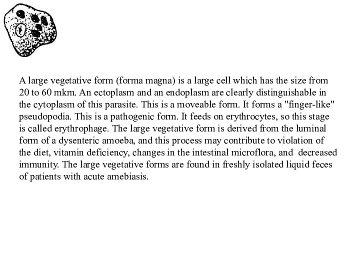
A large vegetative form (forma magna) is a large cell which
A large vegetative form (forma magna) is a large cell which
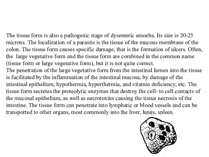
The tissue form is also a pathogenic stage of dysenteric amoeba.
The tissue form is also a pathogenic stage of dysenteric amoeba.
LIFE CYCLE OF ENTAMOEBA HISTOLYTICA
in asymptomatic carriers
The way for invasion
LIFE CYCLE OF ENTAMOEBA HISTOLYTICA
in asymptomatic carriers
The way for invasion
LIFE CYCLE OF ENTAMOEBA HISTOLYTICA
in asymptomatic carriers
These small vegetative forms
LIFE CYCLE OF ENTAMOEBA HISTOLYTICA
in asymptomatic carriers
These small vegetative forms
LIFE CYCLE OF ENTAMOEBA HISTOLYTICA
IN PATIENTS WITH AMEBIASIS
In a number of
LIFE CYCLE OF ENTAMOEBA HISTOLYTICA
IN PATIENTS WITH AMEBIASIS
In a number of
LIFE CYCLE OF ENTAMOEBA HISTOLYTICA
IN PATIENTS WITH AMEBIASIS
A certain part of
LIFE CYCLE OF ENTAMOEBA HISTOLYTICA
IN PATIENTS WITH AMEBIASIS
A certain part of
Patients with amoebic dysentery must be hospitalized. In the absence of
Patients with amoebic dysentery must be hospitalized. In the absence of
Diagnostics of amebiasis
During the acute form of the disease many forma
Diagnostics of amebiasis
During the acute form of the disease many forma
PREVENTION
RECOMMENDATIONS FOR PERSONAL PREVENTION:
use boiled water,
wash hands before eating
PREVENTION
RECOMMENDATIONS FOR PERSONAL PREVENTION:
use boiled water,
wash hands before eating

RECOMMENDATIONS FOR PUBLIC PREVENTION:
closing of access to local water-sources,
import
RECOMMENDATIONS FOR PUBLIC PREVENTION:
closing of access to local water-sources,
import
In phylum protozoa there are facultative parasites. These are Negleria fowleri
In phylum protozoa there are facultative parasites. These are Negleria fowleri
Амёбный менингоэнцефалит (Acanthamoeba keratitis) - острый гнойный менингоэнцефалит, проявляющийся поражение глазного
Амёбный менингоэнцефалит (Acanthamoeba keratitis) - острый гнойный менингоэнцефалит, проявляющийся поражение глазного
CLASS
ZOOMASTIGOPHORA
(FLAGELLATA)
CLASS
ZOOMASTIGOPHORA
(FLAGELLATA)
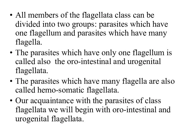
All members of the flagellata class can be divided into two
All members of the flagellata class can be divided into two
Flagellata with one flagellum have different life forms.
А- Metacyclic form; B-
Flagellata with one flagellum have different life forms.
А- Metacyclic form; B-
E - Amastigote (Leishmania) form
The amastigote form is an intracellular spherical
E - Amastigote (Leishmania) form
The amastigote form is an intracellular spherical

D - Promastigote (Leptomonad) form
The promastigote form has an elongated body
D - Promastigote (Leptomonad) form
The promastigote form has an elongated body
C- Epimastigote (crithidial) form
The epimastigote form has an elongated body, single
C- Epimastigote (crithidial) form
The epimastigote form has an elongated body, single
B- Trypomastigote (Trypanosomal) form
The trypomastigote form has an elongated body, single
B- Trypomastigote (Trypanosomal) form
The trypomastigote form has an elongated body, single

А- Metacyclic form
The metacyclic form is morphologically similar to trypomastigote stage
А- Metacyclic form
The metacyclic form is morphologically similar to trypomastigote stage
The causative agents of leishmaniasis
The causative agents of leishmaniasis
Causative agents of leishmaniasis are members of the genus Leihmania species.
All
Causative agents of leishmaniasis are members of the genus Leihmania species. All
The main foci of leishmaniasis.
The main foci of leishmaniasis.

The life cycle of the causative agent of cutaneous leishmaniasis on
The life cycle of the causative agent of cutaneous leishmaniasis on
The life cycle of the causative agent of cutaneous leishmaniasis by
The life cycle of the causative agent of cutaneous leishmaniasis by

At the place of a bite, there develops dry painless ulcer,
At the place of a bite, there develops dry painless ulcer,
THE CUTANEOUS LEISHMANIASIS
THE CUTANEOUS LEISHMANIASIS

The life cycle of the causative agent of visceral leishmaniasis on
The life cycle of the causative agent of visceral leishmaniasis on
The life cycle of the causative agent of visceral leishmaniasis on
The life cycle of the causative agent of visceral leishmaniasis on
Leishmania braziliensis braziliensis
Geographical Distribution: Tropical forests of South America and
Leishmania braziliensis braziliensis
Geographical Distribution: Tropical forests of South America and
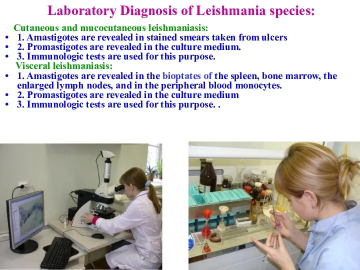
Laboratory Diagnosis of Leishmania species:
Сutaneous and mucocutaneous leishmaniasis:
1. Amastigotes are
Laboratory Diagnosis of Leishmania species:
Сutaneous and mucocutaneous leishmaniasis:
1. Amastigotes are
PREVENTION
RECOMMENDATIONS FOR PUBLIC PREVENTION :
Treatment of infected individuals,
Destruction of specific
PREVENTION
RECOMMENDATIONS FOR PUBLIC PREVENTION :
Treatment of infected individuals,
Destruction of specific

The causative agents
of African sleeping sickness
(African trypanosomiasis)
The causative agents
of African sleeping sickness
(African trypanosomiasis)
Causative agents of African sleeping sickness are members of the species
Causative agents of African sleeping sickness are members of the species
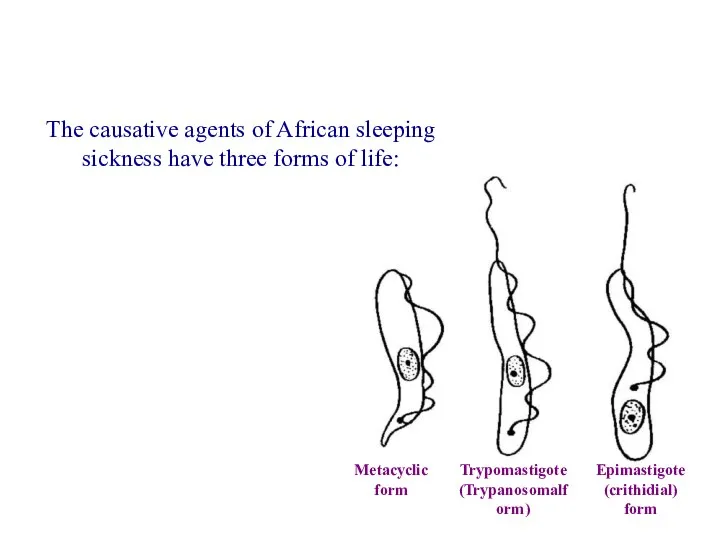
The causative agents of African sleeping sickness have three forms of
The causative agents of African sleeping sickness have three forms of
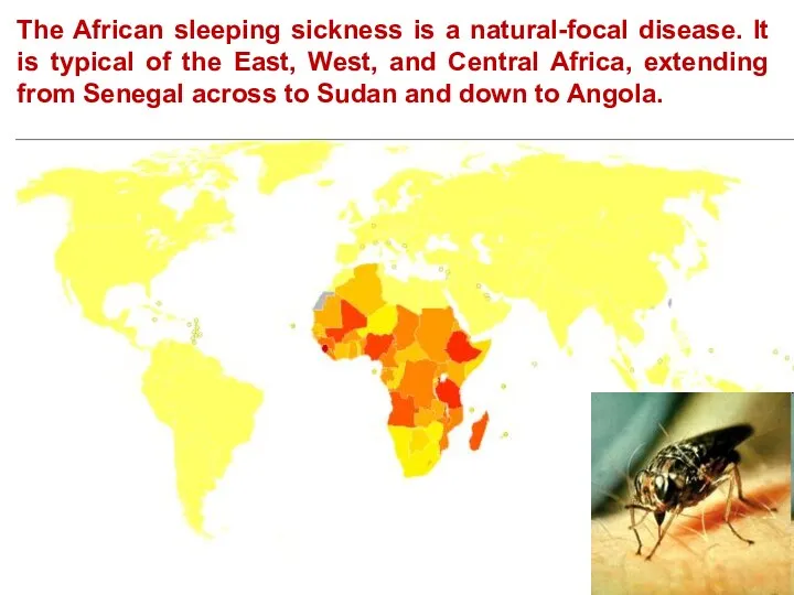
The African sleeping sickness is a natural-focal disease. It is typical
The African sleeping sickness is a natural-focal disease. It is typical

The life cycle of Tripanosoma brucei gambiense
Glossina palpalis
Metacyclic form
Epimastigote (crithidial) form
The
The life cycle of Tripanosoma brucei gambiense
Glossina palpalis
Metacyclic form
Epimastigote (crithidial) form
The
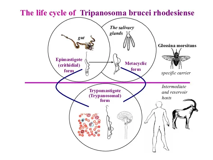
The life cycle of Tripanosoma brucei rhodesiense
Glossina morsitans
Metacyclic form
Epimastigote (crithidial) form
gut
The
The life cycle of Tripanosoma brucei rhodesiense
Glossina morsitans
Metacyclic form
Epimastigote (crithidial) form
gut
The
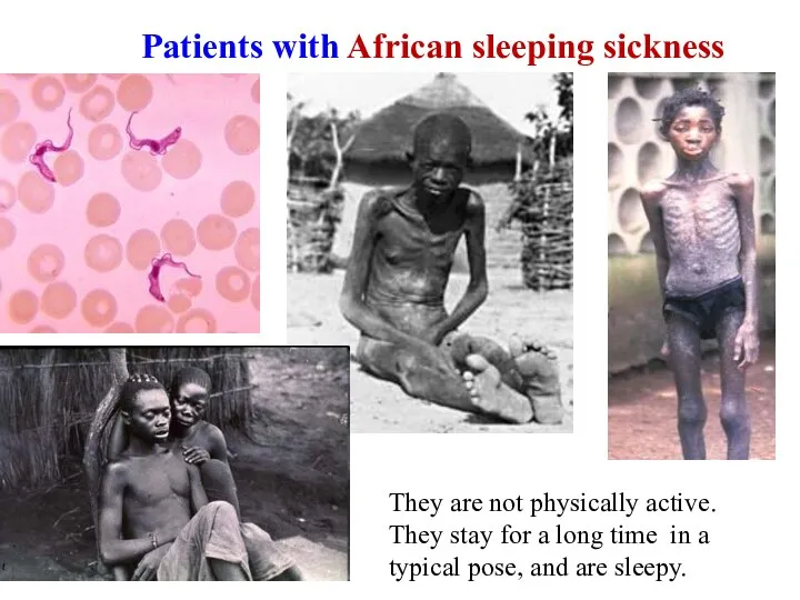
Patients with African sleeping sickness
They are not physically active. They
Patients with African sleeping sickness
They are not physically active. They
African sleeping sickness
African sleeping sickness
THE CAUSATIVE AGENT OF LATIN-AMERICAN TRYPANOSOMIASIS (CHAGOS` DISEASE) IS TRIPANOSOMA CRUSI
THE CAUSATIVE AGENT OF LATIN-AMERICAN TRYPANOSOMIASIS (CHAGOS` DISEASE) IS TRIPANOSOMA CRUSI
The life cycle of Tripanosoma crusi
genus Triatoma
genus Panstrogylus
Metacyclic form
Epimastigote (crithidial) form
gut
Amastigote
The life cycle of Tripanosoma crusi
genus Triatoma
genus Panstrogylus
Metacyclic form
Epimastigote (crithidial) form
gut
Amastigote
Lamblia intestenalis
This species is also called Giardia intestinalis or G.duodenale
Geographical Distribution:
Lamblia intestenalis
This species is also called Giardia intestinalis or G.duodenale
Geographical Distribution:
Manifestations of lambliosis
Manifestations of lambliosis
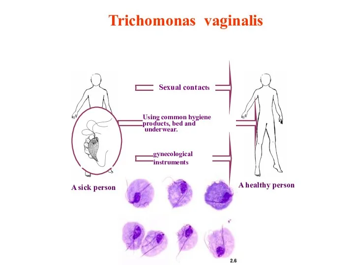
Trichomonas vaginalis
A sick person
A healthy person
Sexual contacts
gynecological instruments
Using common hygiene products,
Trichomonas vaginalis
A sick person
A healthy person
Sexual contacts
gynecological instruments
Using common hygiene products,
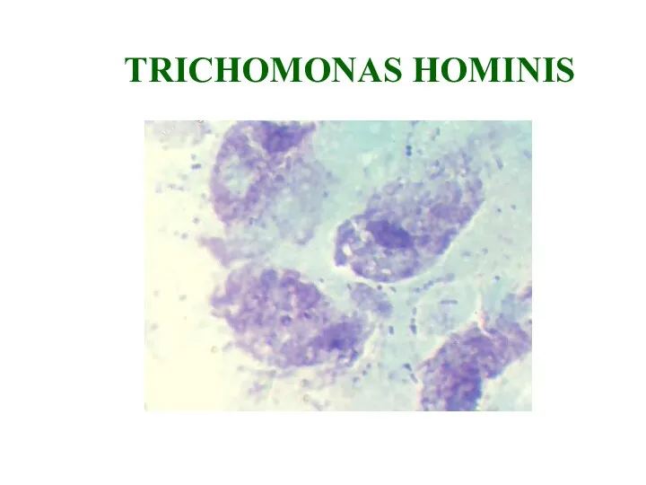
TRICHOMONAS HOMINIS
TRICHOMONAS HOMINIS
 III, IV, VI жұптар – көз алмасын қозғайтын, шығыр, әкеткіш нервтері
 Полиомиелит, энтеровирусные инфекции
 Перитонит. Классификация перитонита
 Нуклеопротеидттер алмасуының бұзылуы. Подагра
 Этиологическая и патогенетическая классификация анемий
 Туберкулез почек, мочевыводящей системы и мужских половых органов
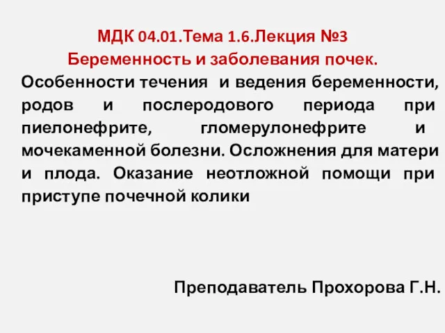 Беременность и заболевания почек
 Патофизиология экстремальных состояний
 Гіпертонічна хвороба
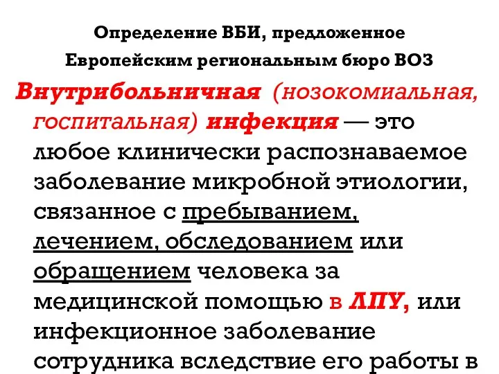 Внутрибольничные инфекции
 Медицинская защита населения и спасателей в чрезвычайных ситуациях
 Телесно-ориентированные техники в психологической работе с паллиативными детьми раннего и младенческого возраста
 Алгоритм действий медицинской сестры при почечной колике
 Тиреотоксикоз. Диффузный токсический зоб. Гипотиреоз
 Иммунитет как механизм регуляции и защиты
 Нейродегенеративные заболевания
 Модель Гиппократа и проблема доверия к профессии врача
 Классификация детей с ОВЗ
 Косметологиядағы криотерапия
 Компания Capsid Illuminesca. Вирусология
 Здоровый образ жизни
 Рациональное питание
 Государственная система управления здравоохранением. Современные формы управления в системе здравоохранения
 Гипс. Фиксация в кювете
 Стерилизация тиімділігін бақылау әдістері
 Местные средства профилактики кариеса
 Потери. Смерть. Горе
 Рак яичников