- The Functional Histology of Respiratory System

Содержание
- 2. The Respiratory System Function of the respiratory system 1.Replinish blood oxygen levels which is needed for
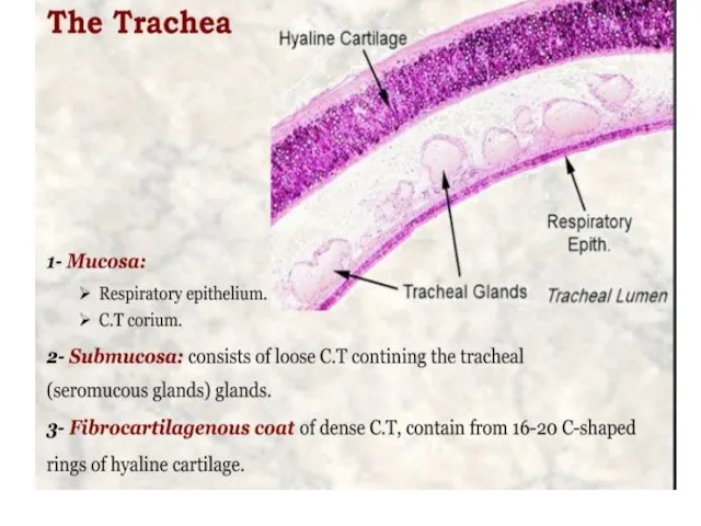
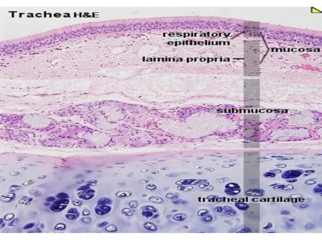
- 3. The Respiratory System consist of 1.Conducting portion, which consists of the nasal cavities, nasopharynx, larynx, trachea,
- 4. Conducting portion ciliated pseudostratified columnar epithelium
- 5. Cells of respiratory epithelium 1.Ciliated columnar cells are the most abundant, each with about 300 cilia
- 7. The nasal cavities (respiratory area) lie within the skull as two cavernous chambers separated by the
- 8. Swell bodies(cavernous bodies) Within the lamina propria of the conchae are large venous plexuses known as
- 9. cells of olfactory epithelium 1.Basal cells are small, spherical. They are the stem cells for the
- 10. bilateral cavities in the frontal, maxillary, ethmoid, and sphenoid bones of the skull
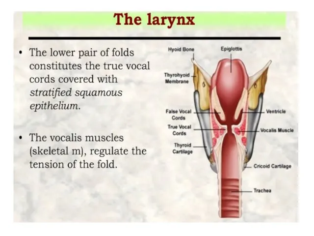
- 11. the thyroid, cricoid, and the inferior arytenoid epiglottis, cuneiform, corniculate, and the superior arytenoid cartilages
- 18. The trachea divides into two primary bronchi that enter the lungs at the hilum, the primary
- 19. BRONCHI 1.The walls of bronchi contain irregular plates of cartilage and circular smooth-muscle fascicles bound together
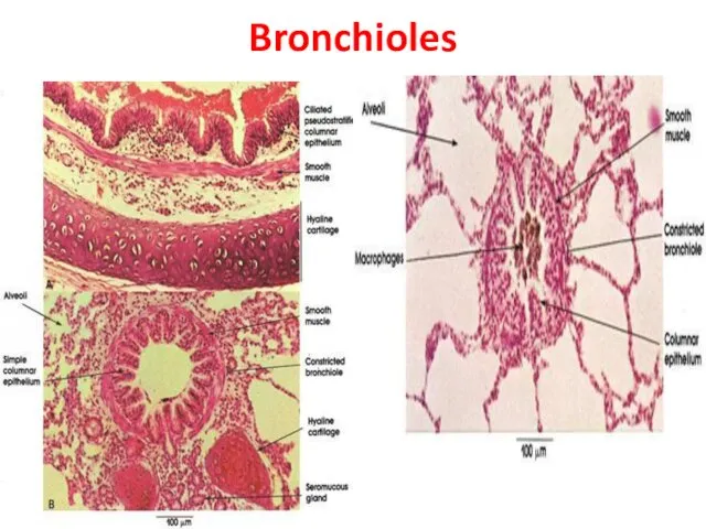
- 20. Bronchioles 1.Bronchioles are the intralobular airways with diameters of 5 mm or less, formed after about
- 21. Bronchioles
- 22. Clara cells 1.These mitotically active cells 2.secrete surfactant components 3.and have various important defensive roles. 4.are
- 23. Function of conducting portion 1.Dry air: moisten it by goblet cells and mucous. 2. Particles: trap
- 24. Respiratory portion The respiratory bronchioles mucosa is structurally identical to that of the terminal bronchioles, except
- 25. Alveolar Ducts and sacs 1.Respiratory bronchioles branch into tubes called alveolar ducts that are completely lined
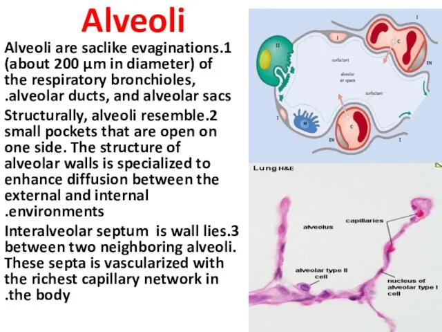
- 26. Alveoli 1.Alveoli are saclike evaginations (about 200 µm in diameter) of the respiratory bronchioles, alveolar ducts,
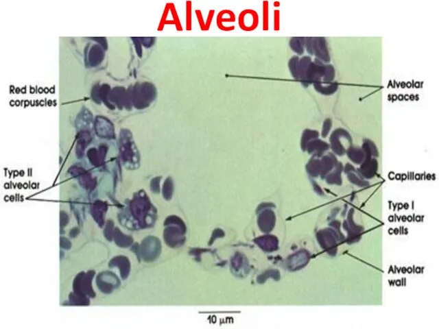
- 27. The cells of alveoli 1.Type I alveolar cells are extremely attenuated cells that line the alveolar
- 28. Alveoli
- 30. Скачать презентацию
The Respiratory System
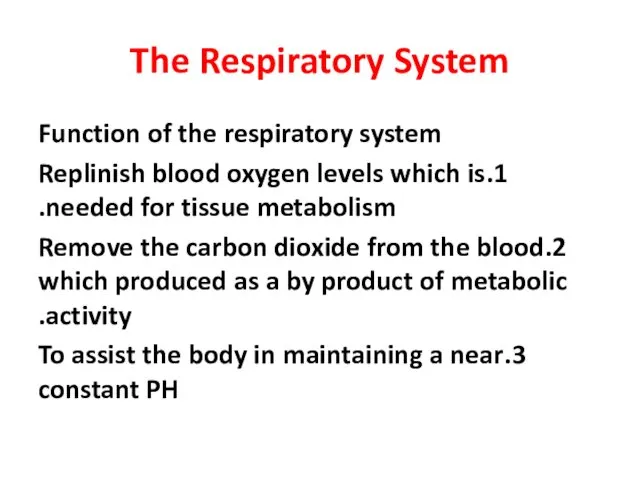
Function of the respiratory system
1.Replinish blood oxygen levels which
The Respiratory System
Function of the respiratory system
1.Replinish blood oxygen levels which
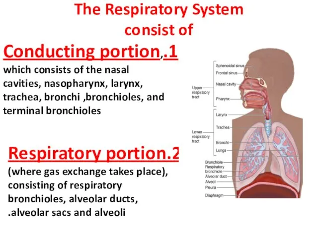
The Respiratory System
consist of
1.Conducting portion, which consists of the nasal cavities,
The Respiratory System
consist of
1.Conducting portion, which consists of the nasal cavities,
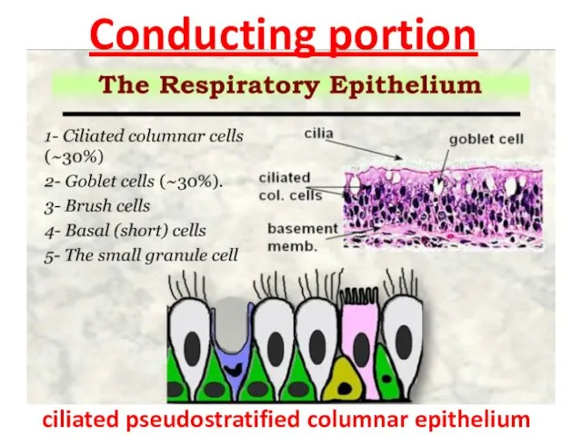
Conducting portion
ciliated pseudostratified columnar epithelium
Conducting portion
ciliated pseudostratified columnar epithelium
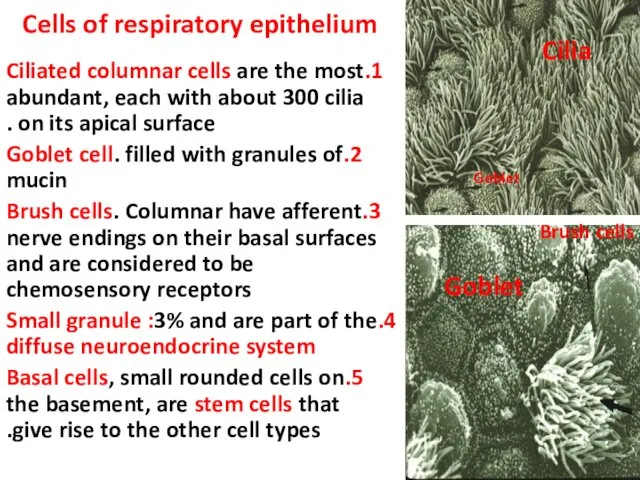
Cells of respiratory epithelium
1.Ciliated columnar cells are the most abundant, each
Cells of respiratory epithelium
1.Ciliated columnar cells are the most abundant, each
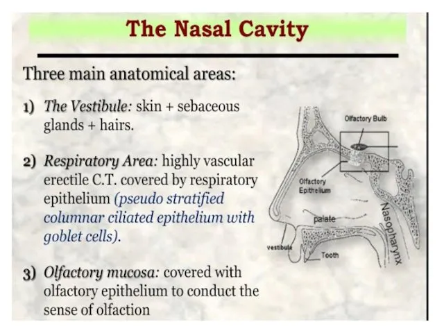
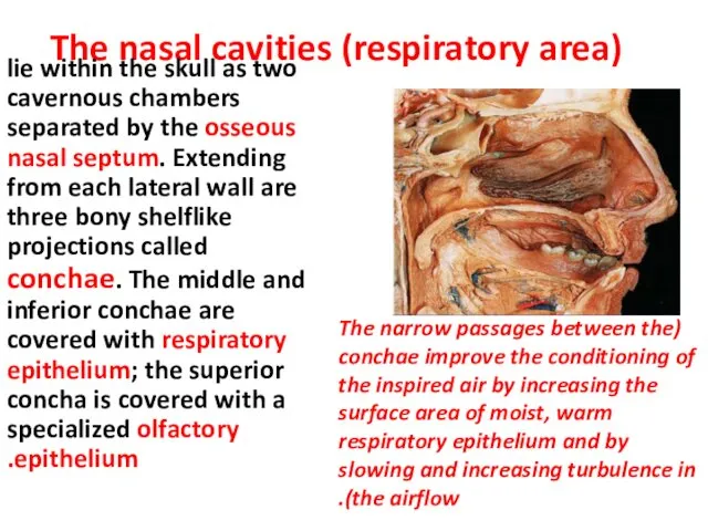
The nasal cavities (respiratory area)
lie within the skull as
The nasal cavities (respiratory area)
lie within the skull as
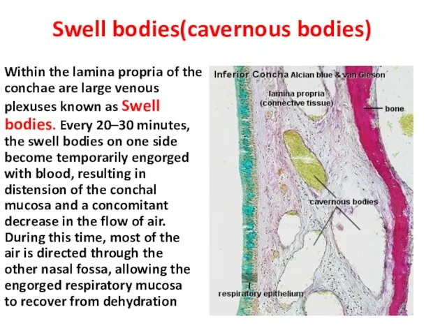
Swell bodies(cavernous bodies)
Within the lamina propria of the conchae are large
Swell bodies(cavernous bodies)
Within the lamina propria of the conchae are large
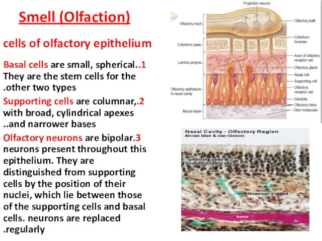
cells of olfactory epithelium
1.Basal cells are small, spherical. They are
cells of olfactory epithelium
1.Basal cells are small, spherical. They are
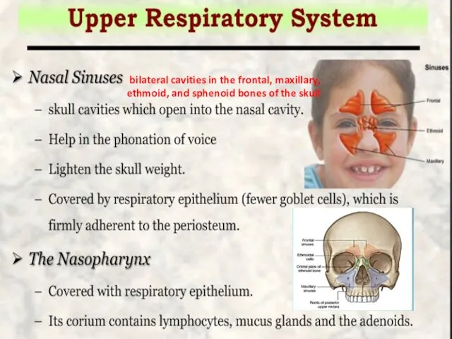
bilateral cavities in the frontal, maxillary, ethmoid, and sphenoid bones of
bilateral cavities in the frontal, maxillary, ethmoid, and sphenoid bones of
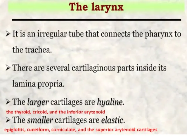
the thyroid, cricoid, and the inferior arytenoid
epiglottis, cuneiform, corniculate, and
the thyroid, cricoid, and the inferior arytenoid
epiglottis, cuneiform, corniculate, and
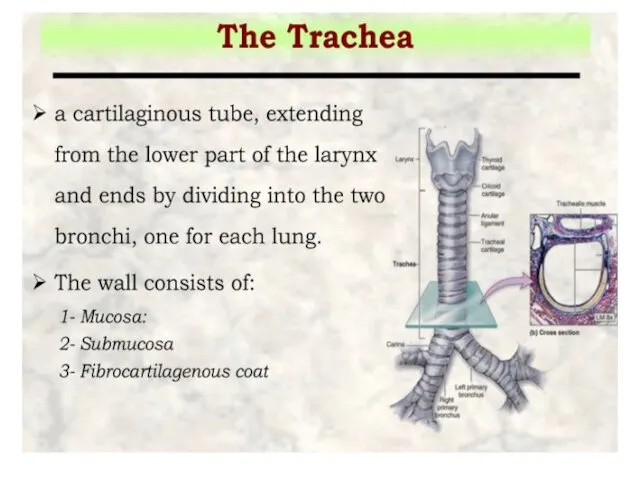
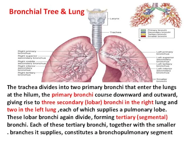
The trachea divides into two primary bronchi that enter the lungs
The trachea divides into two primary bronchi that enter the lungs
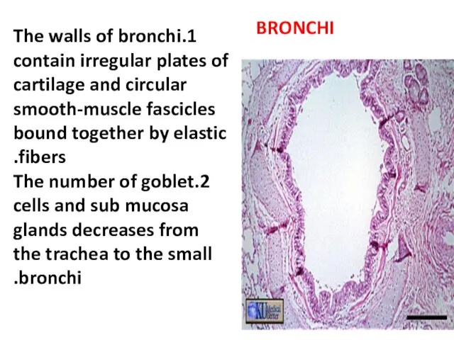
BRONCHI
1.The walls of bronchi contain irregular plates of cartilage and circular
BRONCHI
1.The walls of bronchi contain irregular plates of cartilage and circular
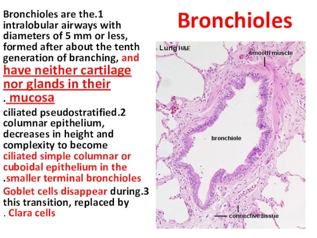
Bronchioles
1.Bronchioles are the intralobular airways with diameters of 5 mm or
Bronchioles
1.Bronchioles are the intralobular airways with diameters of 5 mm or
Bronchioles
Bronchioles
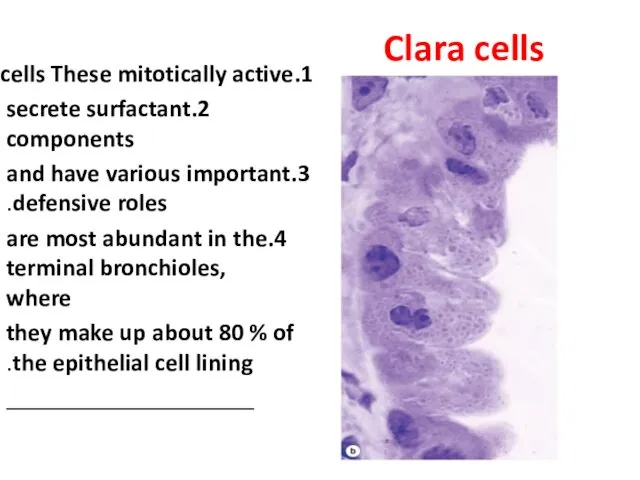
Clara cells
1.These mitotically active cells
2.secrete surfactant components
3.and have various
Clara cells
1.These mitotically active cells
2.secrete surfactant components
3.and have various
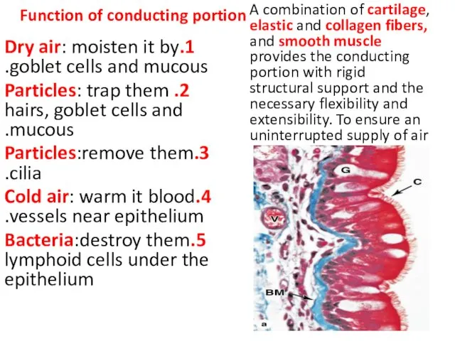
Function of conducting portion
1.Dry air: moisten it by goblet cells
Function of conducting portion
1.Dry air: moisten it by goblet cells
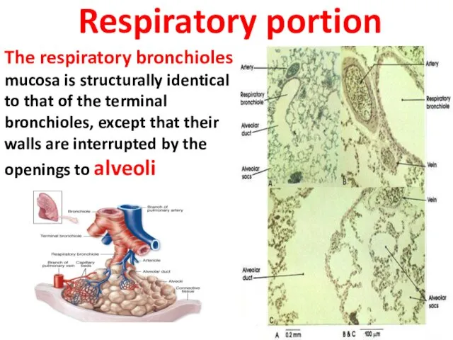
Respiratory portion
The respiratory bronchioles mucosa is structurally identical to that of
Respiratory portion
The respiratory bronchioles mucosa is structurally identical to that of
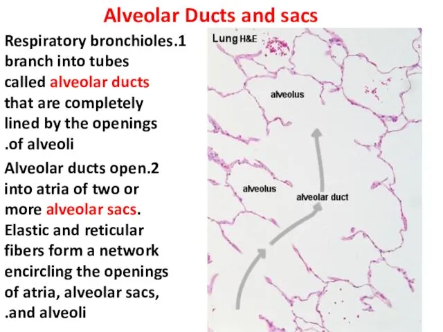
Alveolar Ducts and sacs
1.Respiratory bronchioles branch into tubes called alveolar ducts
Alveolar Ducts and sacs
1.Respiratory bronchioles branch into tubes called alveolar ducts
Alveoli
1.Alveoli are saclike evaginations (about 200 µm in diameter) of the
Alveoli
1.Alveoli are saclike evaginations (about 200 µm in diameter) of the

The cells of alveoli
1.Type I alveolar cells are extremely attenuated cells
The cells of alveoli
1.Type I alveolar cells are extremely attenuated cells
Alveoli
Alveoli
 Физиология сердечно-сосудистой системы. (Лекция 14)
 Медицинские средства защиты
 Қарыншалар гипертрофиясының ЭКГ белгілері
 Шистосомозы. Этиологиясы. Эпидемиологиясы. Патогенезі. Классификациясы. Клиникасы. Диагностикасы. Екшеу диагностикасы. Емі
 Введение в медицинскую информатику. Определения, терминология
 Заманауи эндодонттық аспаптар
 СПИД и его профилактика
 Юношеская дисфункция ВНЧС
 Анемия. Анемияны емдеу және одан сақтану шаралары
 Болезнь крона
 Особенности применения базисных противовоспалительных препаратов при беременности, лактации и у пожилых
 Комплексное лечение зубочелюстных аномалий и деформаций
 Иммунопатология. Патологическая анатомия
 Асқазан-12-елі ішіктің ойық жара аурулары
 Противотуберкулезные и противосифилитические препараты
 Рак яичников
 Мастоидит, түрлері, клиникасы, диагностикасы, емі
 Беттің туа біткен патологиясын алдын алу. Беттің, жақсүйектерінің және ауыз қуысының ағзаларының ақаулары болған кездегі
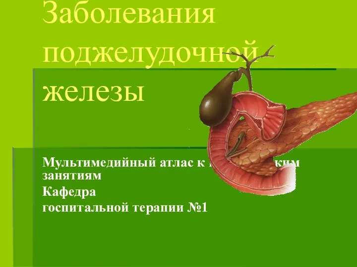 Заболевания поджелудочной железы
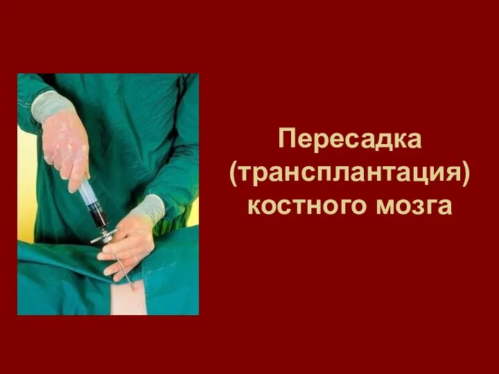 Пересадка (трансплантация) костного мозга
 Наркотики и их влияние на развитие полноценной личности
 Профилактика вирусных инфекций
 Хроническое воспаление. Гранулематозное и специфическое воспаление
 Восстановление двигательного акта (навыка)
 Моральные проблемы суррогатного материнства
 Влияние приобретенного дальтонизма на социальную адаптацию (социализацию) молодежи в России
 Известный наркотик СНЮС
 Фармациядағы менеджменттің функционалдық мазмұны. Басқару шешімдері