- Tooth structure. Modal verbs: can

Содержание
- 2. The tooth has three anatomical parts :
- 3. The crown of a tooth is that part of the tooth which is covered with enamel
- 4. Neck The neck of a tooth is the part of a tooth that is located at
- 5. The root is the part embedded in the jaw. It anchors the tooth in its bony
- 6. Teeth are made of hard tissues that protect the pulp located in the middle. Dental anatomy
- 7. Enamel is the hardest and most highly mineralized substance of the body ; 96% of enamel
- 8. Cementum is a specialized bone like substance covering the root of a tooth. Its coloration is
- 9. Dentin is the substance between enamel or cementum and the pulp chamber. It forms the highest
- 10. The dental pulp (also called "the nerve" of the tooth) is the central part of the
- 11. The crown of a tooth contains the coronal pulp. The coronal pulp is the largest mass
- 12. The radicular pulp is that pulp extending from the cervical region of the crown (where the
- 13. The periapical tissue (or apical periodontium) is located underneath the tooth ; basically, the radicular pulp
- 14. The dental speciality concerned with the study and treatment of dental pulp and periapical tissue infections
- 15. Modal verbs are a part of the larger category called auxiliary verbs which are verbs that
- 16. (+) Subject + Modal Verb + Verb (base form of the infinitive) (-) Subject + Modal
- 18. Скачать презентацию
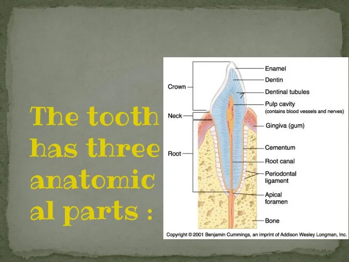
The tooth has three anatomical parts :
The tooth has three anatomical parts :
The crown of a tooth is that part of the tooth
The crown of a tooth is that part of the tooth
Neck
The neck of a tooth is the part of a tooth
Neck
The neck of a tooth is the part of a tooth
The root is the part embedded in the jaw. It anchors
The root is the part embedded in the jaw. It anchors
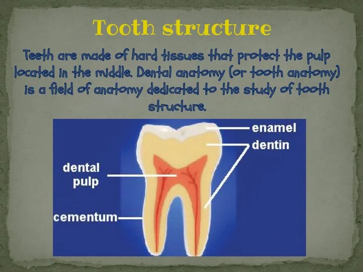
Teeth are made of hard tissues that protect the pulp located
Teeth are made of hard tissues that protect the pulp located
Enamel is the hardest and most highly mineralized substance of the
Enamel is the hardest and most highly mineralized substance of the
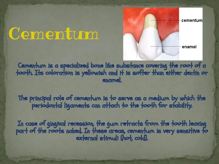
Cementum is a specialized bone like substance covering the root of
Cementum is a specialized bone like substance covering the root of
Dentin is the substance between enamel or cementum and the pulp
Dentin is the substance between enamel or cementum and the pulp
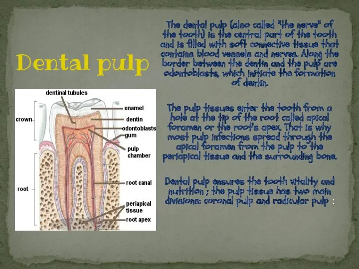
The dental pulp (also called "the nerve" of the tooth) is
The dental pulp (also called "the nerve" of the tooth) is
The crown of a tooth contains the coronal pulp. The coronal
The crown of a tooth contains the coronal pulp. The coronal
The radicular pulp is that pulp extending from the cervical region
The radicular pulp is that pulp extending from the cervical region
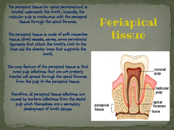
The periapical tissue (or apical periodontium) is located underneath the tooth
The periapical tissue (or apical periodontium) is located underneath the tooth
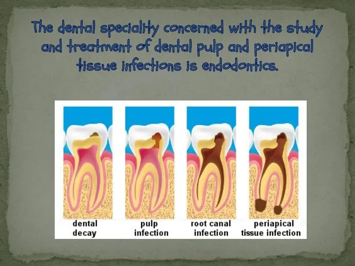
The dental speciality concerned with the study and treatment of dental
The dental speciality concerned with the study and treatment of dental
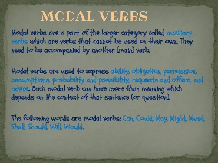
Modal verbs are a part of the larger category called auxiliary
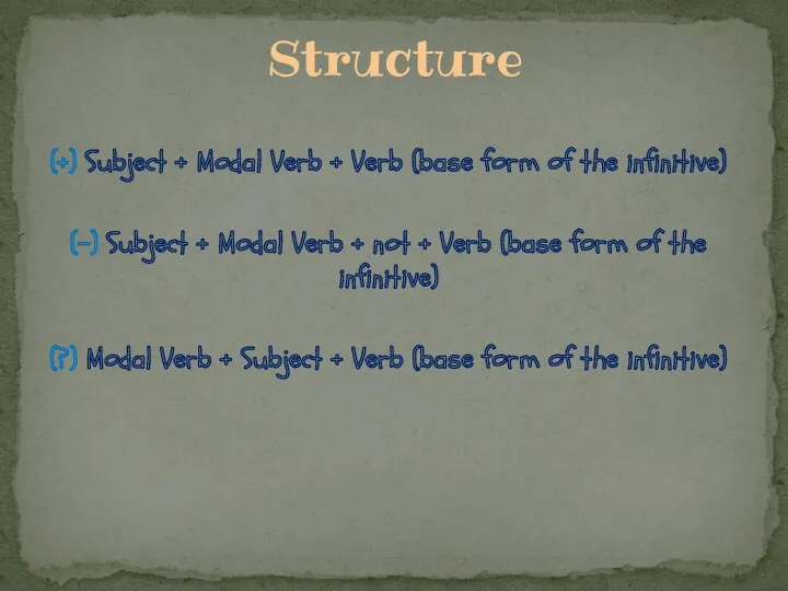
(+) Subject + Modal Verb + Verb (base form of the
(+) Subject + Modal Verb + Verb (base form of the
 Бөртпе синдромымен өтетін аурулар
 Инсектная аллергия
 Видеотрансляция операции Лабиринт IIIB, пластики ДМПП, аорто–коронарного шунтирования
 Фурункул, карбункул, рожа, панариций и др. Хирургическая инфекция мягких тканей
 Санитарноэпидемиологические требования к устройству, содержанию и организации режима работы в дошкольных организациях
 Мова мислення. Свідомість
 Химиотерапевтические средства
 Принципы межличностного общения и консультирования пациентов врачом общей практики. Особенности ведения больных
 Принципы пациент-ориентированного обучения
 Клинико-экономические исследования
 Препараты, используемые в лечении болезни Паркинсона
 Санаторий-профилакторий Романтика в Кемеровской области
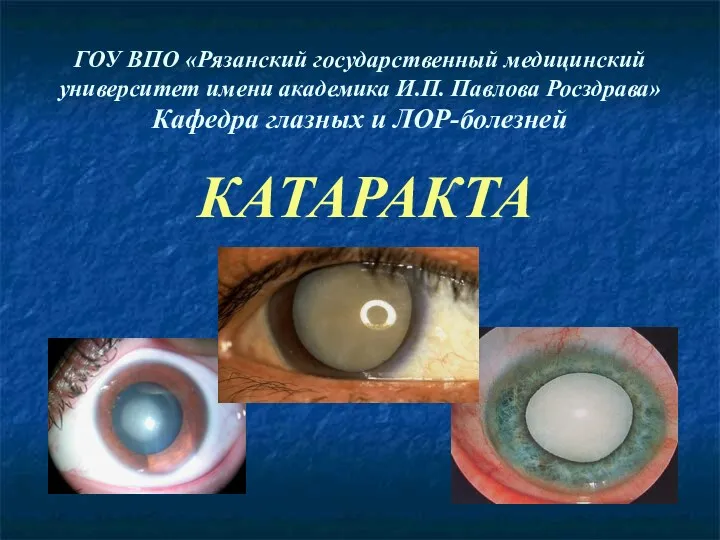 Катаракта – помутнение хрусталика
 Антиангинальные средства
 Современное оснащение стоматологического кабинета
 Спадковість і патологія. Лекція 4
 Антипсихотические средства, антидепрессанты
 Средства, угнетающие холинергические синапсы
 Теоретические и методические основы военной эпидемиологии
 Проблемы семьи инкурабельного пациента
 План клинического исследования больного. Основные методы исследования
 Врожденные пороки развития органов пищеварения
 Терминальное состояние: стадии, клиника, диагностика, критерии оценки тяжести состояния больного
 Профилактика раковых заболеваний
 Средства гигиенического ухода за полостью рта и требования, предъявляемые к ним
 Хирургиялық құралдар. Микрохирургия ұғымы. Тіндерді қосу және ажырату ережелері мен әдістері
 Болезни новорождённых. Болезни кожи. Пупка. Сепсис
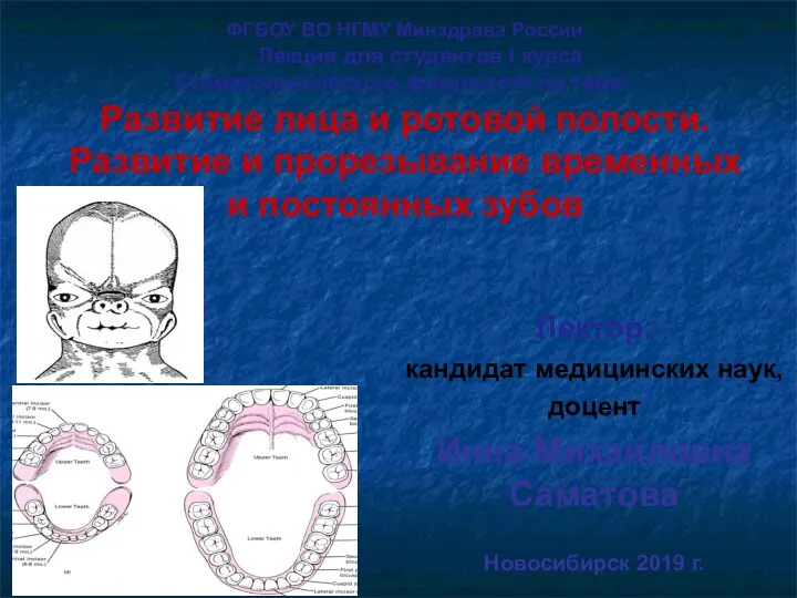 Развитие лица и ротовой полости. Развитие и прорезывание временных и постоянных зубов