- Type of Imaging Modalities In Radiology

Содержание
- 2. Medical imaging is the technique and process of creating visual representations of the interior of a
- 3. As a discipline it is part of biological imaging and incorporates radiology which uses the imaging
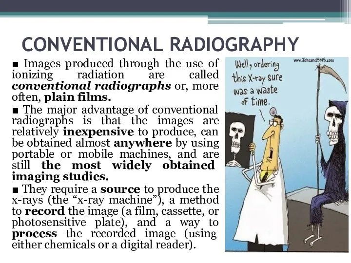
- 4. CONVENTIONAL RADIOGRAPHY ■ Images produced through the use of ionizing radiation are called conventional radiographs or,
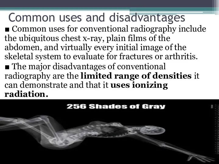
- 5. Common uses and disadvantages ■ Common uses for conventional radiography include the ubiquitous chest x-ray, plain
- 6. CT or CAT scan
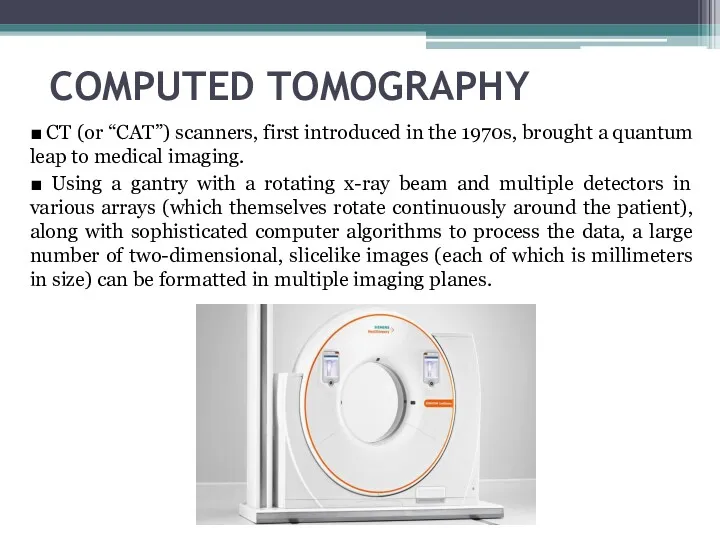
- 7. COMPUTED TOMOGRAPHY ■ CT (or “CAT”) scanners, first introduced in the 1970s, brought a quantum leap
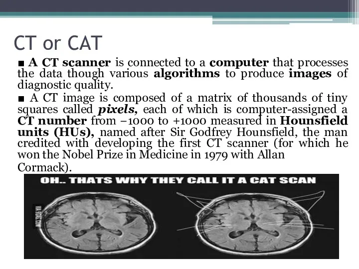
- 8. CT or CAT ■ A CT scanner is connected to a computer that processes the data
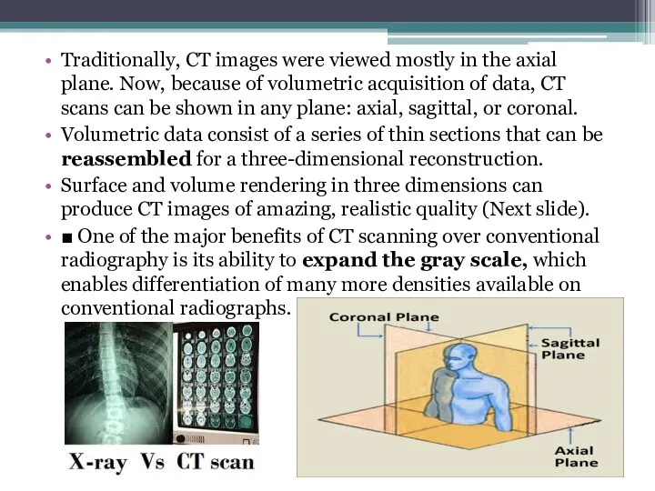
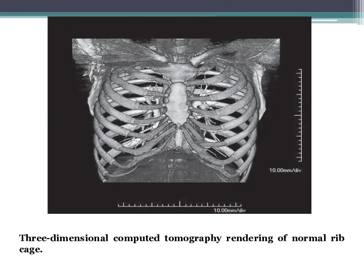
- 9. Traditionally, CT images were viewed mostly in the axial plane. Now, because of volumetric acquisition of
- 10. Three-dimensional computed tomography rendering of normal rib cage.
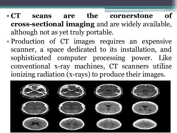
- 11. CT scans are the cornerstone of cross-sectional imaging and are widely available, although not as yet
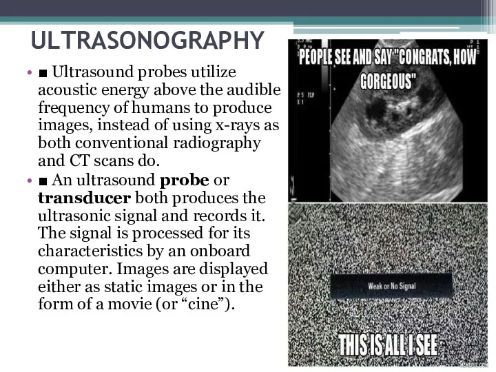
- 13. ULTRASONOGRAPHY ■ Ultrasound probes utilize acoustic energy above the audible frequency of humans to produce images,
- 14. Benefits ■ Ultrasound scanners are relatively inexpensive compared with CT and MRI scanners. They are widely
- 15. ■ Other common uses are evaluation of cystic versus solid breast masses, thyroid nodules, and tendons
- 16. MAGNETIC RESONANCE IMAGING ■ MRI utilizes the potential energy stored in the body’s hydrogen atoms. The
- 17. ■ MRI is widely used in neurologic imaging and is particularly sensitive in imaging soft tissues
- 18. FLUOROSCOPY Fluoroscopy is a modality in which ionizing radiation (x-rays) is used in performing real-time visualization
- 19. ■ In interventional radiology, iodinated contrast is selectively injected into blood vessels or other ducts that
- 20. NUCLEAR MEDICINE ■ A radioactive isotope (radioisotope) is an unstable form of an element that emits
- 21. Radiopharmaceuticals are combinations of radioisotopes attached to a pharmaceutical that has binding properties that allow it
- 22. ■ After the radiopharmaceutical is carried to a tissue or organ in the body, usually via
- 23. ■ Positron emission tomography (PET) is used to produce three-dimensional images that depict the body’s biochemical
- 24. ■ PET scanning is most often used in the diagnosis and treatment follow-up of cancer. It
- 25. ■ Compared with CT and fluoroscopy, nuclear medicine studies, in general, produce less patient exposure. The
- 26. Thank you for your attention !!! This guy partially amputated his finger on a circular saw.
- 28. Скачать презентацию
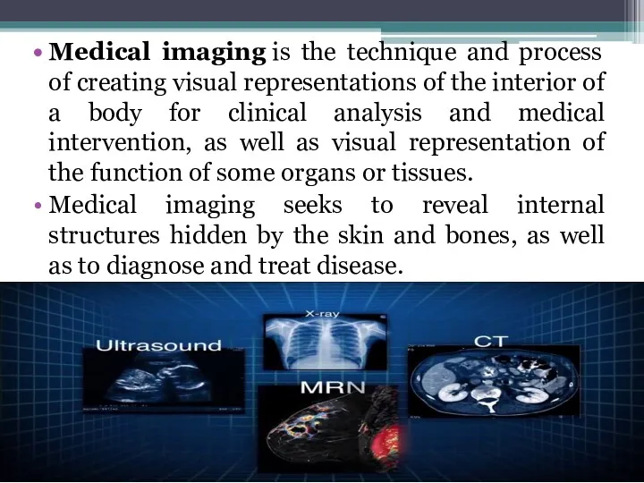
Medical imaging is the technique and process of creating visual representations of
Medical imaging is the technique and process of creating visual representations of
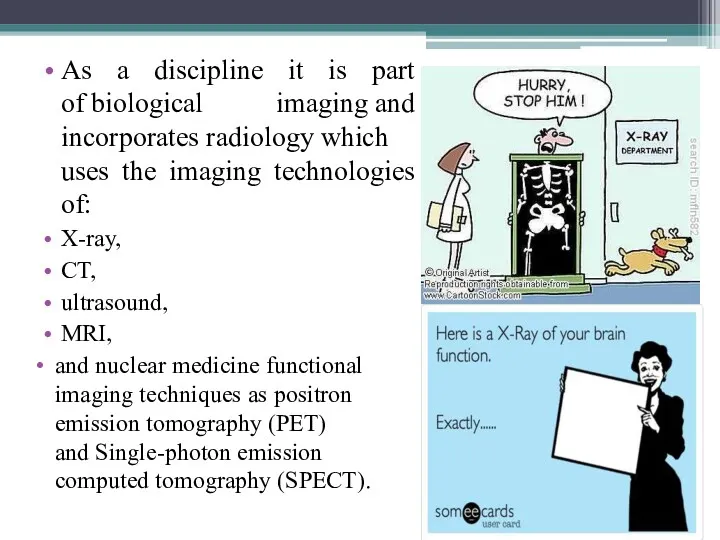
As a discipline it is part of biological imaging and incorporates radiology which uses the
As a discipline it is part of biological imaging and incorporates radiology which uses the
CONVENTIONAL RADIOGRAPHY
■ Images produced through the use of ionizing radiation are
CONVENTIONAL RADIOGRAPHY
■ Images produced through the use of ionizing radiation are
Common uses and disadvantages
■ Common uses for conventional radiography include the
Common uses and disadvantages
■ Common uses for conventional radiography include the
CT or CAT scan
CT or CAT scan
COMPUTED TOMOGRAPHY
■ CT (or “CAT”) scanners, first introduced in the 1970s,
COMPUTED TOMOGRAPHY
■ CT (or “CAT”) scanners, first introduced in the 1970s,
CT or CAT
■ A CT scanner is connected to a computer
CT or CAT
■ A CT scanner is connected to a computer
Traditionally, CT images were viewed mostly in the axial plane. Now,
Traditionally, CT images were viewed mostly in the axial plane. Now,
Three-dimensional computed tomography rendering of normal rib cage.
Three-dimensional computed tomography rendering of normal rib cage.
CT scans are the cornerstone of cross-sectional imaging and are widely
CT scans are the cornerstone of cross-sectional imaging and are widely

ULTRASONOGRAPHY
■ Ultrasound probes utilize acoustic energy above the audible frequency of
ULTRASONOGRAPHY
■ Ultrasound probes utilize acoustic energy above the audible frequency of
Benefits
■ Ultrasound scanners are relatively inexpensive compared with CT and MRI
Benefits
■ Ultrasound scanners are relatively inexpensive compared with CT and MRI
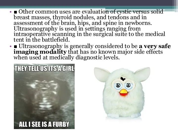
■ Other common uses are evaluation of cystic versus solid breast
■ Other common uses are evaluation of cystic versus solid breast
MAGNETIC RESONANCE IMAGING
■ MRI utilizes the potential energy stored in the
MAGNETIC RESONANCE IMAGING
■ MRI utilizes the potential energy stored in the
■ MRI is widely used in neurologic imaging and is particularly
■ MRI is widely used in neurologic imaging and is particularly
FLUOROSCOPY
Fluoroscopy is a modality in which ionizing radiation (x-rays) is used
FLUOROSCOPY
Fluoroscopy is a modality in which ionizing radiation (x-rays) is used
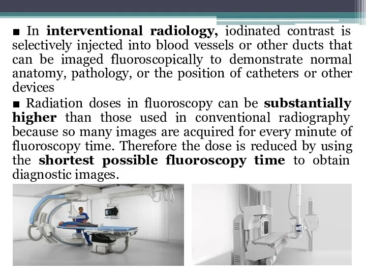
■ In interventional radiology, iodinated contrast is selectively injected into blood
■ In interventional radiology, iodinated contrast is selectively injected into blood

NUCLEAR MEDICINE
■ A radioactive isotope (radioisotope) is an unstable form of
NUCLEAR MEDICINE
■ A radioactive isotope (radioisotope) is an unstable form of
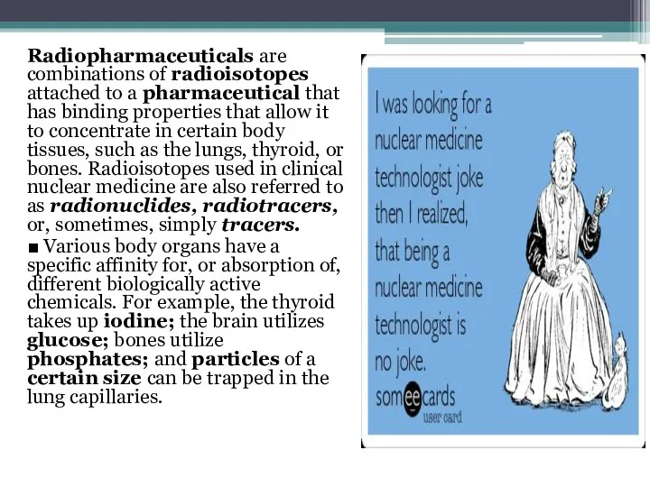
Radiopharmaceuticals are combinations of radioisotopes attached to a pharmaceutical that has
Radiopharmaceuticals are combinations of radioisotopes attached to a pharmaceutical that has
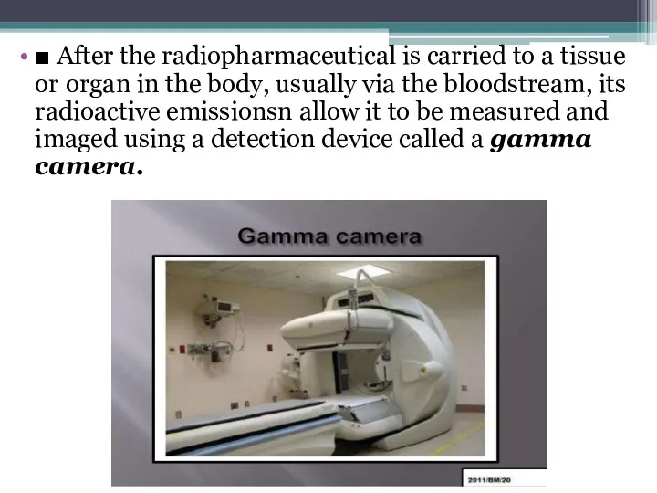
■ After the radiopharmaceutical is carried to a tissue or organ
■ After the radiopharmaceutical is carried to a tissue or organ
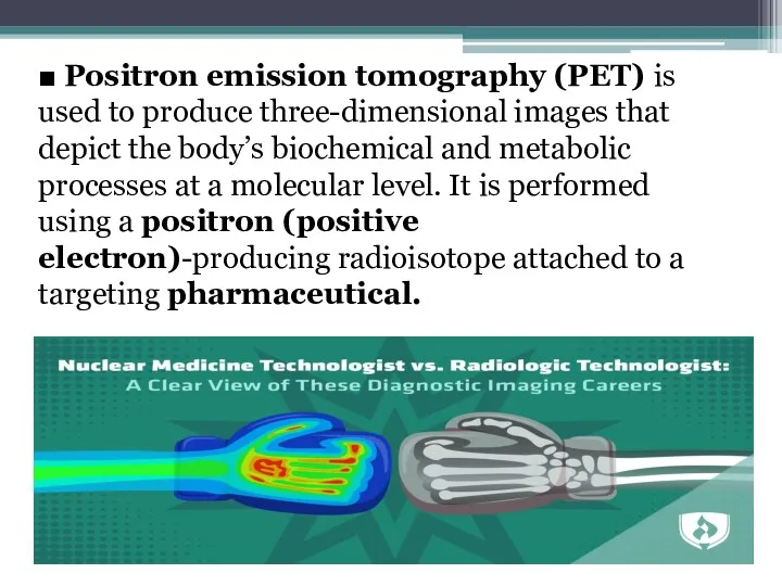
■ Positron emission tomography (PET) is used to produce three-dimensional images
■ Positron emission tomography (PET) is used to produce three-dimensional images
■ PET scanning is most often used in the diagnosis and
■ PET scanning is most often used in the diagnosis and
■ Compared with CT and fluoroscopy, nuclear medicine studies, in general,
■ Compared with CT and fluoroscopy, nuclear medicine studies, in general,
Thank you for your attention !!!
This guy partially amputated his finger
Thank you for your attention !!!
This guy partially amputated his finger
 Общие принципы диагностики злокачественных опухолей
 Синтетические противомикробные средства
 Мұрынның және мұрын қосалқы қуыстарының, жұмсақ және қатты таңдайдың туа пайда болған ақаулары
 Дифтерия. Патогенез дифтерии
 ВКР: Деятельность медицинской сестры по уходу и наблюдению за пациентами при ОРВИ
 Планирование лечения
 Пломбирование зубов композитными пломбировочными материалами
 Топографическая анатомия поясничной области
 Здоровый образ жизни. Личная гигиена
 Сестринский уход при хроническом панкреатите
 Современные подходы к диагностике и лечению вегетососудистой дистонии у пациентов детского возраста
 Гипертоническая болезнь (артериальная гипертензия) - формулировка диагноза
 Лучевое исследование эндокринной системы
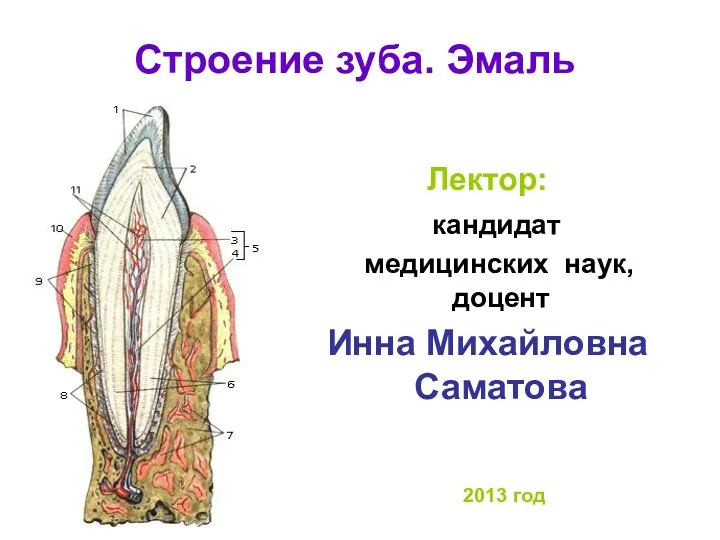 Строение зуба. Эмаль
 Введение в медицинскую информатику
 Острое инфекционное заболевание дифтерия
 Принципы гигиенического нормирования вредных веществ
 Өңештің зақымдануы
 Введение. Методы клинического сестринского обследования пациентов
 Факторы и закономерности роста и развития организма. (Лекция 2)
 Методы полногеномного анализа в медицине. Курс 3 ЦИОП Медицина будущего
 Стерилизация тиімділігін бақылау әдістері
 Ювенильный ревматоидный артрит
 Травма таза
 Стероид резистентті нефротикалық синдром
 Резективные методы лечения. (гингивэктомия, гемисекция, ампутация корня зуба)
 Кожные заболевания у детей и их профилактика
 Бронхит у детей. Острый бронхит, острый обструктивный бронхит, острый бронхиолит