- Yersinia pestis

Содержание
- 2. Yersinia pestis (formerly Pasteurella pestis) is a Gram-negative rod-shapedbacterium belonging to the family Enterobacteriaceae. It is
- 3. Role in Black Death Confirmed presence of Y. pestis would suggest that it was a contributing
- 4. SYMPTOMS Chills General ill feeling (malaise) High fever (39 °C; 102 °F) Muscle cramps Seizures Smooth,
- 5. Cause Bubonic plague is an infection of the lymphatic system, usually resulting from the bite of
- 6. Antigenic structure Flagellar H antigens Somatic antigens Antigens of virulence (v, w) According to the antigen
- 7. Laboratory Diagnosis: Blood is taken for culture and lymph node aspirate for smear and culture. Sputum
- 8. Biochemical Test and Identification of Yersinia pestis
- 9. Microscopy antibody, stain, 200x, magnification Intracellular parasitism of Y. pestis
- 10. Cultural properties the colonial morphology displayed by Gram-negative Yersinia pestis bacteria, which was grown on a
- 11. Antigen Detection: F1 glycoprotein antigen complex may be detected in sputum and aspirated fluid from bubo
- 13. Скачать презентацию
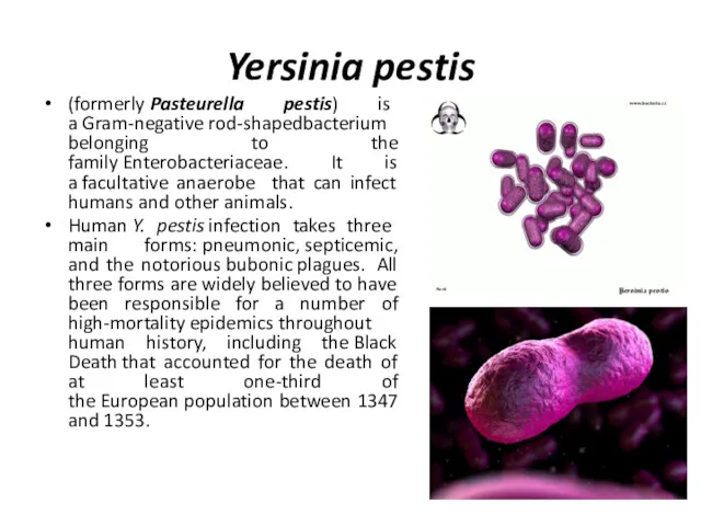
Yersinia pestis
(formerly Pasteurella pestis) is a Gram-negative rod-shapedbacterium belonging to the family Enterobacteriaceae. It is a facultative
Yersinia pestis
(formerly Pasteurella pestis) is a Gram-negative rod-shapedbacterium belonging to the family Enterobacteriaceae. It is a facultative
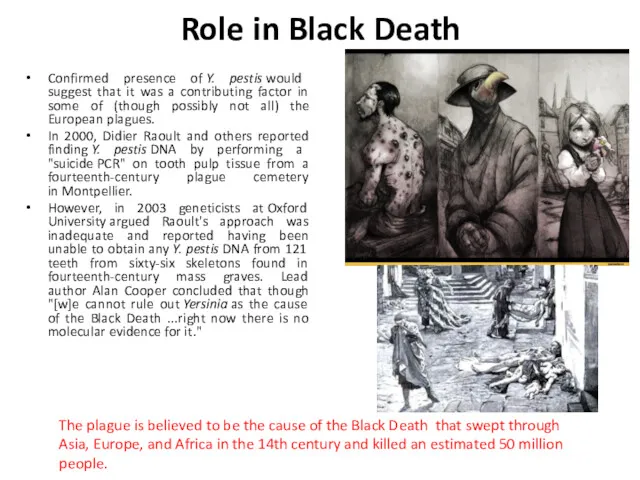
Role in Black Death
Confirmed presence of Y. pestis would suggest that it was
Role in Black Death
Confirmed presence of Y. pestis would suggest that it was
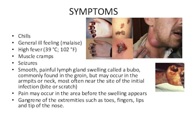
SYMPTOMS
Chills
General ill feeling (malaise)
High fever (39 °C; 102 °F)
Muscle cramps
Seizures
Smooth, painful lymph gland
SYMPTOMS
Chills
General ill feeling (malaise)
High fever (39 °C; 102 °F)
Muscle cramps
Seizures
Smooth, painful lymph gland
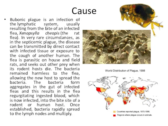
Cause
Bubonic plague is an infection of the lymphatic system, usually resulting from
Cause
Bubonic plague is an infection of the lymphatic system, usually resulting from
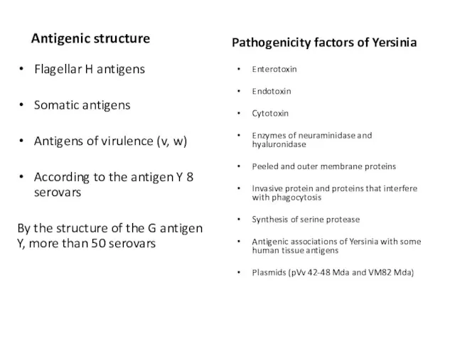
Antigenic structure
Flagellar H antigens
Somatic antigens
Antigens of virulence (v, w)
According to the
Antigenic structure
Flagellar H antigens
Somatic antigens
Antigens of virulence (v, w)
According to the
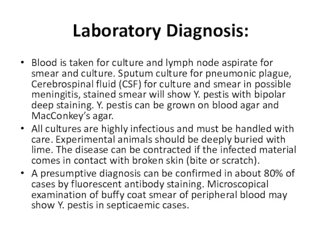
Laboratory Diagnosis:
Blood is taken for culture and lymph node aspirate for
Laboratory Diagnosis:
Blood is taken for culture and lymph node aspirate for
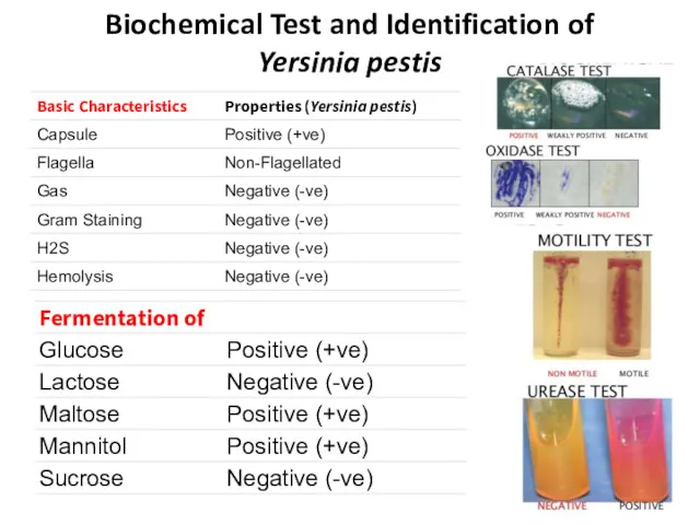
Biochemical Test and Identification of
Yersinia pestis
Biochemical Test and Identification of
Yersinia pestis
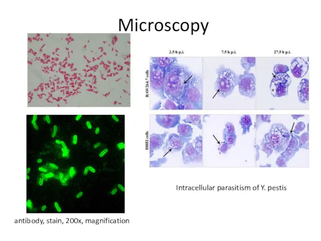
Microscopy
antibody, stain, 200x, magnification
Intracellular parasitism of Y. pestis
Microscopy
antibody, stain, 200x, magnification
Intracellular parasitism of Y. pestis
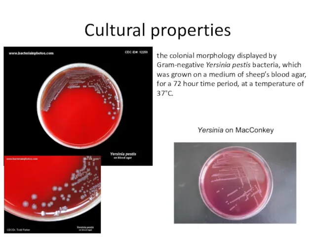
Cultural properties
the colonial morphology displayed by Gram-negative Yersinia pestis bacteria, which was grown
Cultural properties
the colonial morphology displayed by Gram-negative Yersinia pestis bacteria, which was grown
Antigen Detection:
F1 glycoprotein antigen complex may be detected in sputum and
Antigen Detection:
F1 glycoprotein antigen complex may be detected in sputum and
 Виды шприцев и игл
 Ð_еÑ_Ñ_иÑ_идÑ_
 Патофизиология CCC
 Скарлатина кезіндегі шаралардың стандарттары және алгоритмдері
 Экстрагенитальная патология
 ЛФК при переломе позвоночника
 Наследственные заболевания лёгких и пороки развития
 Лекарства. Немного истории
 Влияние солнечной активности на организм человека
 Вопросы врача общей практики в жизни врача стоматолога
 Энпиты и нутриенты. Энтеральное питание
 НР-ассоциированные заболевания желудка (хронический гастрит, язвенная болезнь желудка и двенадцатиперстной кишки, рак желудка)
 Травма грудной клетки у детей
 Сосудистые заболевания спинного мозга
 Санитарное просвещение
 Аномалии сократительной деятельности матки во время родов
 Социальный квест Клад здоровья
 Слабительные средства
 Проблемы регулирования цен на лекарственные препараты в Российской Федерации и возможные пути их решения
 Заболевания век, слезных органов, орбиты и конъюнктивы
 Видеотрансляция операции Лабиринт IIIB
 Средства физической культуры в регулировании работоспособности
 Лекарственные растения: свойства, история использования
 Острый коронарный синдром
 Кардиомиопатиялардың патологиялық анатомиясы. Нәтижесі, асқынуы, өлім себебі
 Изготовление лекарственной формы по рецепту
 Гонорея: возбудитель, симптомы, зоны поражения
 Лучевая диагностика заболеваний пищеварительного тракта