- بسم الله الرحمن الرحيم

Содержание
- 2. أولا نقدم شكر خاص للدكتور : مدحت طه
- 3. The anatomy of eye
- 4. The objectives External Anatomy of the Eye Lacrimal Apparatus of the Eye Anatomy of the Eyeball
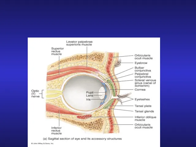
- 5. External Anatomy of the Eye
- 6. Lacrimal Apparatus of the Eye
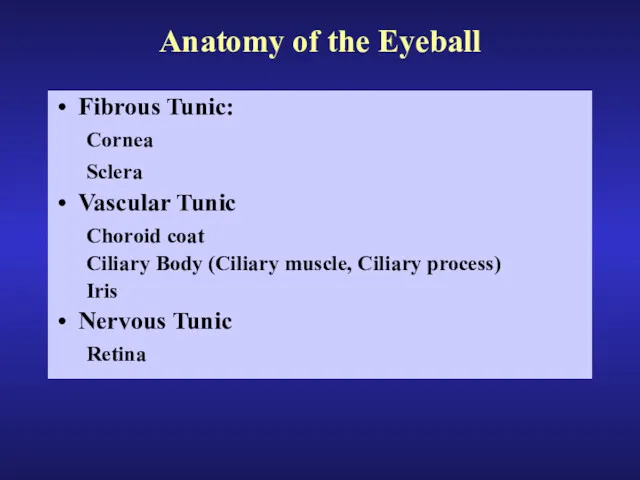
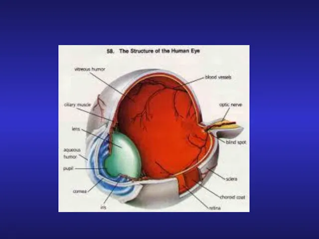
- 7. Anatomy of the Eyeball Fibrous Tunic: Cornea Sclera Vascular Tunic Choroid coat Ciliary Body (Ciliary muscle,
- 8. Layers of eye
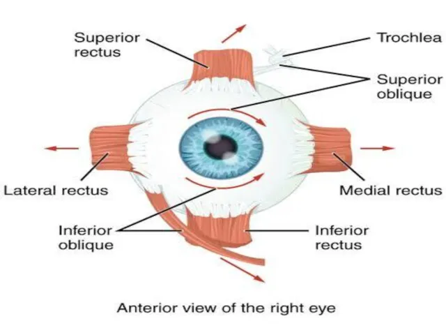
- 11. Muscle of eye
- 13. Internal Anatomy of the Eye
- 14. Detail view of the anterior anatomy of the eye
- 15. Production of Aqueous Humor and Intraocular pressure Ciliary Process: Produces Aqueous Humor 2. Posterior Chamber: Aqueous
- 16. Opthalmoscopic view of the retina showing the location of the Macula to the Optic Disc
- 17. Photomicroscopic view of the Histology of the Eye showing the location of the central fovea
- 18. Intrinsic Eye Muscles and their response to light
- 19. The arteries of eye
- 20. Veins of eye
- 21. Anatomy of Rods and Cones
- 22. The Visual Pathway
- 23. Visual Pathway Cones Bipolar neurons Ganglion cell’s axon forms the optic nerve Optic nerve to the
- 26. Скачать презентацию
أولا
نقدم شكر خاص للدكتور : مدحت طه
أولا
نقدم شكر خاص للدكتور : مدحت طه
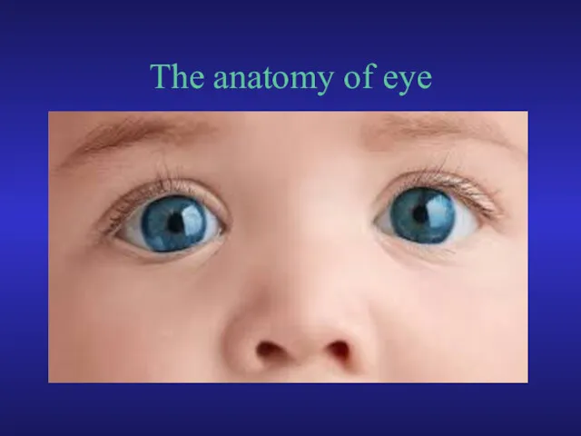
The anatomy of eye
The anatomy of eye

The objectives
External Anatomy of the Eye
Lacrimal Apparatus of the Eye
Anatomy of
The objectives
External Anatomy of the Eye
Lacrimal Apparatus of the Eye
Anatomy of
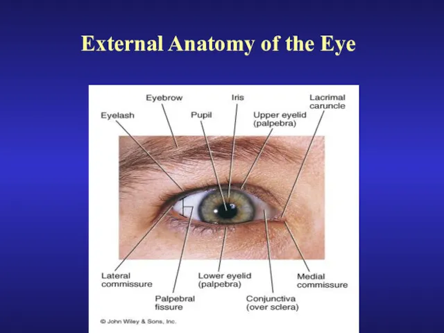
External Anatomy of the Eye
External Anatomy of the Eye
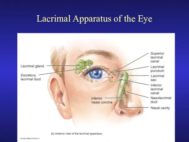
Lacrimal Apparatus of the Eye
Lacrimal Apparatus of the Eye
Anatomy of the Eyeball
Fibrous Tunic:
Cornea
Sclera
Vascular Tunic
Choroid coat
Ciliary Body (Ciliary
Anatomy of the Eyeball
Fibrous Tunic:
Cornea
Sclera
Vascular Tunic
Choroid coat
Ciliary Body (Ciliary
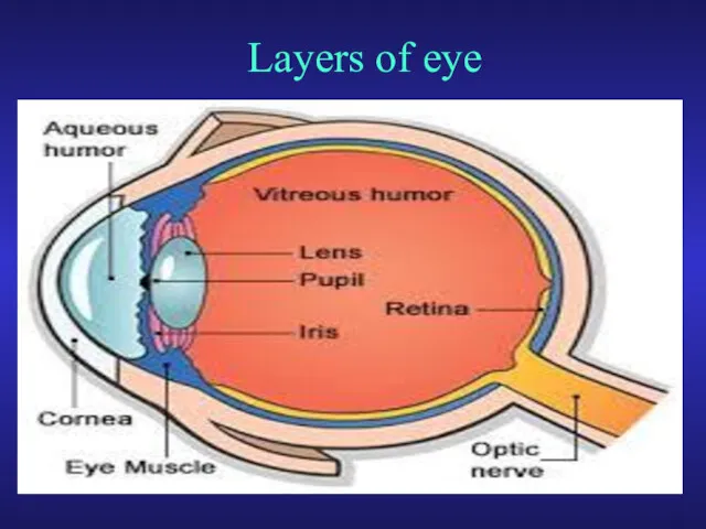
Layers of eye
Layers of eye
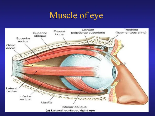
Muscle of eye
Muscle of eye
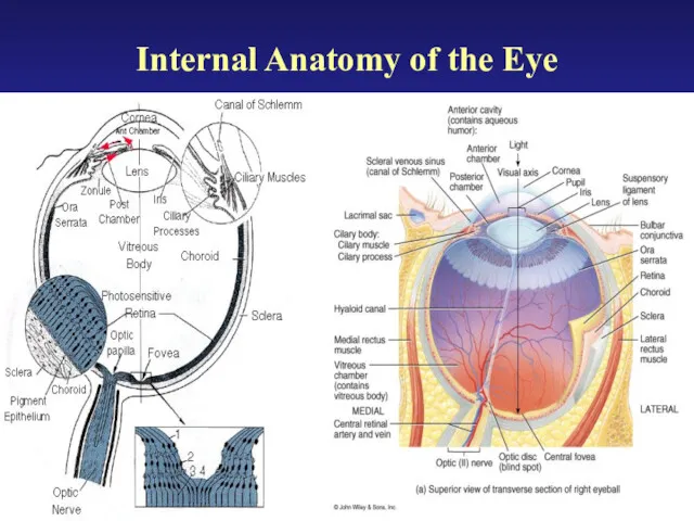
Internal Anatomy of the Eye
Internal Anatomy of the Eye
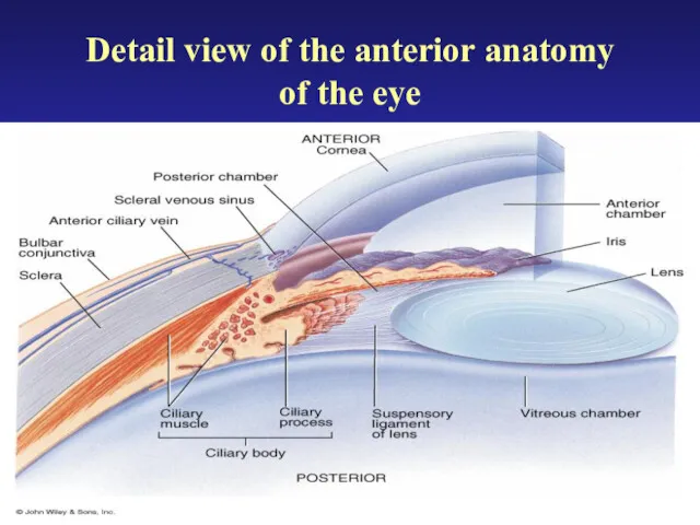
Detail view of the anterior anatomy
of the eye
Detail view of the anterior anatomy
of the eye
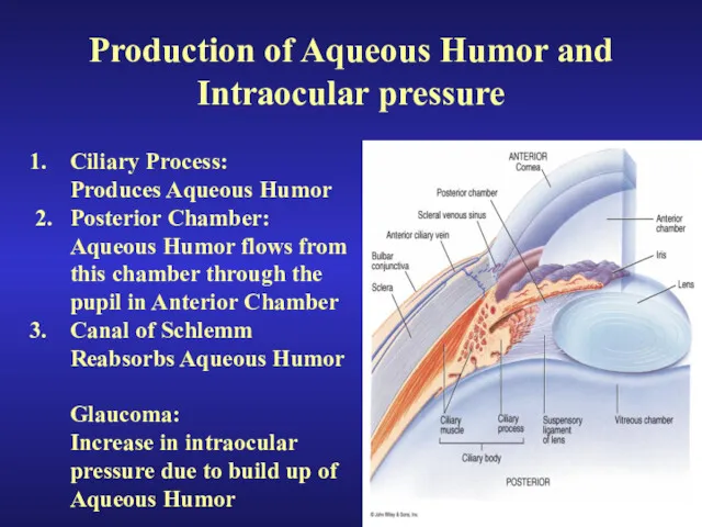
Production of Aqueous Humor and Intraocular pressure
Ciliary Process:
Produces Aqueous Humor
2.
Production of Aqueous Humor and Intraocular pressure
Ciliary Process:
Produces Aqueous Humor
2.
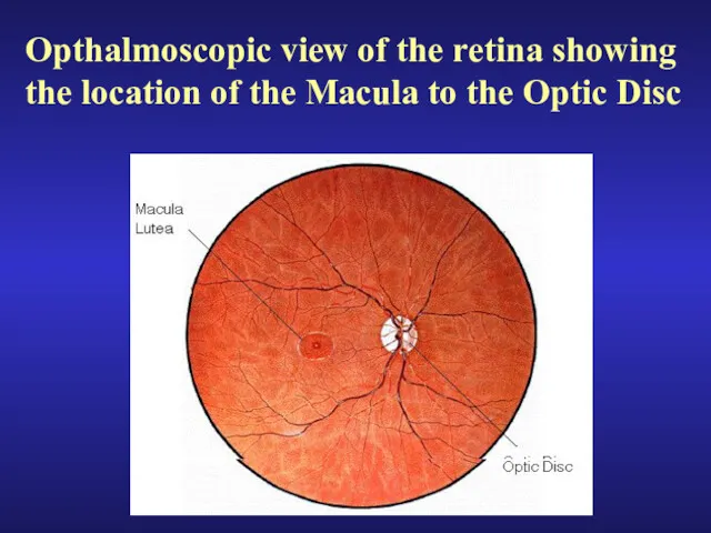
Opthalmoscopic view of the retina showing the location of the Macula
Opthalmoscopic view of the retina showing the location of the Macula
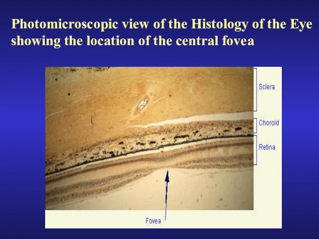
Photomicroscopic view of the Histology of the Eye
showing the location of
Photomicroscopic view of the Histology of the Eye
showing the location of
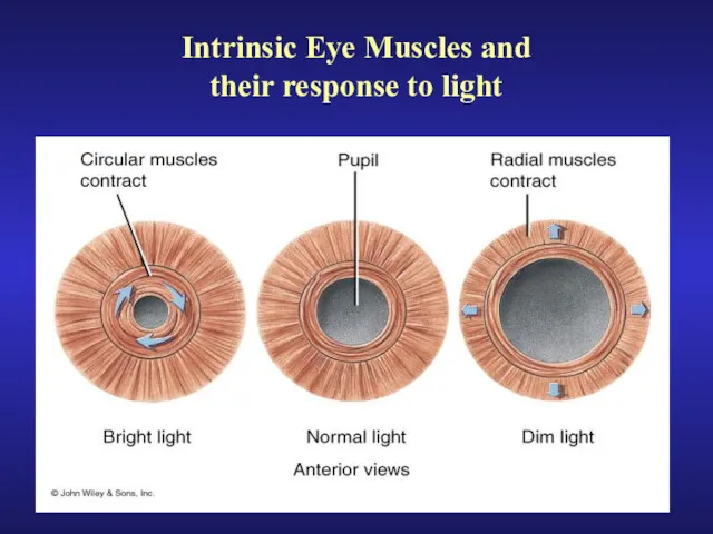
Intrinsic Eye Muscles and
their response to light
Intrinsic Eye Muscles and
their response to light
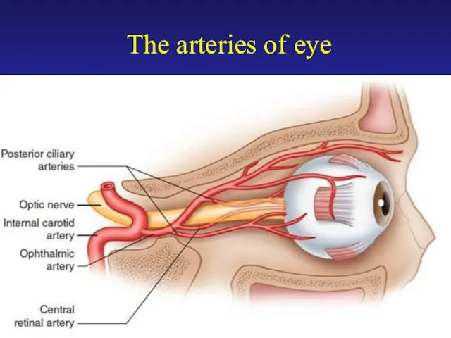
The arteries of eye
The arteries of eye
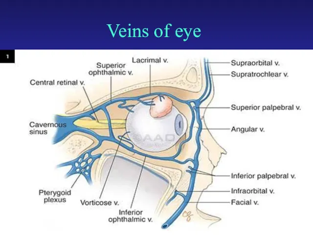
Veins of eye
Veins of eye
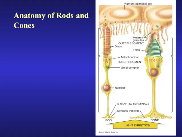
Anatomy of Rods and
Cones
Anatomy of Rods and
Cones
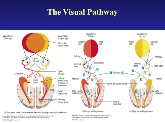
The Visual Pathway
The Visual Pathway
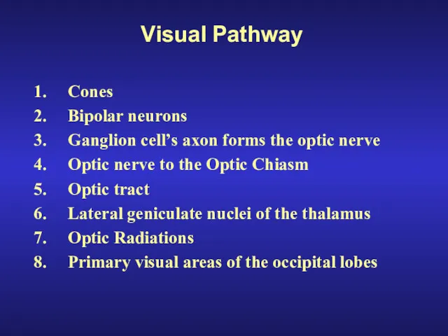
Visual Pathway
Cones
Bipolar neurons
Ganglion cell’s axon forms the optic nerve
Optic nerve to
Visual Pathway
Cones
Bipolar neurons
Ganglion cell’s axon forms the optic nerve
Optic nerve to

 Артикуляционная гимнастика.
 Focusing ground penetrating radar images with vertical offset filtering
 Конспект урока по химии в 8 классе по теме Простые вещества - металлы
 Конспект урока в 7-ом классе по географии Движение воды в океане
 Сабриново. Создание сети домов-пансионатов для пожилых людей
 Smart home. Technologies. Automation and robotics
 Автоледи
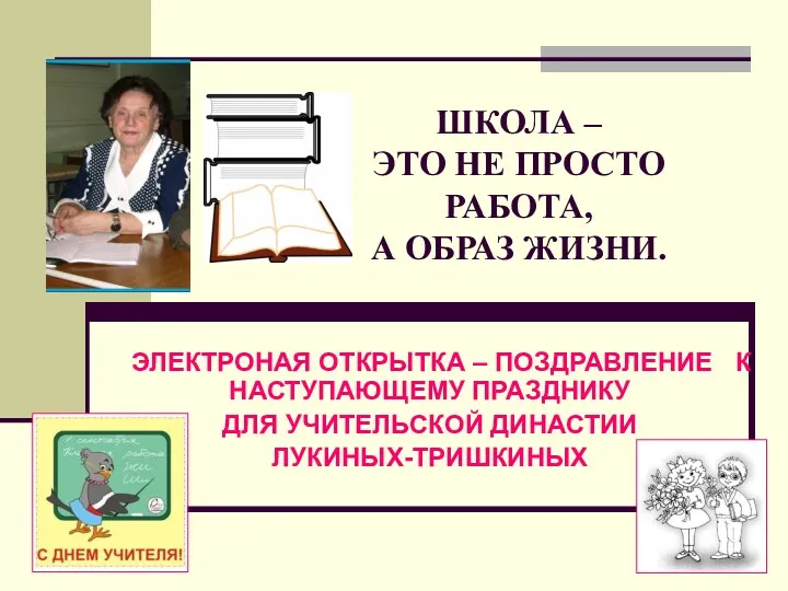 Моя семья - моё богатство
 Шкала самооценки Спилбергера-Ханина
 методическое пособие Разложи по порядку
 Порядковое числительное. 6 класс
 Загадки на звуки (Р и Р`) (логопедические упражнения)
 Развитие туризма в Древнем Китае
 Анализ сегментов рынка земли. Характеристики цен на товарном рынке. Определение типа рынка и выбор методов ценообразования
 Игра Найди 10 мышек
 Дерматомиозит/ полимиозит
 Тест. Планеты Солнечной системы
 Дети военных лет
 Опыт проведения православного фестиваля детского художественного творчества Светлая Пасха
 Михаил Юрьевич Лермонтов Бородино
 презентация на тему Игрушки Диск
 Изменение климата
 Презентация Жанры изобразительного искусства
 Минеральные ресурсы мира
 Теорія Надлюдини Фрідріха Ніцше та її місце у романі Кена Кізі Політ над гніздом зозулі
 Электронная база викторин по безопасности.
 Фотосинтез
 Безопасное колесо. Конкурс юных инспекторов движения