- Brain development

Содержание
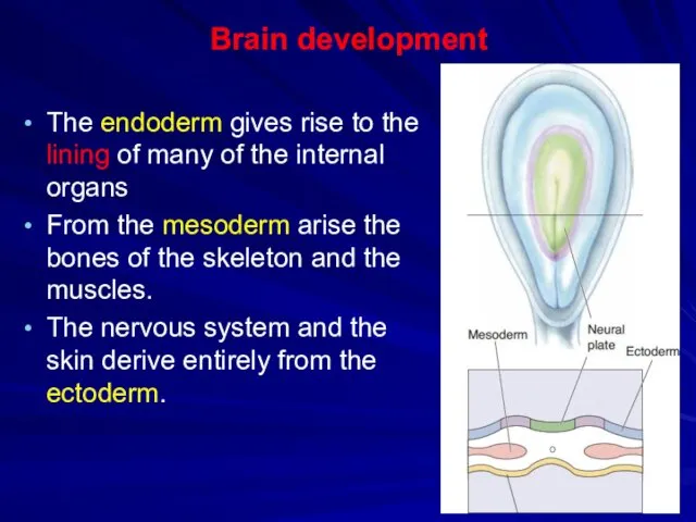
- 2. Brain development The endoderm gives rise to the lining of many of the internal organs From
- 3. The stages of nerves system development Formation of the Neural Tube Formation and differentiation of three
- 4. Formation of the Neural Tube Neurulation is the formation of neural tube from neural plate
- 5. Formation of the Neural Tube At early stage the brain consists only of a flat sheet
- 6. Formation of the Neural Tube The next event is the formation of a groove in the
- 7. Formation of the Neural Tube The walls of the groove (neural folds) subsequently move together and
- 8. Formation of the Neural Tube Some neural ectoderm is pinched off and comes to lie just
- 9. Formation of the Neural Tube The mesoderm at this stage in development forms somites on either
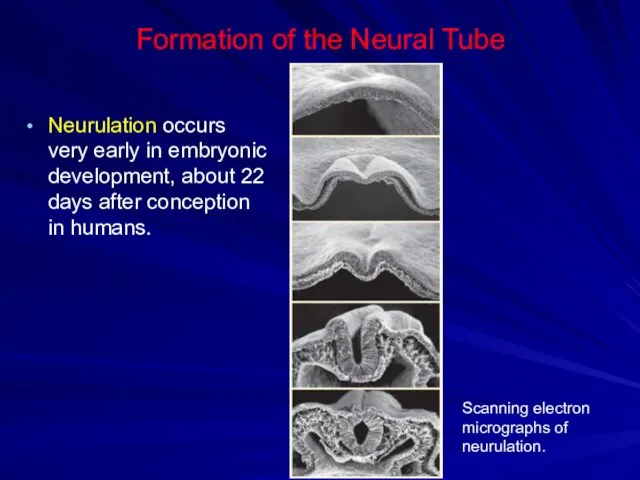
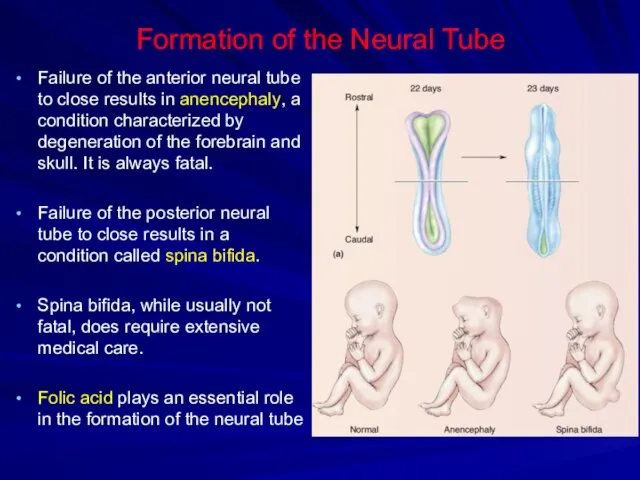
- 10. Formation of the Neural Tube Neurulation occurs very early in embryonic development, about 22 days after
- 11. Formation of the Neural Tube Failure of the anterior neural tube to close results in anencephaly,
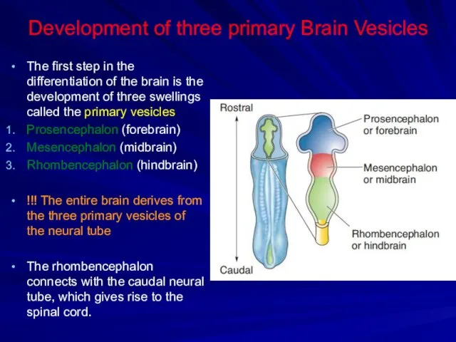
- 12. Development of three primary Brain Vesicles The first step in the differentiation of the brain is
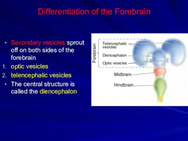
- 13. Differentiation of the Forebrain Secondary vesicles sprout off on both sides of the forebrain optic vesicles
- 14. Differentiation of the Forebrain The optic vesicles grow and invaginate to form the optic stalks and
- 15. Differentiation of the Telencephalon and Diencephalon The telencephalic vesicles together form the telencephalon, consisting of the
- 16. Differentiation of the Telencephalon and Diencephalon The telencephalon continues to develop in four ways. First way
- 17. Differentiation of the Telencephalon and Diencephalon Second way Another pair of vesicles sprout off the ventral
- 18. Differentiation of the Telencephalon and Diencephalon Third way The cells of the walls of the telencephalon
- 19. Differentiation of the Telencephalon and Diencephalon The fluid-filled spaces within the cerebral hemispheres are called the
- 20. Differentiation of the Telencephalon and Diencephalon The walls of the telencephalic vesicles appear swollen due to
- 21. Differentiation of the Telencephalon and Diencephalon The diencephalon differentiates into two structures: the thalamus the hypothalamus
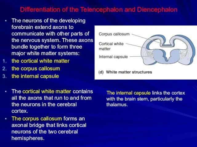
- 22. Differentiation of the Telencephalon and Diencephalon The neurons of the developing forebrain extend axons to communicate
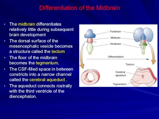
- 23. Differentiation of the Midbrain The midbrain differentiates relatively little during subsequent brain development The dorsal surface
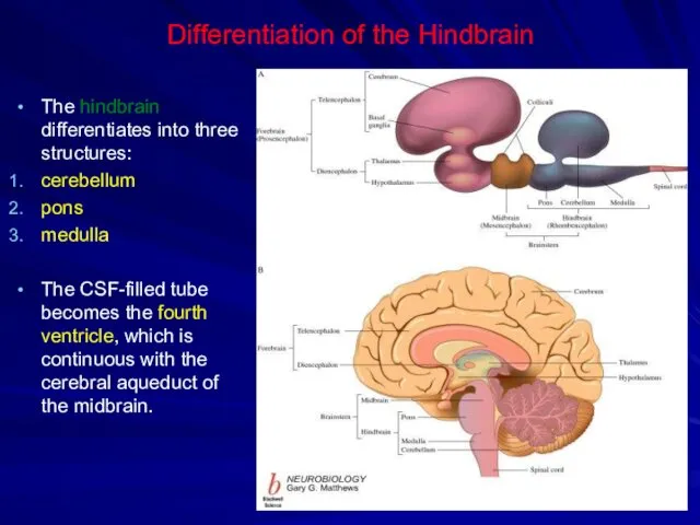
- 24. Differentiation of the Hindbrain The hindbrain differentiates into three structures: cerebellum pons medulla The CSF-filled tube
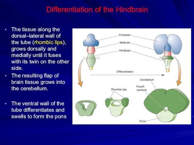
- 25. Differentiation of the Hindbrain The tissue along the dorsal–lateral wall of the tube (rhombic lips), grows
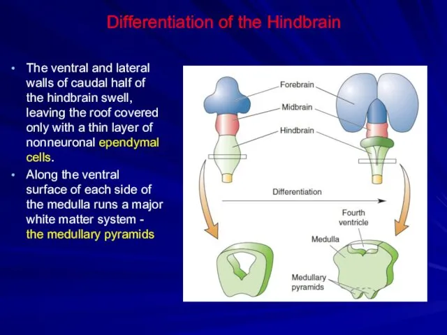
- 26. Differentiation of the Hindbrain The ventral and lateral walls of caudal half of the hindbrain swell,
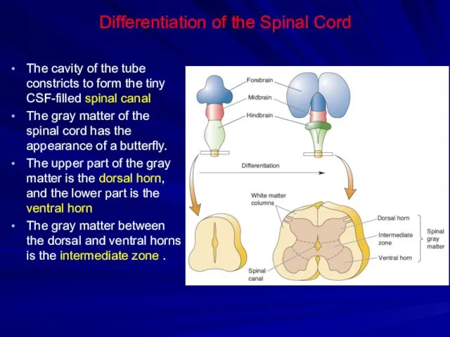
- 27. Differentiation of the Spinal Cord The cavity of the tube constricts to form the tiny CSF-filled
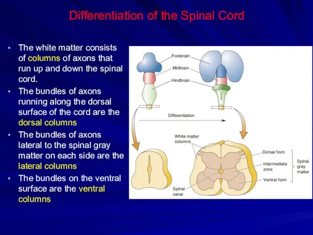
- 28. Differentiation of the Spinal Cord The white matter consists of columns of axons that run up
- 29. Resume of brain development
- 30. Resume of brain development
- 32. Скачать презентацию
Brain development
The endoderm gives rise to the lining of many of
Brain development
The endoderm gives rise to the lining of many of
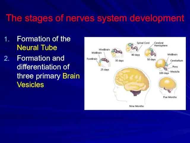
The stages of nerves system development
Formation of the Neural Tube
Formation and
The stages of nerves system development
Formation of the Neural Tube
Formation and
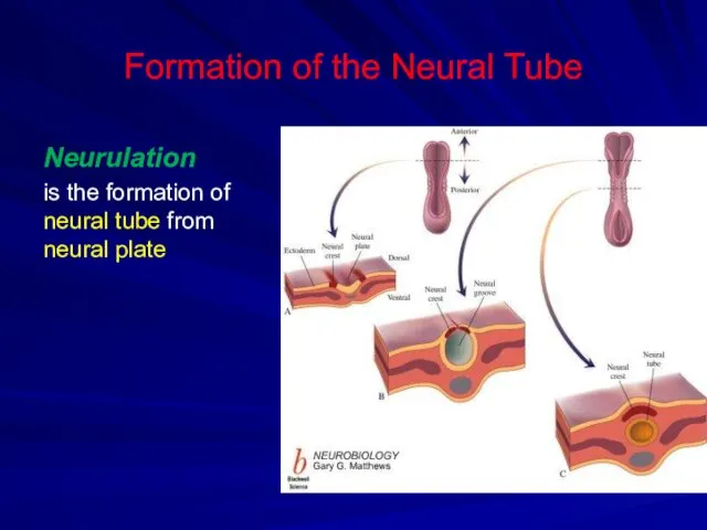
Formation of the Neural Tube
Neurulation
is the formation of neural tube from
Formation of the Neural Tube
Neurulation
is the formation of neural tube from
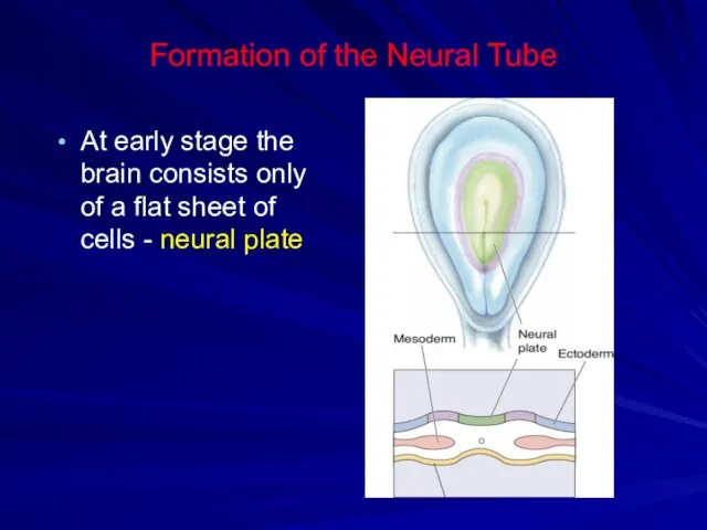
Formation of the Neural Tube
At early stage the brain consists only
Formation of the Neural Tube
At early stage the brain consists only
Formation of the Neural Tube
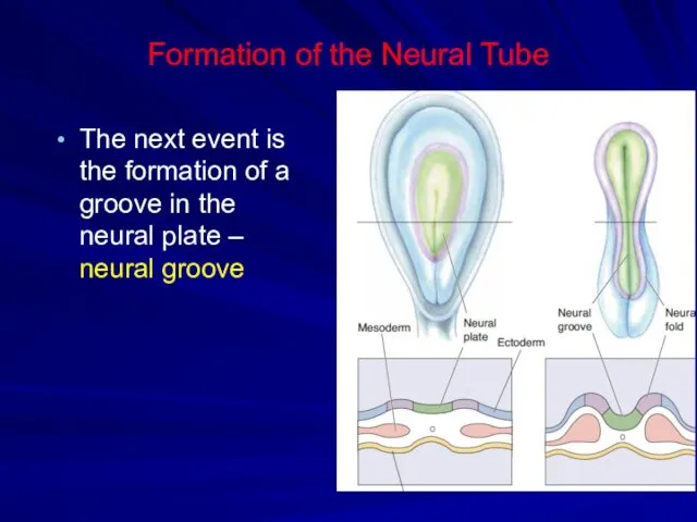
The next event is the formation of
Formation of the Neural Tube
The next event is the formation of
Formation of the Neural Tube
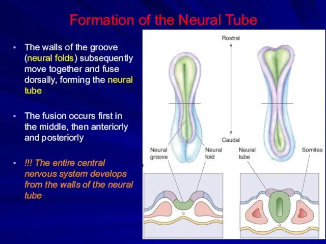
The walls of the groove (neural folds)
Formation of the Neural Tube
The walls of the groove (neural folds)
Formation of the Neural Tube
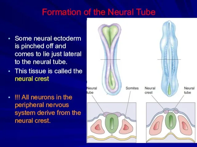
Some neural ectoderm is pinched off and
Formation of the Neural Tube
Some neural ectoderm is pinched off and
Formation of the Neural Tube
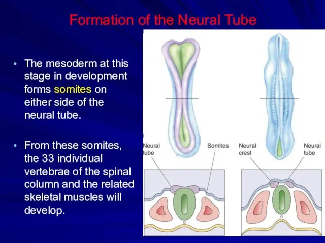
The mesoderm at this stage in development
Formation of the Neural Tube
The mesoderm at this stage in development
Formation of the Neural Tube
Neurulation occurs very early in embryonic development,
Formation of the Neural Tube
Neurulation occurs very early in embryonic development,
Formation of the Neural Tube
Failure of the anterior neural tube to
Formation of the Neural Tube
Failure of the anterior neural tube to
Development of three primary Brain Vesicles
The first step in the differentiation
Development of three primary Brain Vesicles
The first step in the differentiation
Differentiation of the Forebrain
Secondary vesicles sprout off on both sides of
Differentiation of the Forebrain
Secondary vesicles sprout off on both sides of
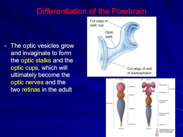
Differentiation of the Forebrain
The optic vesicles grow and invaginate to form
Differentiation of the Forebrain
The optic vesicles grow and invaginate to form
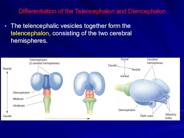
Differentiation of the Telencephalon and Diencephalon
The telencephalic vesicles together form the
Differentiation of the Telencephalon and Diencephalon
The telencephalic vesicles together form the
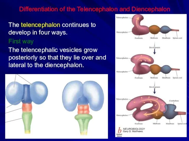
Differentiation of the Telencephalon and Diencephalon
The telencephalon continues to develop in
Differentiation of the Telencephalon and Diencephalon
The telencephalon continues to develop in
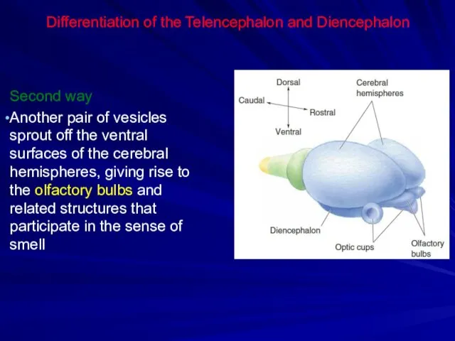
Differentiation of the Telencephalon and Diencephalon
Second way
Another pair of vesicles sprout
Differentiation of the Telencephalon and Diencephalon
Second way
Another pair of vesicles sprout
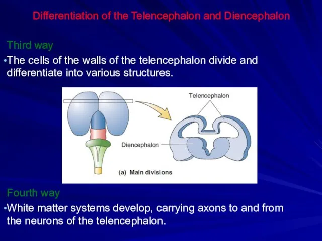
Differentiation of the Telencephalon and Diencephalon
Third way
The cells of the walls
Differentiation of the Telencephalon and Diencephalon
Third way
The cells of the walls
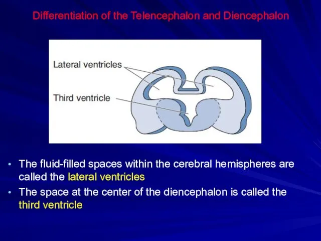
Differentiation of the Telencephalon and Diencephalon
The fluid-filled spaces within the cerebral
Differentiation of the Telencephalon and Diencephalon
The fluid-filled spaces within the cerebral
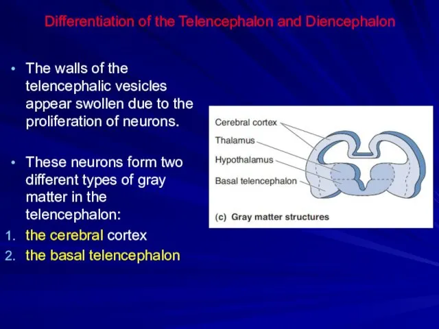
Differentiation of the Telencephalon and Diencephalon
The walls of the telencephalic vesicles
Differentiation of the Telencephalon and Diencephalon
The walls of the telencephalic vesicles
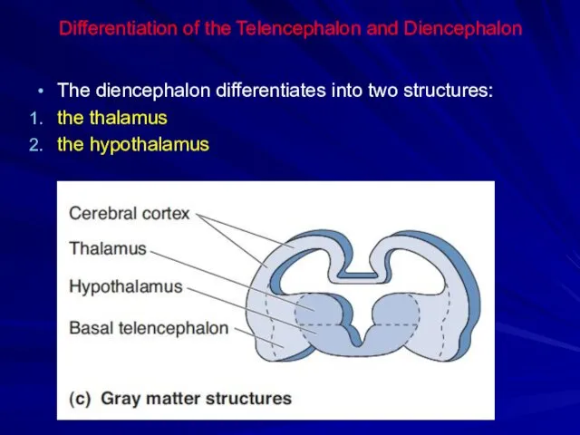
Differentiation of the Telencephalon and Diencephalon
The diencephalon differentiates into two structures:
Differentiation of the Telencephalon and Diencephalon
The diencephalon differentiates into two structures:
Differentiation of the Telencephalon and Diencephalon
The neurons of the developing forebrain
Differentiation of the Telencephalon and Diencephalon
The neurons of the developing forebrain
Differentiation of the Midbrain
The midbrain differentiates relatively little during subsequent brain
Differentiation of the Midbrain
The midbrain differentiates relatively little during subsequent brain
Differentiation of the Hindbrain
The hindbrain differentiates into three structures:
cerebellum
pons
medulla
The CSF-filled
Differentiation of the Hindbrain
The hindbrain differentiates into three structures:
cerebellum
pons
medulla
The CSF-filled
Differentiation of the Hindbrain
The tissue along the dorsal–lateral wall of the
Differentiation of the Hindbrain
The tissue along the dorsal–lateral wall of the
Differentiation of the Hindbrain
The ventral and lateral walls of caudal half
Differentiation of the Hindbrain
The ventral and lateral walls of caudal half
Differentiation of the Spinal Cord
The cavity of the tube constricts to
Differentiation of the Spinal Cord
The cavity of the tube constricts to
Differentiation of the Spinal Cord
The white matter consists of columns of
Differentiation of the Spinal Cord
The white matter consists of columns of
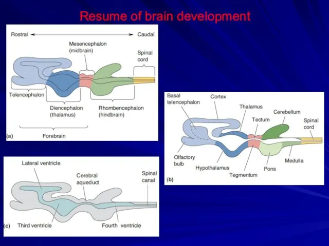
Resume of brain development
Resume of brain development
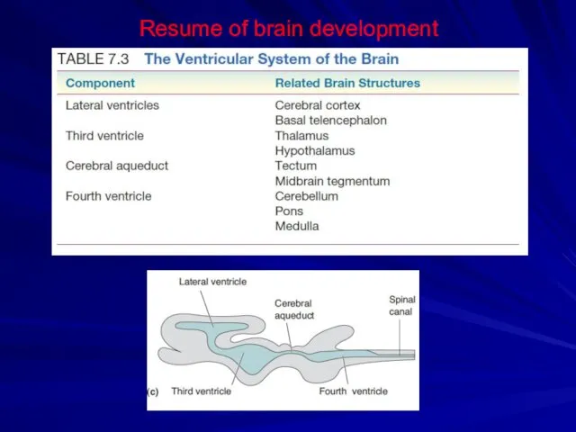
Resume of brain development
Resume of brain development
 Гражданское право. Договор поставки
 Текстовые процессоры и текстовые редакторы
 Презентация Африка
 Әлемдік діндер мәдениеті: буддизм, християндық исламдық
 Системы счисления. Основные понятия систем счисления
 Царство Грибы. Общая характеристика грибов
 Коронавирус COVID-19
 Представление технологии Диск
 Патофизиология красной крови
 Из опыта работы по формированию у дошкольников представлений о правилах дорожного движения (презентация)
 Что такое доброта?
 Автоматизация звука Ш. Артикуляционная гимнастика
 Программа оценки навыков речи и социального взаимодействия для детей с аутизмом и другими нарушениями VB-MAPP
 Влияние сотовых телефонов на здоровье человека
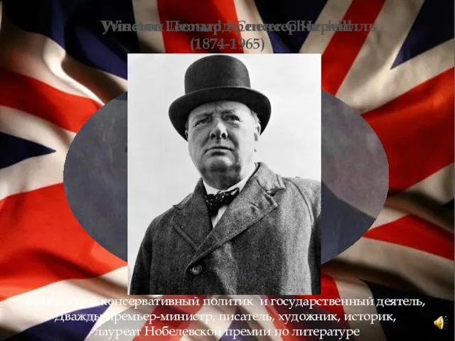 Уинстон Леонард Спенсер Черчилль
 Безопасное поведение детей в сети
 Логический квадрат. Ложные, истинные и неопределенные суждения
 2615Імунофлуорисцентний експрес аналізатор LS-1100
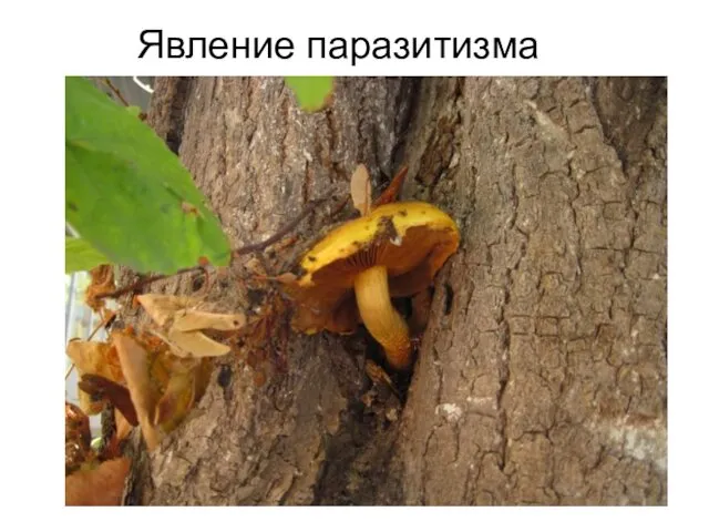 Явление паразитизма
 Музыкальная композиция
 Проектирование водопроводной насосной станции II подъема
 Доколе я в мире, я свет миру
 Алгоритм ветвления. Условный оператор
 Электронное портфолио по предмету Окружающий мир
 презентация проекта по ПДД Нам на улице не страшно
 Школьный спортивный клуб Спасатель
 My favorite paintings
 Родительское собрание Подготовка ребёнка ко 2 классу