- Muscle tissue

Содержание
- 2. Muscle tissue satisfy requirement of the body in movement.
- 3. Classification – The 3 types of muscle tissue: 1. skeletal 2. cardiac 3. smooth groups: Striated
- 4. Why do muscles contract? Muscle cells have contractile proteins - actin and myosin, and some another
- 5. Why do muscles contract? Actin and myosin form myofilaments: Myosin - thick, dark and Anisotropic (A)
- 6. SMOOTH MUSCLE
- 7. Locations: walls of visceral hollow organs (stomach). Functions: involuntary movement -- (peristaltics) (The innervation -- by
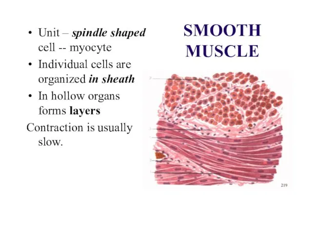
- 8. SMOOTH MUSCLE Unit – spindle shaped cell -- myocyte Individual cells are organized in sheath In
- 9. Origin of smooth muscle Smooth muscle cells arise from mesenchymal cells.
- 10. Striated muscles
- 11. See: regular organization of the myofibrils gives rise to the cross-striation, which characterises skeletal and cardiac
- 12. CARDIAC MUSCLE Locations: heart Function: involuntary, rhythmic contraction Unit – cardiomyocyte (cell)
- 13. Cardiac muscle cells: 3 types: Contractile, Conducting Secretory
- 14. CARDIAC MUSCLE cardiac muscle cells are cylindrical, connect end-by-end, and form “functional fiber”, which often branch
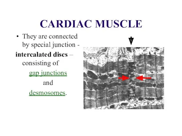
- 15. CARDIAC MUSCLE They are connected by special junction - intercalated discs – consisting of gap junctions
- 17. SKELETAL MUSCLE
- 18. Location Muscles associated with the skeleton (are connected to bones by tendons). Platisma and mimic muscles
- 19. SKELETAL MUSCLE --- is innervated by the somatic nervous system – voluntary!! ---- consists of very
- 20. SKELETAL MUSCLE Skeletal muscle fibers run the full length of a muscle. The average length of
- 21. Nuclei: Skeletal muscle fibres contain many nuclei (up to several hundred ) placed beneath the plasma
- 22. Myofibrils Mechanism of contraction: Sliding filaments model
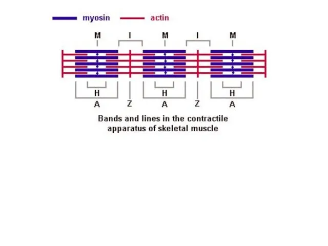
- 23. Myofibrils has some bands and lines depending on the distribution and interconnection of myofilaments -- :
- 26. Sarcomeres -- are parts, smallest contractile units of myofibrils. Sarcomere formula: S = ½ I +
- 27. Sarcomere formula after contraction S = A (- ½ I, - ½ I, - H)
- 28. Mechanism of contraction
- 29. Origin of skeletal muscle The myoblasts of all skeletal muscle fibres originate from the paraxial mesoderm
- 30. 1. Myoblasts undergo frequent divisions and coalesce with the formation of a multinucleated, syncytial muscle fibre
- 32. Скачать презентацию
Muscle tissue satisfy requirement of the body in movement.
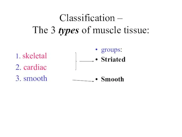
Classification –
The 3 types of muscle tissue:
1. skeletal
2. cardiac
3.
Classification –
The 3 types of muscle tissue:
1. skeletal
2. cardiac
3.
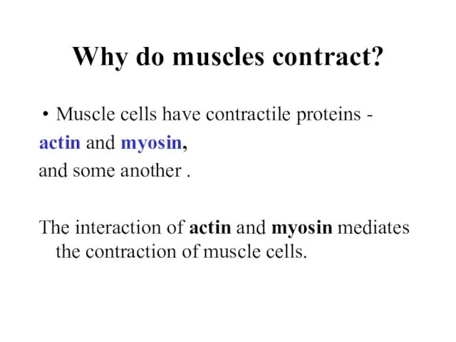
Why do muscles contract?
Muscle cells have contractile proteins -
actin and
Why do muscles contract?
Muscle cells have contractile proteins -
actin and
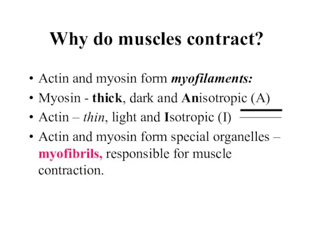
Why do muscles contract?
Actin and myosin form myofilaments:
Myosin - thick, dark
Why do muscles contract?
Actin and myosin form myofilaments:
Myosin - thick, dark

SMOOTH MUSCLE
SMOOTH MUSCLE
Locations: walls of visceral hollow organs
(stomach).
Functions: involuntary movement
Locations: walls of visceral hollow organs
(stomach).
Functions: involuntary movement
SMOOTH MUSCLE
Unit – spindle shaped cell -- myocyte
Individual cells are organized
SMOOTH MUSCLE
Unit – spindle shaped cell -- myocyte
Individual cells are organized
Origin of smooth muscle
Smooth muscle cells arise from mesenchymal cells.
Origin of smooth muscle
Smooth muscle cells arise from mesenchymal cells.
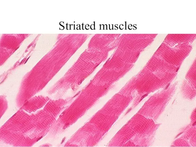
Striated muscles
Striated muscles
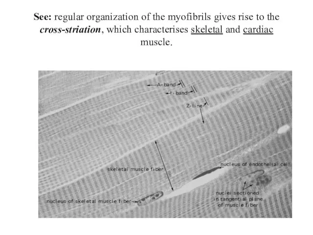
See: regular organization of the myofibrils gives rise to the cross-striation,
See: regular organization of the myofibrils gives rise to the cross-striation,
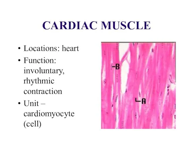
CARDIAC MUSCLE
Locations: heart
Function: involuntary, rhythmic contraction
Unit – cardiomyocyte (cell)
CARDIAC MUSCLE
Locations: heart
Function: involuntary, rhythmic contraction
Unit – cardiomyocyte (cell)
Cardiac muscle cells:
3 types:
Contractile,
Conducting
Secretory
Cardiac muscle cells:
3 types:
Contractile,
Conducting
Secretory
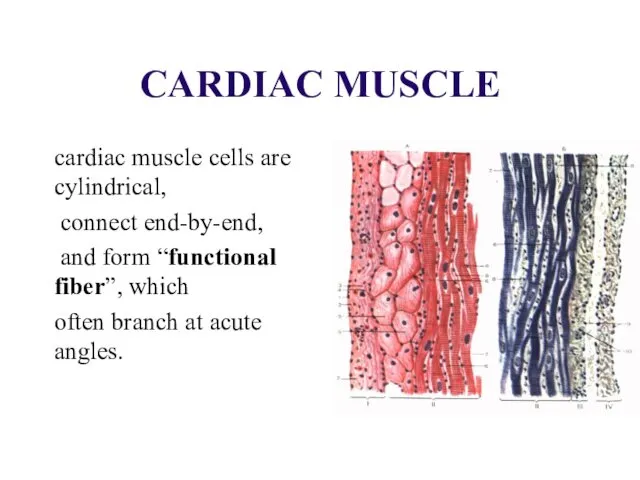
CARDIAC MUSCLE
cardiac muscle cells are cylindrical,
connect end-by-end,
and
CARDIAC MUSCLE
cardiac muscle cells are cylindrical,
connect end-by-end,
and
CARDIAC MUSCLE
They are connected by special junction -
intercalated discs
CARDIAC MUSCLE
They are connected by special junction -
intercalated discs
SKELETAL MUSCLE
SKELETAL MUSCLE
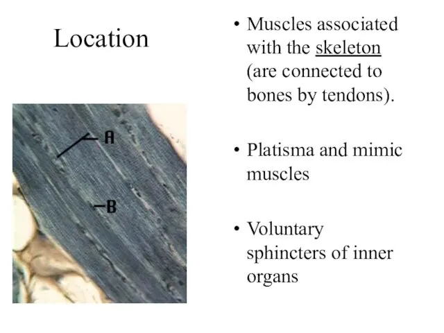
Location
Muscles associated with the skeleton (are connected to bones by tendons).
Location
Muscles associated with the skeleton (are connected to bones by tendons).
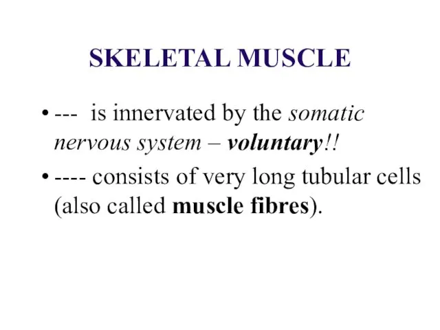
SKELETAL MUSCLE
--- is innervated by the somatic nervous system – voluntary!!
SKELETAL MUSCLE
--- is innervated by the somatic nervous system – voluntary!!
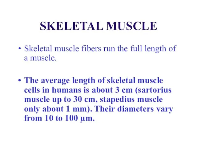
SKELETAL MUSCLE
Skeletal muscle fibers run the full length of a muscle.
SKELETAL MUSCLE
Skeletal muscle fibers run the full length of a muscle.
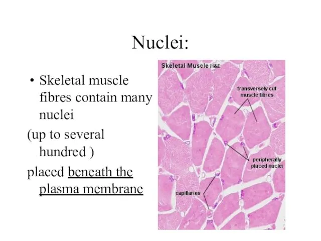
Nuclei:
Skeletal muscle fibres contain many nuclei
(up to several hundred )
placed
Nuclei:
Skeletal muscle fibres contain many nuclei
(up to several hundred )
placed
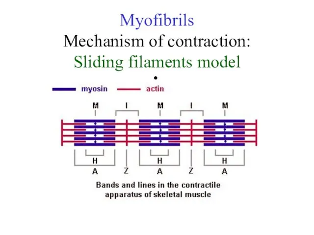
Myofibrils
Mechanism of contraction:
Sliding filaments model
Myofibrils
Mechanism of contraction:
Sliding filaments model
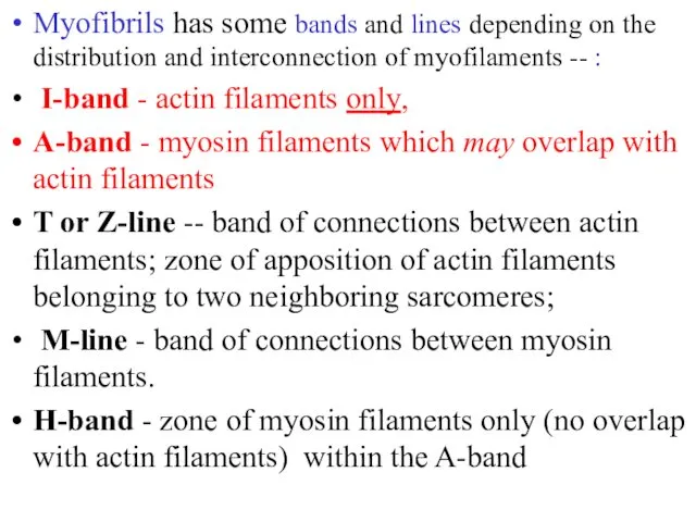
Myofibrils has some bands and lines depending on the distribution and
Myofibrils has some bands and lines depending on the distribution and

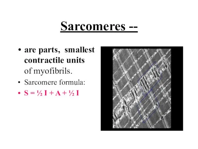
Sarcomeres --
are parts, smallest contractile units of myofibrils.
Sarcomere formula:
S =
Sarcomeres --
are parts, smallest contractile units of myofibrils.
Sarcomere formula:
S =
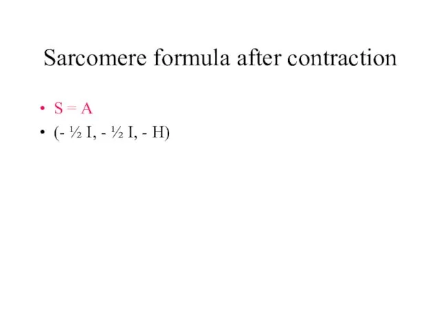
Sarcomere formula after contraction
S = A
(- ½ I, - ½ I,
Sarcomere formula after contraction
S = A
(- ½ I, - ½ I,
Mechanism of contraction
Mechanism of contraction
Origin of skeletal muscle
The myoblasts of all skeletal muscle fibres originate
Origin of skeletal muscle
The myoblasts of all skeletal muscle fibres originate
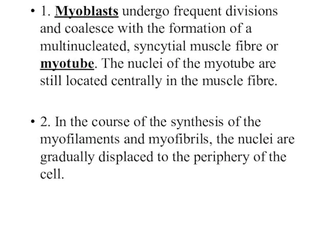
1. Myoblasts undergo frequent divisions and coalesce with the formation of
1. Myoblasts undergo frequent divisions and coalesce with the formation of
 Красота в искусстве и жизни
 Пп Егoр
 10 важных навыков для работы будущего
 Синтетические противомикробные средства
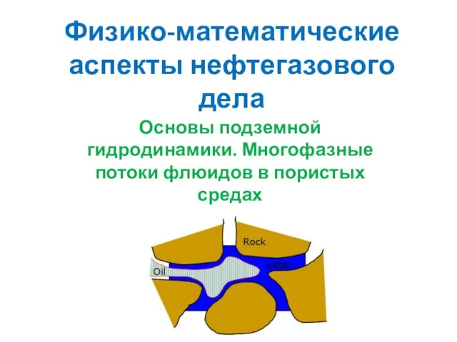 Физико-математические аспекты нефтегазового дела
 Презентация День Победы
 20231025_prezentatsiya_po_obshchestvoznaniyu_na_temu_chelovek_i_ego_deyatelnost_6_klass
 Говорящие фамилии в произведениях
 Тобольский гений России.- презентация
 Сварочные материалы
 Танцы народов мира.
 Модерн в архитектуре
 Измеряй на свой аршин.
 20200108_prezentatsiya_k_zanyatiyu
 Проектирование и эксплуатация газонефтепроводов (часть 1)
 Программирование на языке Паскаль. Символьные строки
 Государственные скрининговые программы по раннему выявлению онкопатологии репродуктивной системы
 Traditions of America
 Камины и печи. Балконы, эркеры. Окна, двери
 Всемирный день борьбы со СПИДом
 Class Lobosa – amoebas, amibes Order
 Сверление. Механизированное и ручное оборудование для сверления
 Технологическое производство и методы получение белково-витаминных концентратов
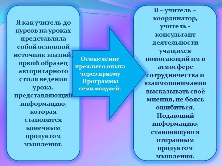 презентация для самообразования учителей о Кембриджской Программе внедрения семи модулей, как Новых подходах в воспитании и обучении детей.
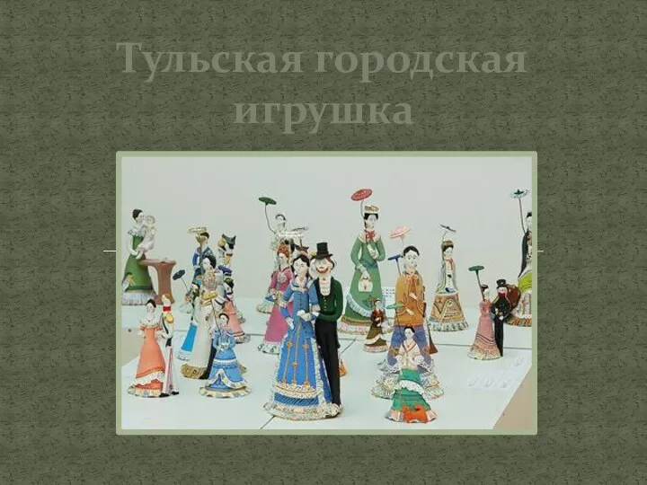 Тульская городская игрушка
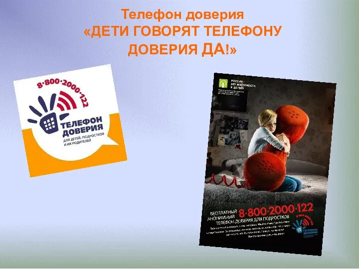 Презентация ДЕТИ ГОВОРЯТ ТЕЛЕФОНУ ДОВЕРИЯ ДА! май 2014
 Обобщение опыта работы по социально-коммуникативному развитию дошкольников.
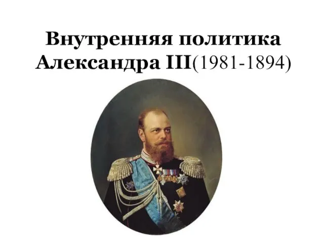 Внутренняя политика Александра III (1981-1894)