- Т_Internal Lecture 1 Resp

Содержание
- 2. Examination of respiratory system History taking Enviromental Usability of the horse Enviromental conditio in stable Food
- 3. Examination of respiratory system General examination Heart rate, breath rate, lymph nodes, membrane mucus, temperature Detail
- 4. Upper respiratory tract disease Rhinitis Necrosis conchae Polyps Ethmoid hematoma Nasal neoplasma Sinusitis Pharyngitis Guttural pouch
- 5. Nasal cavity do prezentacji użyto zdjęć z Atlas of Equine Endoscopy N.M.Slovis wyd Mosby
- 6. Rhinitis Cause: Virus infections-Infuenza, rhinovirus, herpesvirus, arteritis virus, adenovirus, reovirus, Bacterial infections -Streptococcus sp., glanders (Psudomonas
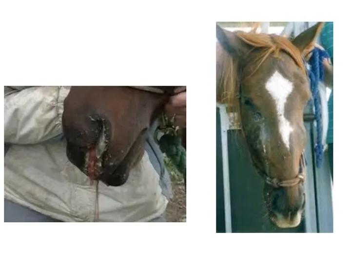
- 7. Foreign body in the nasal cavity do prezentacji użyto zdjęć z Atlas of Equine Endoscopy N.M.Slovis
- 8. Rhinitis Clinical pathology Virology Bacteriology Mycology Mainly to exclude or confirm infectious disease. In some cases
- 9. Necrosis conche Cause Bacterial or fungal infections. Clinical signs Muco-purulent, sometimes blood tinged, odorous discharge uni/bilateral.
- 10. Fungal plaques typical of infection of the nasal cavities with Aspergillus spp. do prezentacji użyto zdjęć
- 11. Polyps Cause Chronic inflamation of nasal mucous membranes of any cause Clinical sign Sero-muco-purulent nasal discharge
- 12. Polyps do prezentacji użyto zdjęć z Atlas of Equine Endoscopy N.M.Slovis wyd Mosby
- 13. Nasal neoplasma Cause Neoplasia- myxoma, fibroma, chondroma, osteochondroma, carcinoma, melanoma Clinical signs Uni/bilateral nasal discharge, sero-muco-purulent,
- 14. Ethmoid conchae do prezentacji użyto zdjęć z Atlas of Equine Endoscopy N.M.Slovis wyd Mosby
- 15. Ethmoidal hematoma Cause Neoplasia? Chronic infections, circulatory defect Clinical signs At the beginning usually unilaterally nasal
- 16. Ethmoidal haematoma do prezentacji użyto zdjęć z Atlas of Equine Endoscopy N.M.Slovis wyd Mosby
- 17. Sinuses
- 18. Sinusitis Cause Usually secondary to rhinitis, tooth problems, defects of sinus communication with nasal cavity Clinical
- 21. Pharyngitis Cause Viral infections- influenza, herpesvirus, adenovirus, arteritis virus, Bacterial infection-mainly Streptococcus spp. Physical trauma-stomach tube,
- 22. Pharyngitis Clinical signs decreased appetite, difficult swallowing, cough, increased temperature of swollen, painful throat and local
- 23. Pharyngitis do prezentacji użyto zdjęć z Atlas of Equine Endoscopy N.M.Slovis wyd Mosby Pharyngeal lymphoid hyperplasia
- 24. Pharyngeal paralysis do prezentacji użyto zdjęć z Atlas of Equine Endoscopy N.M.Slovis wyd Mosby
- 25. Guttural pouch do prezentacji użyto zdjęć z Atlas of Equine Endoscopy N.M.Slovis wyd Mosby
- 26. Guttural pouches do prezentacji użyto zdjęć z Atlas of Equine Endoscopy N.M.Slovis wyd Mosby Stylohyoid bone
- 27. Guttural pouch mycosis Cause: Fungal infections- Aspergillus fumigatus often with bacterial contamination- Pseudomonas aeruginosa. Primary lesion
- 28. Mycosis of the guttural pouches do prezentacji użyto zdjęć z Atlas of Equine Endoscopy N.M.Slovis wyd
- 29. Guttural pouch mycosis Clinical pathology Endoscopy Mycolgy Bacteriology Hematology Treatment Local washing with antifungal drugs (econazol,
- 30. Guttural pouch empyema Cause: mainly Streptococcus spp. Infections, Clinical signs: Uni/bilateral muco-purulent nasal discharge, more obvious
- 31. Purulent inflamation of the guttural pouch do prezentacji użyto zdjęć z Atlas of Equine Endoscopy N.M.Slovis
- 32. Guttural pouch chondroids Cause Inspissated guttural pouch exudate forms stones Clinical signs Swelling of guttural pouch
- 33. Chondroids of the gutural pouch do prezentacji użyto zdjęć z Atlas of Equine Endoscopy N.M.Slovis wyd
- 34. Guttural pouch tympany Cause Congenital defects of guttural pouch operculum Clinical signs Swelling of guttural pouch
- 35. Larynx do prezentacji użyto zdjęć z Atlas of Equine Endoscopy N.M.Slovis wyd Mosby
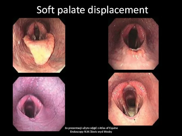
- 36. Soft palate displacement Cause Paresis of soft palate due to some neurological deficit, swelling of soft
- 37. Soft palate displacement do prezentacji użyto zdjęć z Atlas of Equine Endoscopy N.M.Slovis wyd Mosby
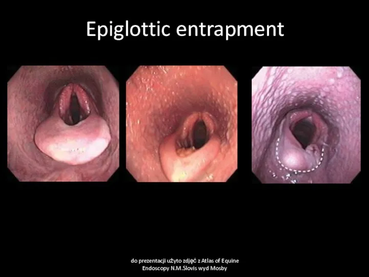
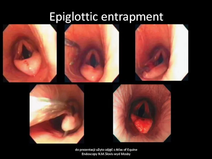
- 38. Aryepiglottic fold displacement (epiglottic entratment) Cause Edema of soft tissue close to epiglottis. Congenital shortening of
- 39. Epiglottic entrapment do prezentacji użyto zdjęć z Atlas of Equine Endoscopy N.M.Slovis wyd Mosby
- 40. Epiglottic entrapment do prezentacji użyto zdjęć z Atlas of Equine Endoscopy N.M.Slovis wyd Mosby
- 41. Laryngitis Cause Viral infections- influenza, herpesvirus, adenovirus, arteritis virus, Bacterial infection-mainly Streptococcus spp. Physical trauma-stomach tube,
- 42. Laryngeal edema Cause Acute inflamation, allergy, irritant substances, surgery at larynx region Clinical signs Abnormal respiratory
- 43. Larynx neoplasms Cause Neoplasia-papilloma, carcinoma, adenoma, fibroma, chondroma Clinical signs: Nasal discharge- muco-purulent, blood tinged, often
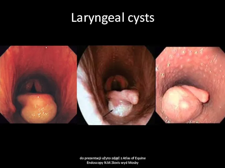
- 44. Larygeal cysts Cause Usually congenital cyst Clinical signs: abnormal laryngeal respiratory sound, dyspnea, cough Clinical pathology
- 45. Laryngeal cysts do prezentacji użyto zdjęć z Atlas of Equine Endoscopy N.M.Slovis wyd Mosby
- 46. Laryngeal hemiplegia Cause Recurrent laryngeal nerve paralysis due to general neuropathy, inherited, poisonings, local swelling, fungal
- 47. Laryngeal hemiplegia do prezentacji użyto zdjęć z Atlas of Equine Endoscopy N.M.Slovis wyd Mosby
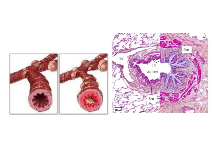
- 48. Trachea do prezentacji użyto zdjęć z Atlas of Equine Endoscopy N.M.Slovis wyd Mosby Bifurcation of the
- 49. Right bronchus do prezentacji użyto zdjęć z Atlas of Equine Endoscopy N.M.Slovis wyd Mosby
- 50. Left bronchus do prezentacji użyto zdjęć z Atlas of Equine Endoscopy N.M.Slovis wyd Mosby
- 51. Tracheitis and bronchitis Cause Infection equine influenza, equine herpes virus, equine viral arteritis, streptococcal infections, other
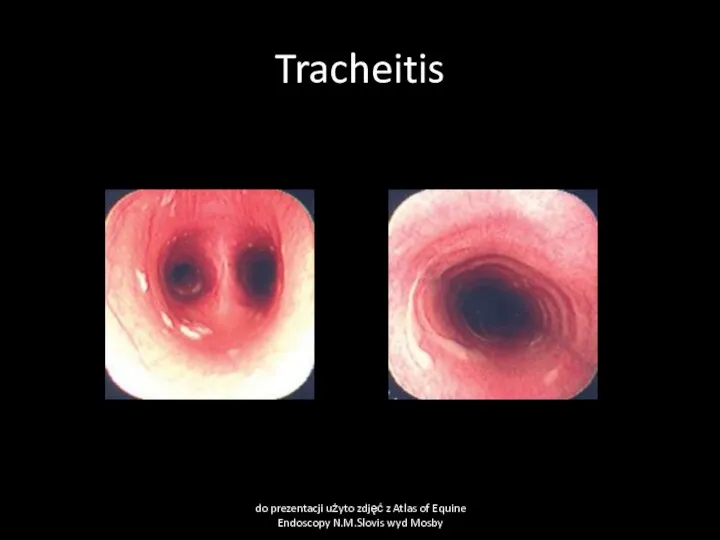
- 52. Tracheitis do prezentacji użyto zdjęć z Atlas of Equine Endoscopy N.M.Slovis wyd Mosby
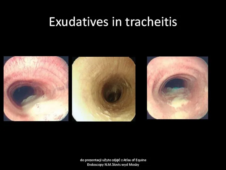
- 53. Exudatives in tracheitis do prezentacji użyto zdjęć z Atlas of Equine Endoscopy N.M.Slovis wyd Mosby
- 54. Tracheitis and bronchitis Clinical pathology bacteriological examination of tracheal wash or tharcheal aspirates, cytology, X-ray, thorax
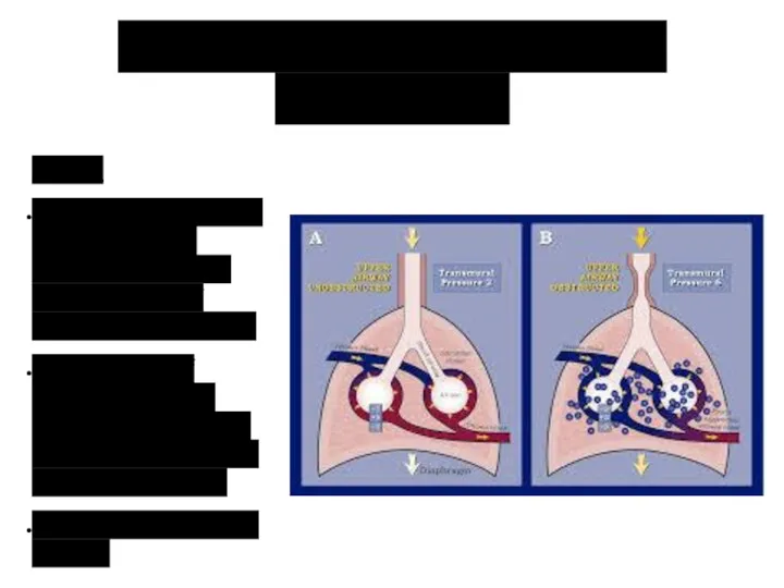
- 55. Diseases of lungs Exercise-induced pulmonary hemorrhage Recurrect airway obstruction
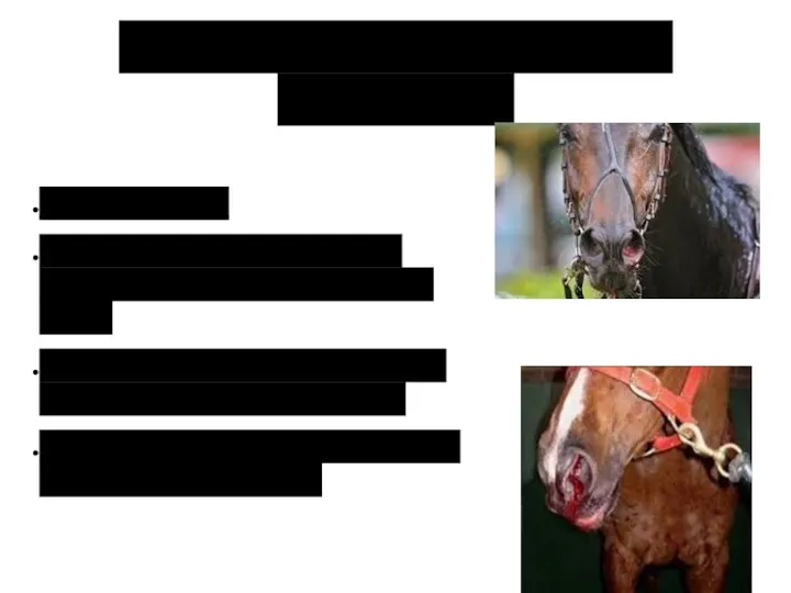
- 56. Exercise-induced pulmonary hemorrhage Cause High pulmonary blood pressure during sternuous exercise cause rupture of pulmonary capillares.
- 57. Exercise-induced pulmonary hemorrhage Clinical signs: May be found in >80% racing horces but clinically observed in
- 58. Exercise-induced pulmonary hemorrhage Clinical pathology Macrophages with digested red blood cells (hemosiderin) in sample of tracheal
- 59. Exercise induced pulmonary hemorrhage do prezentacji użyto zdjęć z Atlas of Equine Endoscopy N.M.Slovis wyd Mosby
- 60. Exercise- induced pulmonary hemorrhage Treatment Rest, Treat respiratory disease if present. Furosemide before sternuous exercise may
- 61. Recurrect Airway Obstruction (Heaves) Cause Dusty stable environment, viral infections, air pollution by Aspergillus fumigatus Actinomyces
- 63. Lungs emphysema
- 64. Recurrect Airway Obstruction (Heaves) Clinical signs: Older than 7 years horses most common affected. At the
- 66. Recurrect Airway Obstruction (Heaves) Clinical pathology Endoscopy examination ( chronic inflamation of bronchi and tracheal mucosa
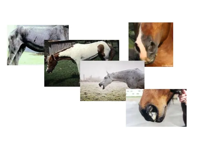
- 67. Heaves do prezentacji użyto zdjęć z Atlas of Equine Endoscopy N.M.Slovis wyd Mosby
- 69. Скачать презентацию
Examination of respiratory system
History taking
Enviromental
Usability of the horse
Enviromental conditio in
Examination of respiratory system
History taking
Enviromental
Usability of the horse
Enviromental conditio in
Examination of respiratory system
General examination
Heart rate, breath rate, lymph nodes, membrane
Examination of respiratory system
General examination
Heart rate, breath rate, lymph nodes, membrane

Upper respiratory tract disease
Rhinitis
Necrosis conchae
Polyps
Ethmoid hematoma
Nasal neoplasma
Sinusitis
Pharyngitis
Guttural pouch empyema
Guttural pouch empyema
Guttural
Upper respiratory tract disease
Rhinitis
Necrosis conchae
Polyps
Ethmoid hematoma
Nasal neoplasma
Sinusitis
Pharyngitis
Guttural pouch empyema
Guttural pouch empyema
Guttural
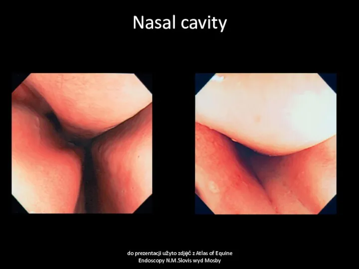
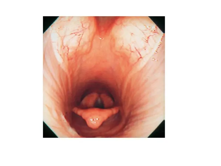
Nasal cavity
do prezentacji użyto zdjęć z Atlas of Equine Endoscopy N.M.Slovis
Nasal cavity
do prezentacji użyto zdjęć z Atlas of Equine Endoscopy N.M.Slovis

Rhinitis
Cause:
Virus infections-Infuenza, rhinovirus, herpesvirus, arteritis virus, adenovirus, reovirus,
Bacterial infections -Streptococcus sp.,
Rhinitis
Cause:
Virus infections-Infuenza, rhinovirus, herpesvirus, arteritis virus, adenovirus, reovirus,
Bacterial infections -Streptococcus sp.,
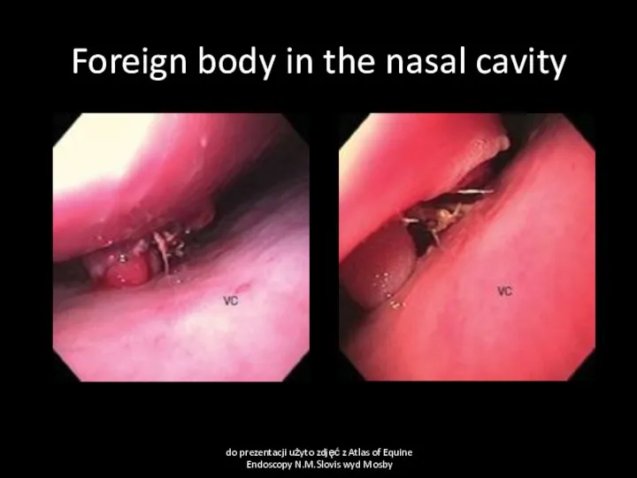
Foreign body in the nasal cavity
do prezentacji użyto zdjęć z Atlas
Foreign body in the nasal cavity
do prezentacji użyto zdjęć z Atlas

Rhinitis
Clinical pathology
Virology
Bacteriology
Mycology
Mainly to exclude or confirm infectious disease.
In some cases endoscopy
Rhinitis
Clinical pathology
Virology
Bacteriology
Mycology
Mainly to exclude or confirm infectious disease.
In some cases endoscopy

Necrosis conche
Cause
Bacterial or fungal infections.
Clinical signs
Muco-purulent, sometimes blood tinged, odorous
Necrosis conche
Cause
Bacterial or fungal infections.
Clinical signs
Muco-purulent, sometimes blood tinged, odorous
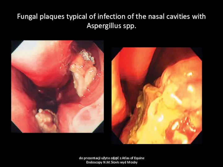
Fungal plaques typical of infection of the nasal cavities with Aspergillus
Fungal plaques typical of infection of the nasal cavities with Aspergillus

Polyps
Cause
Chronic inflamation of nasal mucous membranes of any cause
Clinical sign
Sero-muco-purulent nasal
Polyps
Cause
Chronic inflamation of nasal mucous membranes of any cause
Clinical sign
Sero-muco-purulent nasal
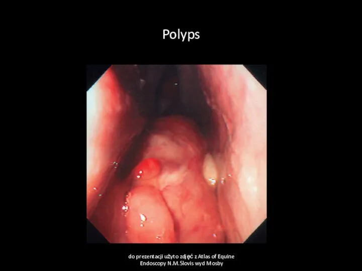
Polyps
do prezentacji użyto zdjęć z Atlas of Equine Endoscopy N.M.Slovis wyd
Polyps
do prezentacji użyto zdjęć z Atlas of Equine Endoscopy N.M.Slovis wyd

Nasal neoplasma
Cause
Neoplasia- myxoma, fibroma, chondroma, osteochondroma, carcinoma, melanoma
Clinical signs
Uni/bilateral nasal discharge,
Nasal neoplasma
Cause
Neoplasia- myxoma, fibroma, chondroma, osteochondroma, carcinoma, melanoma
Clinical signs
Uni/bilateral nasal discharge,
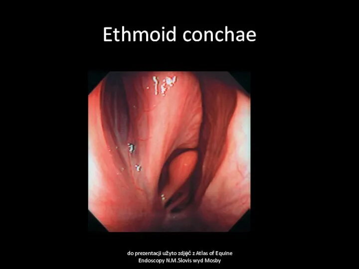
Ethmoid conchae
do prezentacji użyto zdjęć z Atlas of Equine Endoscopy N.M.Slovis
Ethmoid conchae
do prezentacji użyto zdjęć z Atlas of Equine Endoscopy N.M.Slovis

Ethmoidal hematoma
Cause
Neoplasia? Chronic infections, circulatory defect
Clinical signs
At the beginning usually unilaterally
Ethmoidal hematoma
Cause
Neoplasia? Chronic infections, circulatory defect
Clinical signs
At the beginning usually unilaterally
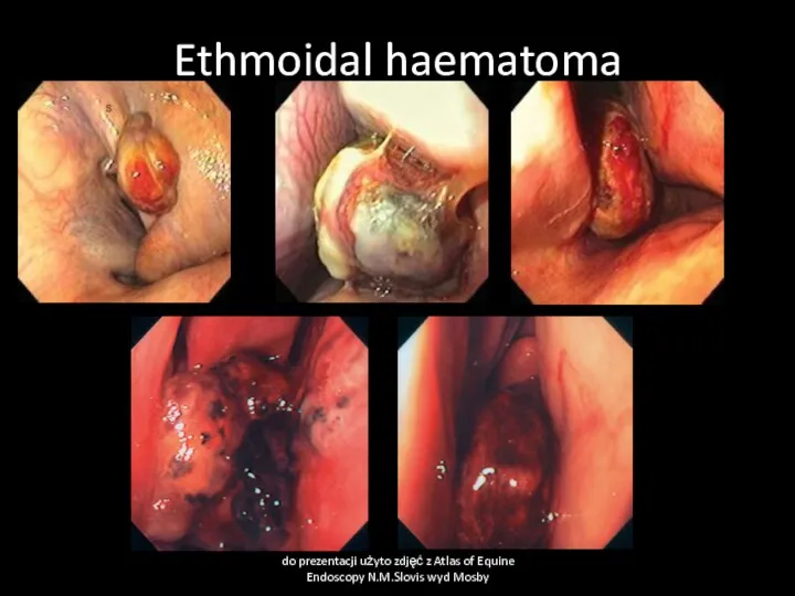
Ethmoidal haematoma
do prezentacji użyto zdjęć z Atlas of Equine Endoscopy N.M.Slovis
Ethmoidal haematoma
do prezentacji użyto zdjęć z Atlas of Equine Endoscopy N.M.Slovis
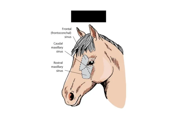
Sinuses
Sinuses

Sinusitis
Cause
Usually secondary to rhinitis, tooth problems, defects of sinus communication with
Sinusitis
Cause
Usually secondary to rhinitis, tooth problems, defects of sinus communication with

Pharyngitis
Cause
Viral infections- influenza, herpesvirus, adenovirus, arteritis virus,
Bacterial infection-mainly Streptococcus spp.
Physical trauma-stomach
Pharyngitis
Cause
Viral infections- influenza, herpesvirus, adenovirus, arteritis virus,
Bacterial infection-mainly Streptococcus spp.
Physical trauma-stomach

Pharyngitis
Clinical signs
decreased appetite, difficult swallowing, cough, increased temperature of swollen, painful
Pharyngitis
Clinical signs
decreased appetite, difficult swallowing, cough, increased temperature of swollen, painful
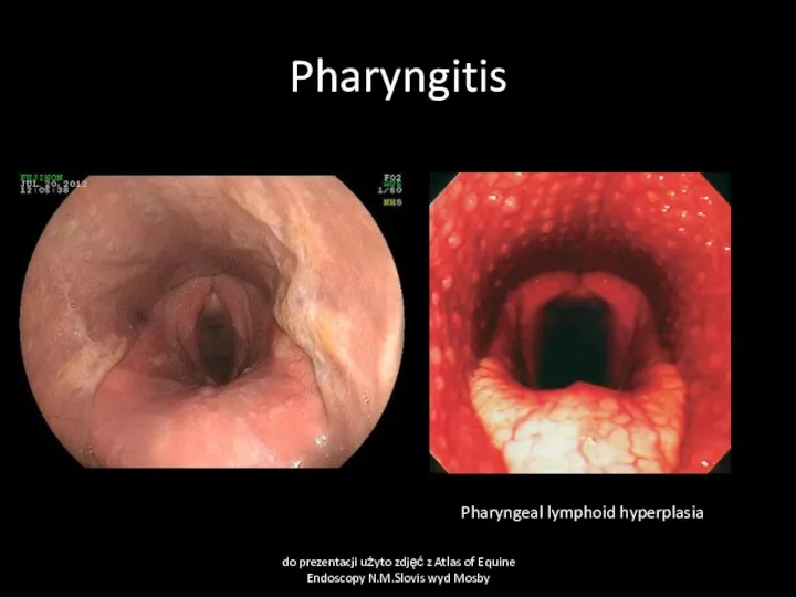
Pharyngitis
do prezentacji użyto zdjęć z Atlas of Equine Endoscopy N.M.Slovis wyd
Pharyngitis
do prezentacji użyto zdjęć z Atlas of Equine Endoscopy N.M.Slovis wyd
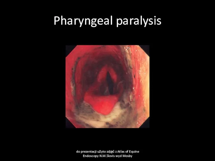
Pharyngeal paralysis
do prezentacji użyto zdjęć z Atlas of Equine Endoscopy N.M.Slovis
Pharyngeal paralysis
do prezentacji użyto zdjęć z Atlas of Equine Endoscopy N.M.Slovis
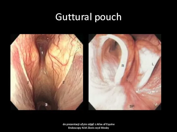
Guttural pouch
do prezentacji użyto zdjęć z Atlas of Equine Endoscopy N.M.Slovis
Guttural pouch
do prezentacji użyto zdjęć z Atlas of Equine Endoscopy N.M.Slovis
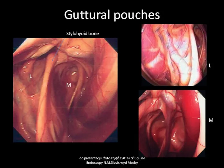
Guttural pouches
do prezentacji użyto zdjęć z Atlas of Equine Endoscopy N.M.Slovis
Guttural pouches
do prezentacji użyto zdjęć z Atlas of Equine Endoscopy N.M.Slovis

Guttural pouch mycosis
Cause:
Fungal infections- Aspergillus fumigatus often with bacterial contamination- Pseudomonas
Guttural pouch mycosis
Cause:
Fungal infections- Aspergillus fumigatus often with bacterial contamination- Pseudomonas
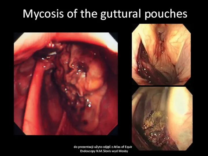
Mycosis of the guttural pouches
do prezentacji użyto zdjęć z Atlas of
Mycosis of the guttural pouches
do prezentacji użyto zdjęć z Atlas of

Guttural pouch mycosis
Clinical pathology
Endoscopy
Mycolgy
Bacteriology
Hematology
Treatment
Local washing with antifungal drugs (econazol, eniconazol, myconazol,
Guttural pouch mycosis
Clinical pathology
Endoscopy
Mycolgy
Bacteriology
Hematology
Treatment
Local washing with antifungal drugs (econazol, eniconazol, myconazol,

Guttural pouch empyema
Cause:
mainly Streptococcus spp. Infections,
Clinical signs:
Uni/bilateral muco-purulent nasal discharge, more
Guttural pouch empyema
Cause:
mainly Streptococcus spp. Infections,
Clinical signs:
Uni/bilateral muco-purulent nasal discharge, more
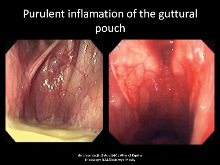
Purulent inflamation of the guttural pouch
do prezentacji użyto zdjęć z Atlas
Purulent inflamation of the guttural pouch
do prezentacji użyto zdjęć z Atlas

Guttural pouch chondroids
Cause
Inspissated guttural pouch exudate forms stones
Clinical signs
Swelling of guttural
Guttural pouch chondroids
Cause
Inspissated guttural pouch exudate forms stones
Clinical signs
Swelling of guttural
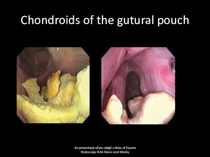
Chondroids of the gutural pouch
do prezentacji użyto zdjęć z Atlas of
Chondroids of the gutural pouch
do prezentacji użyto zdjęć z Atlas of
Guttural pouch tympany
Cause
Congenital defects of guttural pouch operculum
Clinical signs
Swelling of guttural
Guttural pouch tympany
Cause
Congenital defects of guttural pouch operculum
Clinical signs
Swelling of guttural
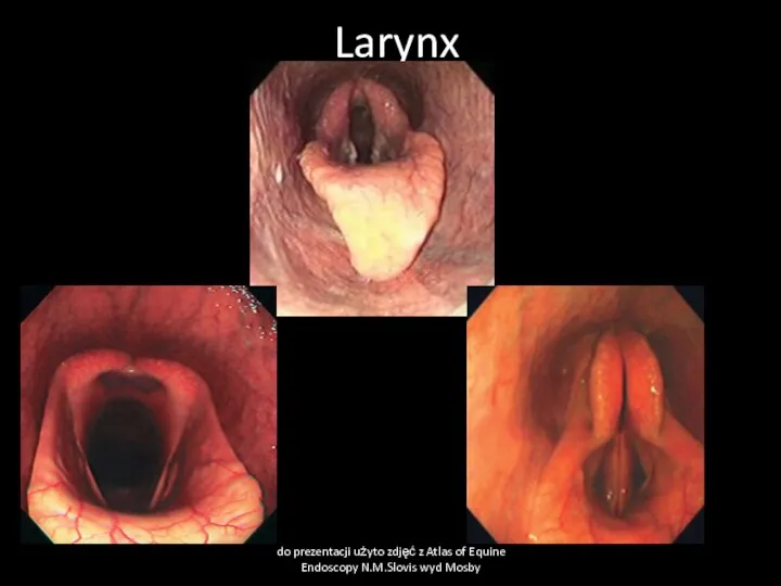
Larynx
do prezentacji użyto zdjęć z Atlas of Equine Endoscopy N.M.Slovis wyd
Larynx
do prezentacji użyto zdjęć z Atlas of Equine Endoscopy N.M.Slovis wyd

Soft palate displacement
Cause
Paresis of soft palate due to some neurological deficit,
Soft palate displacement
Cause
Paresis of soft palate due to some neurological deficit,
Soft palate displacement
do prezentacji użyto zdjęć z Atlas of Equine Endoscopy
Soft palate displacement
do prezentacji użyto zdjęć z Atlas of Equine Endoscopy

Aryepiglottic fold displacement
(epiglottic entratment)
Cause
Edema of soft tissue close to epiglottis. Congenital
Aryepiglottic fold displacement
(epiglottic entratment)
Cause
Edema of soft tissue close to epiglottis. Congenital
Epiglottic entrapment
do prezentacji użyto zdjęć z Atlas of Equine Endoscopy N.M.Slovis
Epiglottic entrapment
do prezentacji użyto zdjęć z Atlas of Equine Endoscopy N.M.Slovis
Epiglottic entrapment
do prezentacji użyto zdjęć z Atlas of Equine Endoscopy N.M.Slovis
Epiglottic entrapment
do prezentacji użyto zdjęć z Atlas of Equine Endoscopy N.M.Slovis

Laryngitis
Cause
Viral infections- influenza, herpesvirus, adenovirus, arteritis virus,
Bacterial infection-mainly Streptococcus spp.
Physical trauma-stomach
Laryngitis
Cause
Viral infections- influenza, herpesvirus, adenovirus, arteritis virus,
Bacterial infection-mainly Streptococcus spp.
Physical trauma-stomach

Laryngeal edema
Cause
Acute inflamation, allergy, irritant substances, surgery at larynx region
Clinical signs
Abnormal
Laryngeal edema
Cause
Acute inflamation, allergy, irritant substances, surgery at larynx region
Clinical signs
Abnormal

Larynx neoplasms
Cause
Neoplasia-papilloma, carcinoma, adenoma, fibroma, chondroma
Clinical signs:
Nasal discharge- muco-purulent, blood tinged,
Larynx neoplasms
Cause
Neoplasia-papilloma, carcinoma, adenoma, fibroma, chondroma
Clinical signs:
Nasal discharge- muco-purulent, blood tinged,

Larygeal cysts
Cause
Usually congenital cyst
Clinical signs:
abnormal laryngeal respiratory sound, dyspnea, cough
Clinical pathology
Endoscopy
Treatment
surgery,
Larygeal cysts
Cause
Usually congenital cyst
Clinical signs:
abnormal laryngeal respiratory sound, dyspnea, cough
Clinical pathology
Endoscopy
Treatment
surgery,
Laryngeal cysts
do prezentacji użyto zdjęć z Atlas of Equine Endoscopy N.M.Slovis
Laryngeal cysts
do prezentacji użyto zdjęć z Atlas of Equine Endoscopy N.M.Slovis

Laryngeal hemiplegia
Cause
Recurrent laryngeal nerve paralysis due to general neuropathy, inherited, poisonings,
Laryngeal hemiplegia
Cause
Recurrent laryngeal nerve paralysis due to general neuropathy, inherited, poisonings,
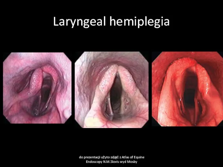
Laryngeal hemiplegia
do prezentacji użyto zdjęć z Atlas of Equine Endoscopy N.M.Slovis
Laryngeal hemiplegia
do prezentacji użyto zdjęć z Atlas of Equine Endoscopy N.M.Slovis
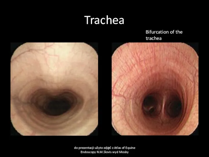
Trachea
do prezentacji użyto zdjęć z Atlas of Equine Endoscopy N.M.Slovis wyd
Trachea
do prezentacji użyto zdjęć z Atlas of Equine Endoscopy N.M.Slovis wyd
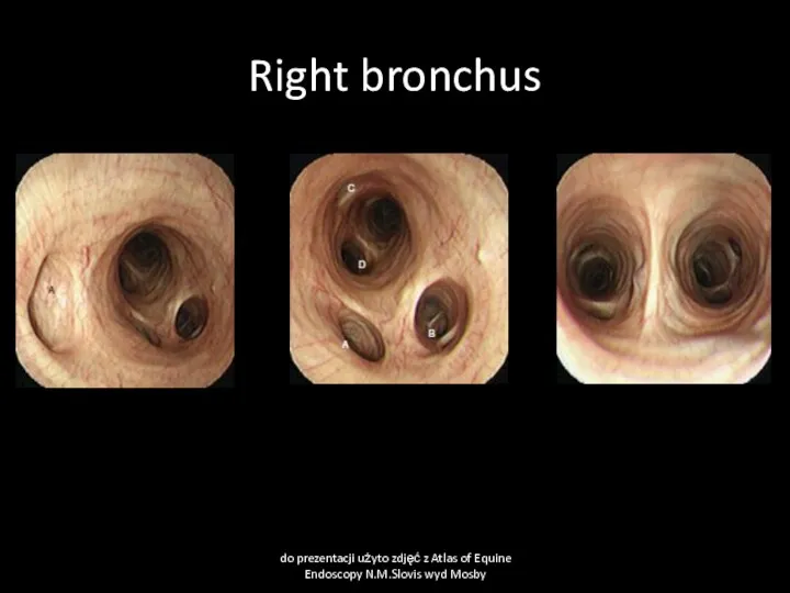
Right bronchus
do prezentacji użyto zdjęć z Atlas of Equine Endoscopy N.M.Slovis
Right bronchus
do prezentacji użyto zdjęć z Atlas of Equine Endoscopy N.M.Slovis
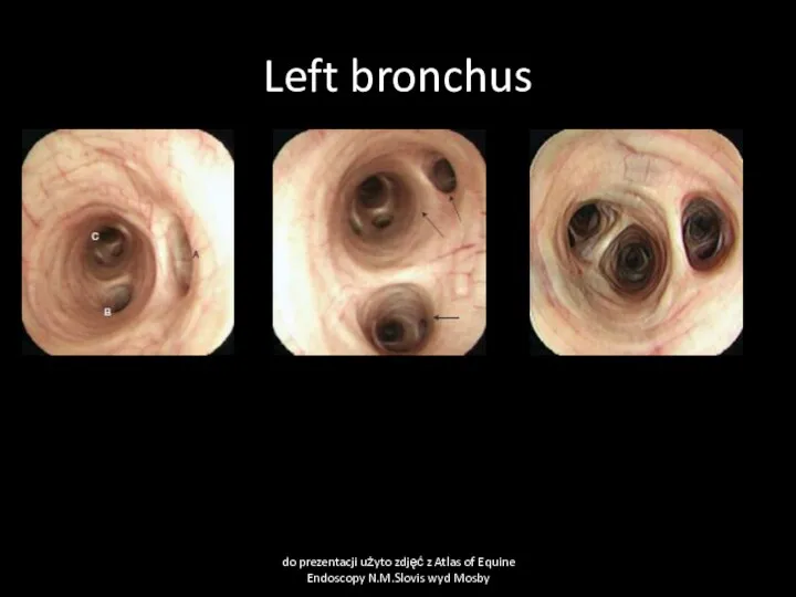
Left bronchus
do prezentacji użyto zdjęć z Atlas of Equine Endoscopy N.M.Slovis
Left bronchus
do prezentacji użyto zdjęć z Atlas of Equine Endoscopy N.M.Slovis

Tracheitis and bronchitis
Cause
Infection equine influenza, equine herpes virus, equine viral arteritis,
Tracheitis and bronchitis
Cause
Infection equine influenza, equine herpes virus, equine viral arteritis,
Tracheitis
do prezentacji użyto zdjęć z Atlas of Equine Endoscopy N.M.Slovis wyd
Tracheitis
do prezentacji użyto zdjęć z Atlas of Equine Endoscopy N.M.Slovis wyd
Exudatives in tracheitis
do prezentacji użyto zdjęć z Atlas of Equine Endoscopy
Exudatives in tracheitis
do prezentacji użyto zdjęć z Atlas of Equine Endoscopy

Tracheitis and bronchitis
Clinical pathology
bacteriological examination of tracheal wash or tharcheal aspirates,
Tracheitis and bronchitis
Clinical pathology
bacteriological examination of tracheal wash or tharcheal aspirates,

Diseases of lungs
Exercise-induced pulmonary hemorrhage
Recurrect airway obstruction
Diseases of lungs
Exercise-induced pulmonary hemorrhage
Recurrect airway obstruction
Exercise-induced pulmonary hemorrhage
Cause
High pulmonary blood pressure during sternuous exercise cause rupture
Exercise-induced pulmonary hemorrhage
Cause
High pulmonary blood pressure during sternuous exercise cause rupture
Exercise-induced pulmonary hemorrhage
Clinical signs:
May be found in >80% racing horces but
Exercise-induced pulmonary hemorrhage
Clinical signs:
May be found in >80% racing horces but

Exercise-induced pulmonary hemorrhage
Clinical pathology
Macrophages with digested red blood cells (hemosiderin) in
Exercise-induced pulmonary hemorrhage
Clinical pathology
Macrophages with digested red blood cells (hemosiderin) in
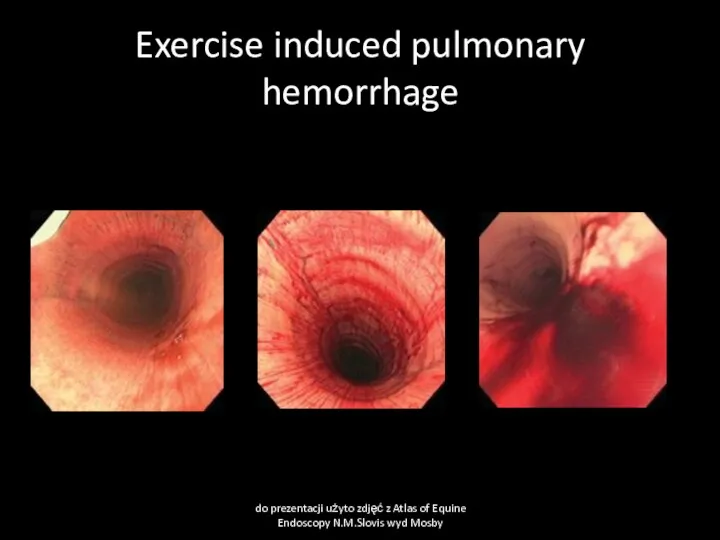
Exercise induced pulmonary hemorrhage
do prezentacji użyto zdjęć z Atlas of Equine
Exercise induced pulmonary hemorrhage
do prezentacji użyto zdjęć z Atlas of Equine

Exercise- induced pulmonary hemorrhage
Treatment
Rest,
Treat respiratory disease if present.
Furosemide before sternuous exercise
Exercise- induced pulmonary hemorrhage
Treatment
Rest,
Treat respiratory disease if present.
Furosemide before sternuous exercise

Recurrect Airway Obstruction (Heaves)
Cause
Dusty stable environment, viral infections, air pollution by
Recurrect Airway Obstruction (Heaves)
Cause
Dusty stable environment, viral infections, air pollution by
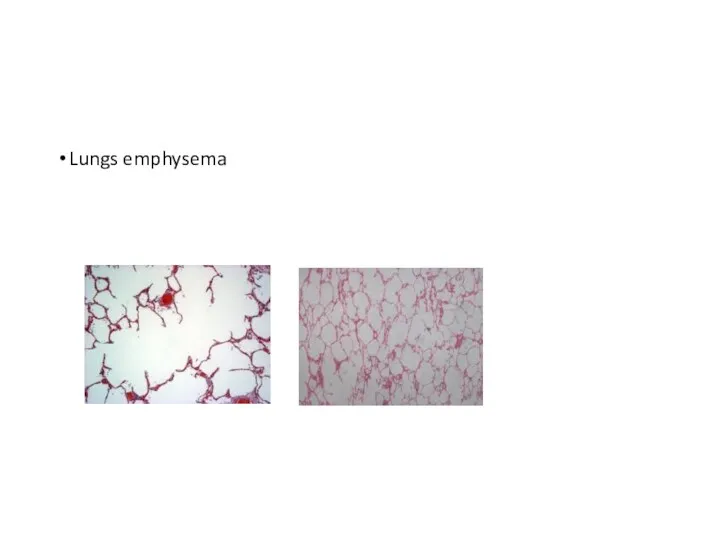
Lungs emphysema
Lungs emphysema

Recurrect Airway Obstruction (Heaves)
Clinical signs:
Older than 7 years horses most common
Recurrect Airway Obstruction (Heaves)
Clinical signs:
Older than 7 years horses most common

Recurrect Airway Obstruction (Heaves)
Clinical pathology
Endoscopy examination ( chronic inflamation of bronchi
Recurrect Airway Obstruction (Heaves)
Clinical pathology
Endoscopy examination ( chronic inflamation of bronchi
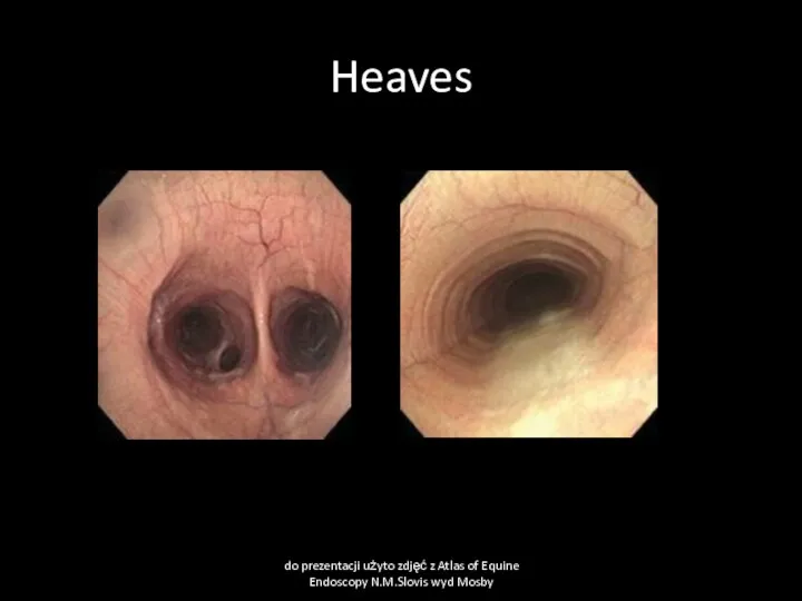
Heaves
do prezentacji użyto zdjęć z Atlas of Equine Endoscopy N.M.Slovis wyd
Heaves
do prezentacji użyto zdjęć z Atlas of Equine Endoscopy N.M.Slovis wyd
 Święci i patroni
 Сказка о том, как Лягушонок научился гудеть, как пароход. Постановка и автоматизация звука [Ы]
 Inventronics. Информация о компании
 Старинная ярмарка.
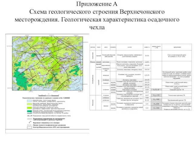 Схема геологического строения Верхнечонского месторождения
 Установка маркшейдерских и геодезических знаков и реперов
 Глава над разделенным Телом. Марк Эфесский и православное понимание единства Церкви
 Непредельные углеводороды. Алкены
 Аутоиммунный гепатит
 Творчество Ф.М. Достоевского (1825-1881)
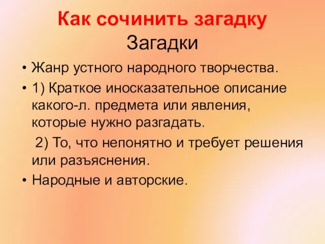 Как сочинить загадку
 Строение ядра клетки
 Мастерская Деда Мороза
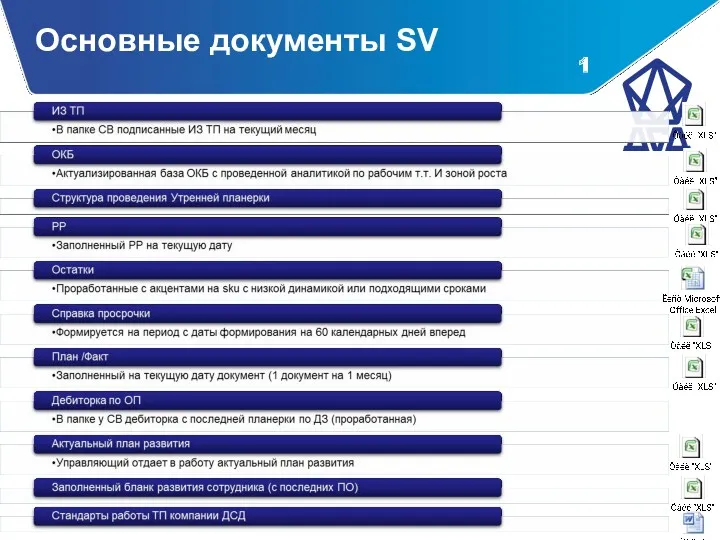 документы SV
 Северо-Восточная Русь в XII – начале XIII веков
 Назначение и состав технологического оборудования стартового комплекса. Лекция 11
 Конституционные суды в системе правосудия РФ
 Токарные станки
 Координаты векторов. Скалярное произведение векторов
 Туркестанская область
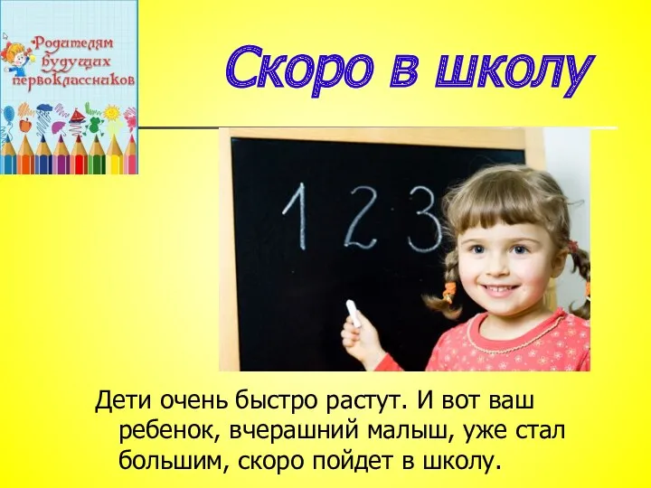 Родительское собрание Подготовка в школе
 Механизация сельского хозяйства. Технология производства продукции растениеводства
 Теория систем и системные исследования в энергетике
 TPR метод в обучении иностранному языку
 Медициналық этика
 It takes many kinds to make the world
 Филогенез систем органов позвоночных животных
 Форма организации труда. Средства производства строительных работ