- A Tour of the Cell

Содержание
- 2. Overview: The Fundamental Units of Life All organisms are made of cells The cell is the
- 3. Fig. 6-1
- 4. Concept 6.1: To study cells, biologists use microscopes and the tools of biochemistry Though usually too
- 5. Microscopy Scientists use microscopes to visualize cells too small to see with the naked eye In
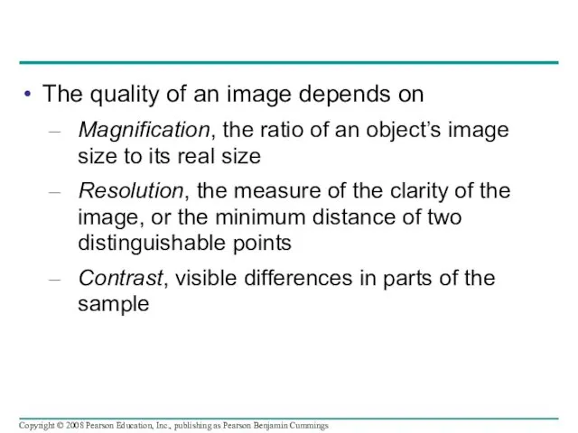
- 6. The quality of an image depends on Magnification, the ratio of an object’s image size to
- 7. Fig. 6-2 10 m 1 m 0.1 m 1 cm 1 mm 100 µm 10 µm
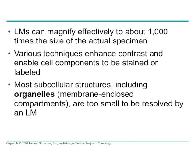
- 8. LMs can magnify effectively to about 1,000 times the size of the actual specimen Various techniques
- 9. Fig. 6-3 TECHNIQUE RESULTS (a) Brightfield (unstained specimen) (b) Brightfield (stained specimen) 50 µm (c) Phase-contrast
- 10. Fig. 6-3ab (a) Brightfield (unstained specimen) (b) Brightfield (stained specimen) TECHNIQUE RESULTS 50 µm
- 11. Fig. 6-3cd (c) Phase-contrast (d) Differential-interference- contrast (Nomarski) TECHNIQUE RESULTS
- 12. Fig. 6-3e (e) Fluorescence TECHNIQUE RESULTS 50 µm
- 13. Fig. 6-3f (f) Confocal TECHNIQUE RESULTS 50 µm
- 14. Two basic types of electron microscopes (EMs) are used to study subcellular structures Scanning electron microscopes
- 15. Fig. 6-4 (a) Scanning electron microscopy (SEM) TECHNIQUE RESULTS (b) Transmission electron microscopy (TEM) Cilia Longitudinal
- 16. Cell Fractionation Cell fractionation takes cells apart and separates the major organelles from one another Ultracentrifuges
- 17. Fig. 6-5 Homogenization TECHNIQUE Homogenate Tissue cells 1,000 g (1,000 times the force of gravity) 10
- 18. Fig. 6-5a Homogenization Homogenate Differential centrifugation Tissue cells TECHNIQUE
- 19. Fig. 6-5b 1,000 g (1,000 times the force of gravity) 10 min Supernatant poured into next
- 20. Concept 6.2: Eukaryotic cells have internal membranes that compartmentalize their functions The basic structural and functional
- 21. Comparing Prokaryotic and Eukaryotic Cells Basic features of all cells: Plasma membrane Semifluid substance called cytosol
- 22. Prokaryotic cells are characterized by having No nucleus DNA in an unbound region called the nucleoid
- 23. Fig. 6-6 Fimbriae Nucleoid Ribosomes Plasma membrane Cell wall Capsule Flagella Bacterial chromosome (a) A typical
- 24. Eukaryotic cells are characterized by having DNA in a nucleus that is bounded by a membranous
- 25. The plasma membrane is a selective barrier that allows sufficient passage of oxygen, nutrients, and waste
- 26. Fig. 6-7 TEM of a plasma membrane (a) (b) Structure of the plasma membrane Outside of
- 27. The logistics of carrying out cellular metabolism sets limits on the size of cells The surface
- 28. Fig. 6-8 Surface area increases while total volume remains constant 5 1 1 6 150 750
- 29. A Panoramic View of the Eukaryotic Cell A eukaryotic cell has internal membranes that partition the
- 30. Fig. 6-9a ENDOPLASMIC RETICULUM (ER) Smooth ER Rough ER Flagellum Centrosome CYTOSKELETON: Microfilaments Intermediate filaments Microtubules
- 31. Fig. 6-9b NUCLEUS Nuclear envelope Nucleolus Chromatin Rough endoplasmic reticulum Smooth endoplasmic reticulum Ribosomes Central vacuole
- 32. Concept 6.3: The eukaryotic cell’s genetic instructions are housed in the nucleus and carried out by
- 33. The Nucleus: Information Central The nucleus contains most of the cell’s genes and is usually the
- 34. Fig. 6-10 Nucleolus Nucleus Rough ER Nuclear lamina (TEM) Close-up of nuclear envelope 1 µm 1
- 35. Pores regulate the entry and exit of molecules from the nucleus The shape of the nucleus
- 36. In the nucleus, DNA and proteins form genetic material called chromatin Chromatin condenses to form discrete
- 37. Ribosomes: Protein Factories Ribosomes are particles made of ribosomal RNA and protein Ribosomes carry out protein
- 38. Fig. 6-11 Cytosol Endoplasmic reticulum (ER) Free ribosomes Bound ribosomes Large subunit Small subunit Diagram of
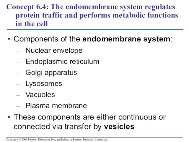
- 39. Concept 6.4: The endomembrane system regulates protein traffic and performs metabolic functions in the cell Components
- 40. The Endoplasmic Reticulum: Biosynthetic Factory The endoplasmic reticulum (ER) accounts for more than half of the
- 41. Fig. 6-12 Smooth ER Rough ER Nuclear envelope Transitional ER Rough ER Smooth ER Transport vesicle
- 42. Functions of Smooth ER The smooth ER Synthesizes lipids Metabolizes carbohydrates Detoxifies poison Stores calcium Copyright
- 43. Functions of Rough ER The rough ER Has bound ribosomes, which secrete glycoproteins (proteins covalently bonded
- 44. The Golgi apparatus consists of flattened membranous sacs called cisternae Functions of the Golgi apparatus: Modifies
- 45. Fig. 6-13 cis face (“receiving” side of Golgi apparatus) Cisternae trans face (“shipping” side of Golgi
- 46. Lysosomes: Digestive Compartments A lysosome is a membranous sac of hydrolytic enzymes that can digest macromolecules
- 47. Some types of cell can engulf another cell by phagocytosis; this forms a food vacuole A
- 48. Fig. 6-14 Nucleus 1 µm Lysosome Digestive enzymes Lysosome Plasma membrane Food vacuole (a) Phagocytosis Digestion
- 49. Fig. 6-14a Nucleus 1 µm Lysosome Lysosome Digestive enzymes Plasma membrane Food vacuole Digestion (a) Phagocytosis
- 50. Fig. 6-14b Vesicle containing two damaged organelles Mitochondrion fragment Peroxisome fragment Peroxisome Lysosome Digestion Mitochondrion Vesicle
- 51. Vacuoles: Diverse Maintenance Compartments A plant cell or fungal cell may have one or several vacuoles
- 52. Food vacuoles are formed by phagocytosis Contractile vacuoles, found in many freshwater protists, pump excess water
- 53. Fig. 6-15 Central vacuole Cytosol Central vacuole Nucleus Cell wall Chloroplast 5 µm
- 54. The Endomembrane System: A Review The endomembrane system is a complex and dynamic player in the
- 55. Fig. 6-16-1 Smooth ER Nucleus Rough ER Plasma membrane
- 56. Fig. 6-16-2 Smooth ER Nucleus Rough ER Plasma membrane cis Golgi trans Golgi
- 57. Fig. 6-16-3 Smooth ER Nucleus Rough ER Plasma membrane cis Golgi trans Golgi
- 58. Concept 6.5: Mitochondria and chloroplasts change energy from one form to another Mitochondria are the sites
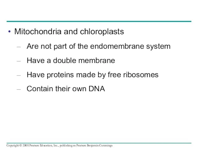
- 59. Mitochondria and chloroplasts Are not part of the endomembrane system Have a double membrane Have proteins
- 60. Mitochondria: Chemical Energy Conversion Mitochondria are in nearly all eukaryotic cells They have a smooth outer
- 61. Fig. 6-17 Free ribosomes in the mitochondrial matrix Intermembrane space Outer membrane Inner membrane Cristae Matrix
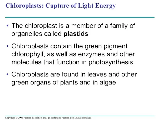
- 62. Chloroplasts: Capture of Light Energy The chloroplast is a member of a family of organelles called
- 63. Chloroplast structure includes: Thylakoids, membranous sacs, stacked to form a granum Stroma, the internal fluid Copyright
- 64. Fig. 6-18 Ribosomes Thylakoid Stroma Granum Inner and outer membranes 1 µm
- 65. Peroxisomes: Oxidation Peroxisomes are specialized metabolic compartments bounded by a single membrane Peroxisomes produce hydrogen peroxide
- 66. Fig. 6-19 1 µm Chloroplast Peroxisome Mitochondrion
- 67. Concept 6.6: The cytoskeleton is a network of fibers that organizes structures and activities in the
- 68. Fig. 6-20 Microtubule Microfilaments 0.25 µm
- 69. Roles of the Cytoskeleton: Support, Motility, and Regulation The cytoskeleton helps to support the cell and
- 70. Fig. 6-21 Vesicle ATP Receptor for motor protein Microtubule of cytoskeleton Motor protein (ATP powered) (a)
- 71. Components of the Cytoskeleton Three main types of fibers make up the cytoskeleton: Microtubules are the
- 72. Table 6-1 10 µm 10 µm 10 µm Column of tubulin dimers Tubulin dimer Actin subunit
- 73. Table 6-1a 10 µm Column of tubulin dimers Tubulin dimer α β 25 nm
- 74. Table 6-1b Actin subunit 10 µm 7 nm
- 75. Table 6-1c 5 µm Keratin proteins Fibrous subunit (keratins coiled together) 8–12 nm
- 76. Microtubules Microtubules are hollow rods about 25 nm in diameter and about 200 nm to 25
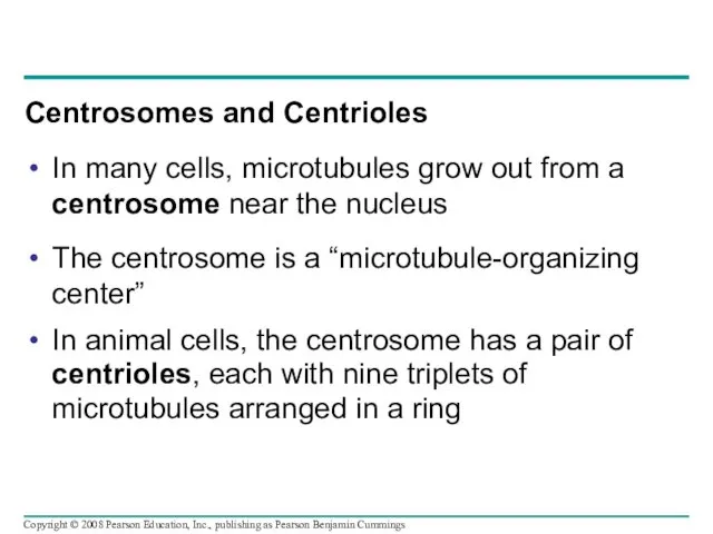
- 77. Centrosomes and Centrioles In many cells, microtubules grow out from a centrosome near the nucleus The
- 78. Fig. 6-22 Centrosome Microtubule Centrioles 0.25 µm Longitudinal section of one centriole Microtubules Cross section of
- 79. Cilia and Flagella Microtubules control the beating of cilia and flagella, locomotor appendages of some cells
- 80. Fig. 6-23 5 µm Direction of swimming (a) Motion of flagella Direction of organism’s movement Power
- 81. Cilia and flagella share a common ultrastructure: A core of microtubules sheathed by the plasma membrane
- 82. Fig. 6-24 0.1 µm Triplet (c) Cross section of basal body (a) Longitudinal section of cilium
- 83. How dynein “walking” moves flagella and cilia: Dynein arms alternately grab, move, and release the outer
- 84. Fig. 6-25 Microtubule doublets Dynein protein ATP ATP (a) Effect of unrestrained dynein movement Cross-linking proteins
- 85. Fig. 6-25a Microtubule doublets Dynein protein (a) Effect of unrestrained dynein movement ATP
- 86. Fig. 6-25b Cross-linking proteins inside outer doublets Anchorage in cell ATP (b) Effect of cross-linking proteins
- 87. Microfilaments (Actin Filaments) Microfilaments are solid rods about 7 nm in diameter, built as a twisted
- 88. Fig. 6-26 Microvillus Plasma membrane Microfilaments (actin filaments) Intermediate filaments 0.25 µm
- 89. Microfilaments that function in cellular motility contain the protein myosin in addition to actin In muscle
- 90. Fig. 6-27 Muscle cell Actin filament Myosin filament Myosin arm (a) Myosin motors in muscle cell
- 91. Fig, 6-27a Muscle cell Actin filament Myosin filament Myosin arm (a) Myosin motors in muscle cell
- 92. Fig. 6-27bc Cortex (outer cytoplasm): gel with actin network Inner cytoplasm: sol with actin subunits Extending
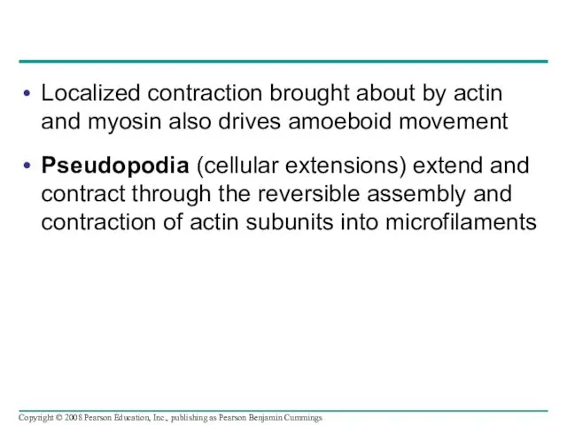
- 93. Localized contraction brought about by actin and myosin also drives amoeboid movement Pseudopodia (cellular extensions) extend
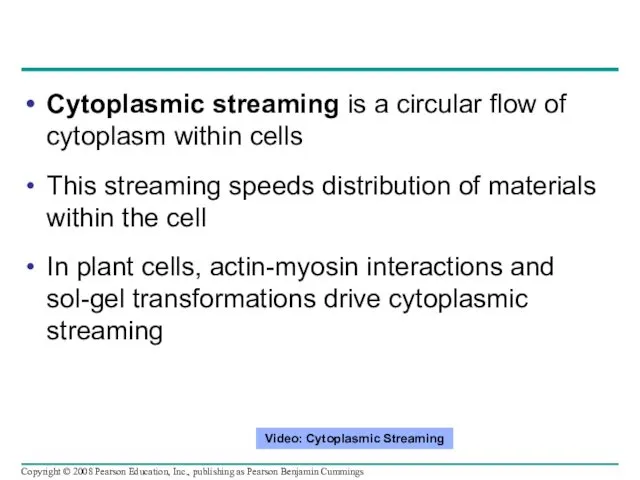
- 94. Cytoplasmic streaming is a circular flow of cytoplasm within cells This streaming speeds distribution of materials
- 95. Intermediate Filaments Intermediate filaments range in diameter from 8–12 nanometers, larger than microfilaments but smaller than
- 96. Concept 6.7: Extracellular components and connections between cells help coordinate cellular activities Most cells synthesize and
- 97. Cell Walls of Plants The cell wall is an extracellular structure that distinguishes plant cells from
- 98. Plant cell walls may have multiple layers: Primary cell wall: relatively thin and flexible Middle lamella:
- 99. Fig. 6-28 Secondary cell wall Primary cell wall Middle lamella Central vacuole Cytosol Plasma membrane Plant
- 100. Fig. 6-29 10 µm Distribution of cellulose synthase over time Distribution of microtubules over time RESULTS
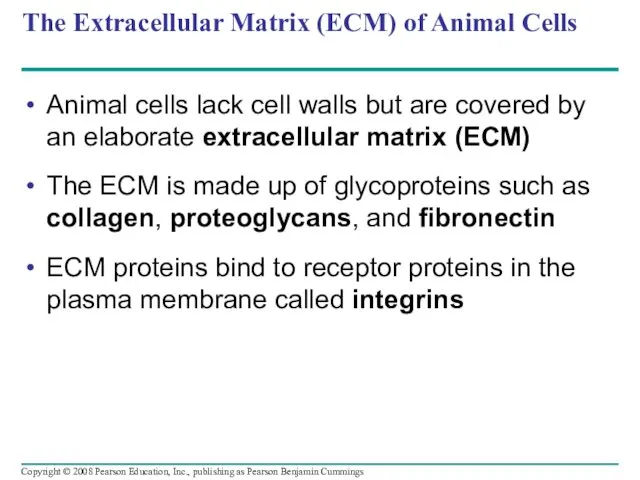
- 101. The Extracellular Matrix (ECM) of Animal Cells Animal cells lack cell walls but are covered by
- 102. Fig. 6-30 EXTRACELLULAR FLUID Collagen Fibronectin Plasma membrane Micro- filaments CYTOPLASM Integrins Proteoglycan complex Polysaccharide molecule
- 103. Fig. 6-30a Collagen Fibronectin Plasma membrane Proteoglycan complex Integrins CYTOPLASM Micro-filaments EXTRACELLULAR FLUID
- 104. Fig. 6-30b Polysaccharide molecule Carbo-hydrates Core protein Proteoglycan molecule Proteoglycan complex
- 105. Functions of the ECM: Support Adhesion Movement Regulation Copyright © 2008 Pearson Education, Inc., publishing as
- 106. Intercellular Junctions Neighboring cells in tissues, organs, or organ systems often adhere, interact, and communicate through
- 107. Plasmodesmata in Plant Cells Plasmodesmata are channels that perforate plant cell walls Through plasmodesmata, water and
- 108. Fig. 6-31 Interior of cell Interior of cell 0.5 µm Plasmodesmata Plasma membranes Cell walls
- 109. Tight Junctions, Desmosomes, and Gap Junctions in Animal Cells At tight junctions, membranes of neighboring cells
- 110. Fig. 6-32 Tight junction 0.5 µm 1 µm Desmosome Gap junction Extracellular matrix 0.1 µm Plasma
- 111. Fig. 6-32a Tight junctions prevent fluid from moving across a layer of cells Tight junction Intermediate
- 112. Fig. 6-32b Tight junction 0.5 µm
- 113. Fig. 6-32c Desmosome 1 µm
- 114. Fig. 6-32d Gap junction 0.1 µm
- 115. The Cell: A Living Unit Greater Than the Sum of Its Parts Cells rely on the
- 116. Fig. 6-33 5 µm
- 117. Fig. 6-UN1 Cell Component Structure Function Houses chromosomes, made of chromatin (DNA, the genetic material, and
- 118. Fig. 6-UN1a Cell Component Structure Function Concept 6.3 The eukaryotic cell’s genetic instructions are housed in
- 119. Fig. 6-UN1b Cell Component Structure Function Concept 6.4 The endomembrane system regulates protein traffic and performs
- 120. Fig. 6-UN1c Cell Component Concept 6.5 Mitochondria and chloro- plasts change energy from one form to
- 121. Fig. 6-UN2
- 122. Fig. 6-UN3
- 123. You should now be able to: Distinguish between the following pairs of terms: magnification and resolution;
- 125. Скачать презентацию
Overview: The Fundamental Units of Life
All organisms are made of cells
The
Overview: The Fundamental Units of Life
All organisms are made of cells
The
Fig. 6-1
Fig. 6-1
Concept 6.1: To study cells, biologists use microscopes and the tools
Concept 6.1: To study cells, biologists use microscopes and the tools
Microscopy
Scientists use microscopes to visualize cells too small to see with
Microscopy
Scientists use microscopes to visualize cells too small to see with
The quality of an image depends on
Magnification, the ratio of an
The quality of an image depends on
Magnification, the ratio of an
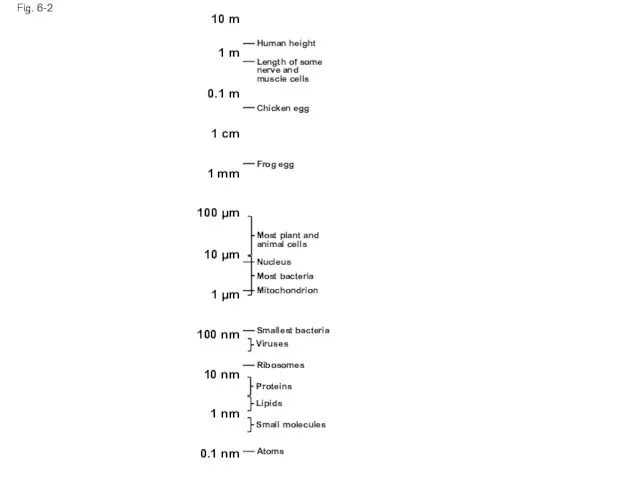
Fig. 6-2
10 m
1 m
0.1 m
1 cm
1 mm
100 µm
10 µm
1 µm
100 nm
10
Fig. 6-2
10 m
1 m
0.1 m
1 cm
1 mm
100 µm
10 µm
1 µm
100 nm
10
LMs can magnify effectively to about 1,000 times the size of
LMs can magnify effectively to about 1,000 times the size of
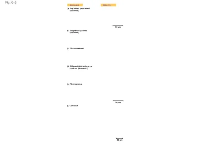
Fig. 6-3
TECHNIQUE
RESULTS
(a) Brightfield (unstained
specimen)
(b) Brightfield (stained
specimen)
50 µm
(c) Phase-contrast
(d) Differential-interference-
Fig. 6-3
TECHNIQUE
RESULTS
(a) Brightfield (unstained
specimen)
(b) Brightfield (stained
specimen)
50 µm
(c) Phase-contrast
(d) Differential-interference-
Fig. 6-3ab
(a) Brightfield (unstained
specimen)
(b) Brightfield (stained
specimen)
TECHNIQUE
RESULTS
50 µm
Fig. 6-3ab
(a) Brightfield (unstained
specimen)
(b) Brightfield (stained
specimen)
TECHNIQUE
RESULTS
50 µm
Fig. 6-3cd
(c) Phase-contrast
(d) Differential-interference-
contrast (Nomarski)
TECHNIQUE
RESULTS
Fig. 6-3cd
(c) Phase-contrast
(d) Differential-interference-
contrast (Nomarski)
TECHNIQUE
RESULTS
Fig. 6-3e
(e) Fluorescence
TECHNIQUE
RESULTS
50 µm
Fig. 6-3e
(e) Fluorescence
TECHNIQUE
RESULTS
50 µm
Fig. 6-3f
(f) Confocal
TECHNIQUE
RESULTS
50 µm
Fig. 6-3f
(f) Confocal
TECHNIQUE
RESULTS
50 µm
Two basic types of electron microscopes (EMs) are used to study
Two basic types of electron microscopes (EMs) are used to study
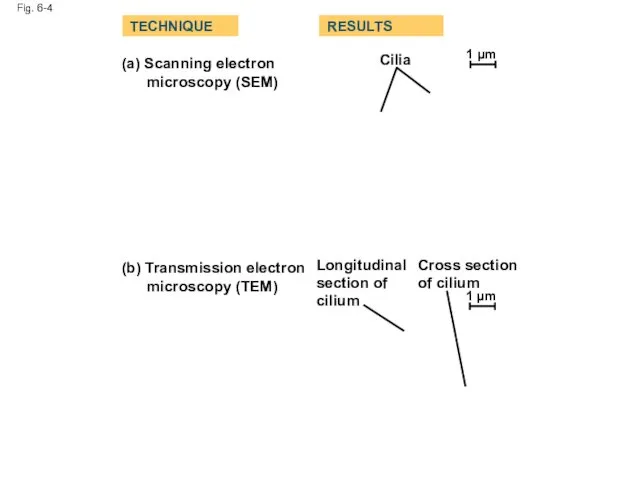
Fig. 6-4
(a) Scanning electron
microscopy (SEM)
TECHNIQUE
RESULTS
(b) Transmission electron
microscopy (TEM)
Cilia
Longitudinal
section of
cilium
Cross
Fig. 6-4
(a) Scanning electron
microscopy (SEM)
TECHNIQUE
RESULTS
(b) Transmission electron
microscopy (TEM)
Cilia
Longitudinal
section of
cilium
Cross
Cell Fractionation
Cell fractionation takes cells apart and separates the major organelles
Cell Fractionation
Cell fractionation takes cells apart and separates the major organelles
Fig. 6-5
Homogenization
TECHNIQUE
Homogenate
Tissue
cells
1,000 g
(1,000 times the
force of gravity)
10 min
Differential centrifugation
Supernatant poured
into next
Fig. 6-5
Homogenization
TECHNIQUE
Homogenate
Tissue
cells
1,000 g
(1,000 times the
force of gravity)
10 min
Differential centrifugation
Supernatant poured
into next
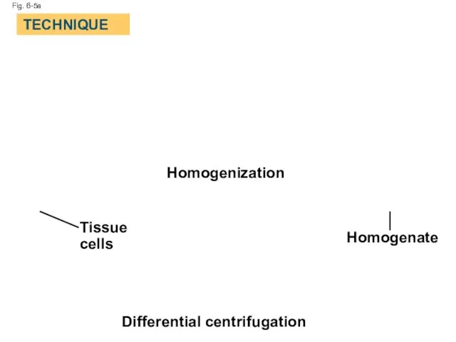
Fig. 6-5a
Homogenization
Homogenate
Differential centrifugation
Tissue
cells
TECHNIQUE
Fig. 6-5a
Homogenization
Homogenate
Differential centrifugation
Tissue
cells
TECHNIQUE
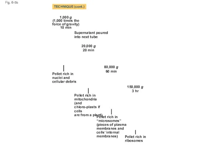
Fig. 6-5b
1,000 g
(1,000 times the force of gravity)
10 min
Supernatant poured into
Fig. 6-5b
1,000 g
(1,000 times the force of gravity)
10 min
Supernatant poured into
Concept 6.2: Eukaryotic cells have internal membranes that compartmentalize their functions
The
Concept 6.2: Eukaryotic cells have internal membranes that compartmentalize their functions
The
Comparing Prokaryotic and Eukaryotic Cells
Basic features of all cells:
Plasma membrane
Semifluid
Comparing Prokaryotic and Eukaryotic Cells
Basic features of all cells:
Plasma membrane
Semifluid
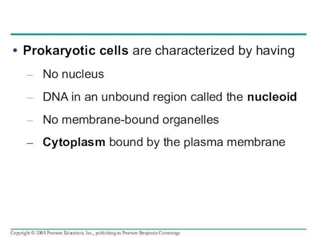
Prokaryotic cells are characterized by having
No nucleus
DNA in an unbound region
Prokaryotic cells are characterized by having
No nucleus
DNA in an unbound region
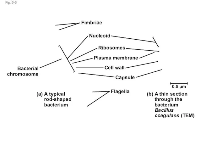
Fig. 6-6
Fimbriae
Nucleoid
Ribosomes
Plasma membrane
Cell wall
Capsule
Flagella
Bacterial
chromosome
(a)
A typical rod-shaped bacterium
(b)
A thin section through the
Fig. 6-6
Fimbriae
Nucleoid
Ribosomes
Plasma membrane
Cell wall
Capsule
Flagella
Bacterial
chromosome
(a)
A typical rod-shaped bacterium
(b)
A thin section through the
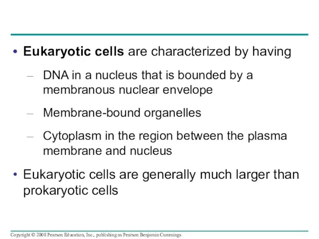
Eukaryotic cells are characterized by having
DNA in a nucleus that is
Eukaryotic cells are characterized by having
DNA in a nucleus that is
The plasma membrane is a selective barrier that allows sufficient passage
The plasma membrane is a selective barrier that allows sufficient passage
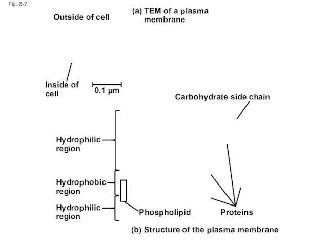
Fig. 6-7
TEM of a plasma
membrane
(a)
(b) Structure of the plasma membrane
Outside of
Fig. 6-7
TEM of a plasma
membrane
(a)
(b) Structure of the plasma membrane
Outside of
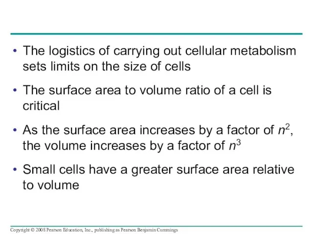
The logistics of carrying out cellular metabolism sets limits on the
The logistics of carrying out cellular metabolism sets limits on the
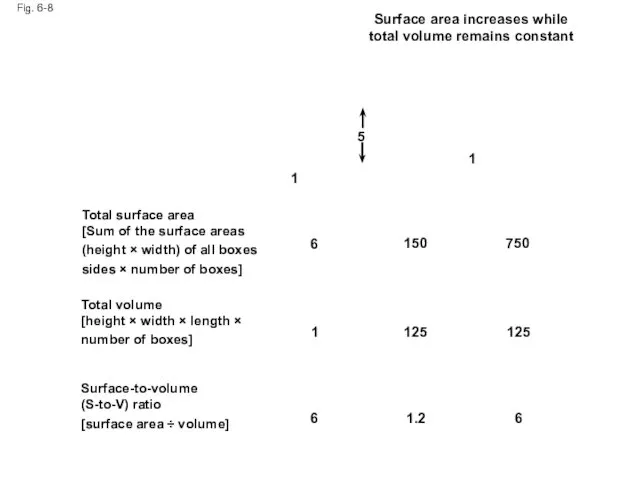
Fig. 6-8
Surface area increases while
total volume remains constant
5
1
1
6
150
750
125
125
1
6
6
1.2
Total surface area
[Sum of
Fig. 6-8
Surface area increases while
total volume remains constant
5
1
1
6
150
750
125
125
1
6
6
1.2
Total surface area
[Sum of
A Panoramic View of the Eukaryotic Cell
A eukaryotic cell has internal
A Panoramic View of the Eukaryotic Cell
A eukaryotic cell has internal
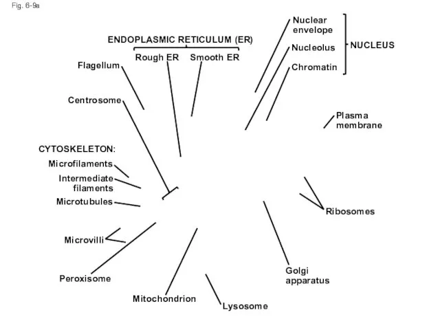
Fig. 6-9a
ENDOPLASMIC RETICULUM (ER)
Smooth ER
Rough ER
Flagellum
Centrosome
CYTOSKELETON:
Microfilaments
Intermediate
filaments
Microtubules
Microvilli
Peroxisome
Mitochondrion
Lysosome
Golgi
apparatus
Ribosomes
Plasma membrane
Nuclear
envelope
Nucleolus
Chromatin
NUCLEUS
Fig. 6-9a
ENDOPLASMIC RETICULUM (ER)
Smooth ER
Rough ER
Flagellum
Centrosome
CYTOSKELETON:
Microfilaments
Intermediate
filaments
Microtubules
Microvilli
Peroxisome
Mitochondrion
Lysosome
Golgi
apparatus
Ribosomes
Plasma membrane
Nuclear
envelope
Nucleolus
Chromatin
NUCLEUS
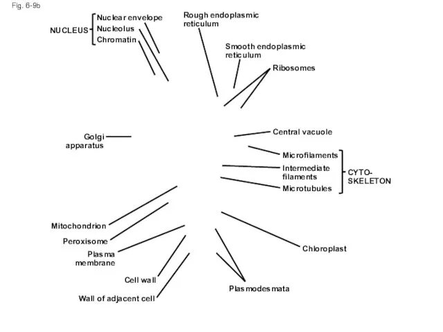
Fig. 6-9b
NUCLEUS
Nuclear envelope
Nucleolus
Chromatin
Rough endoplasmic reticulum
Smooth endoplasmic reticulum
Ribosomes
Central vacuole
Microfilaments
Intermediate filaments
Microtubules
CYTO-
SKELETON
Chloroplast
Plasmodesmata
Wall of adjacent
Fig. 6-9b
NUCLEUS
Nuclear envelope
Nucleolus
Chromatin
Rough endoplasmic reticulum
Smooth endoplasmic reticulum
Ribosomes
Central vacuole
Microfilaments
Intermediate filaments
Microtubules
CYTO-
SKELETON
Chloroplast
Plasmodesmata
Wall of adjacent
Concept 6.3: The eukaryotic cell’s genetic instructions are housed in the
Concept 6.3: The eukaryotic cell’s genetic instructions are housed in the
The Nucleus: Information Central
The nucleus contains most of the cell’s genes
The Nucleus: Information Central
The nucleus contains most of the cell’s genes
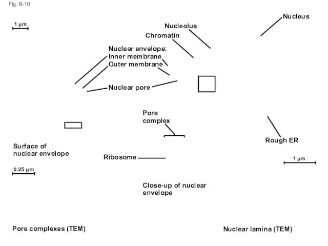
Fig. 6-10
Nucleolus
Nucleus
Rough ER
Nuclear lamina (TEM)
Close-up of nuclear envelope
1 µm
1 µm
0.25 µm
Ribosome
Pore
Fig. 6-10
Nucleolus
Nucleus
Rough ER
Nuclear lamina (TEM)
Close-up of nuclear envelope
1 µm
1 µm
0.25 µm
Ribosome
Pore
Pores regulate the entry and exit of molecules from the nucleus
The
Pores regulate the entry and exit of molecules from the nucleus
The
In the nucleus, DNA and proteins form genetic material called chromatin
In the nucleus, DNA and proteins form genetic material called chromatin
Ribosomes: Protein Factories
Ribosomes are particles made of ribosomal RNA and protein
Ribosomes
Ribosomes: Protein Factories
Ribosomes are particles made of ribosomal RNA and protein
Ribosomes
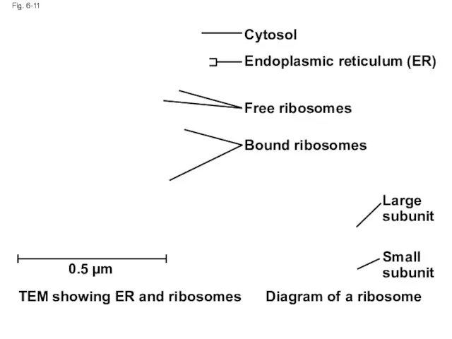
Fig. 6-11
Cytosol
Endoplasmic reticulum (ER)
Free ribosomes
Bound ribosomes
Large subunit
Small subunit
Diagram of a ribosome
TEM
Fig. 6-11
Cytosol
Endoplasmic reticulum (ER)
Free ribosomes
Bound ribosomes
Large subunit
Small subunit
Diagram of a ribosome
TEM
Concept 6.4: The endomembrane system regulates protein traffic and performs metabolic
Concept 6.4: The endomembrane system regulates protein traffic and performs metabolic
The Endoplasmic Reticulum: Biosynthetic Factory
The endoplasmic reticulum (ER) accounts for more
The Endoplasmic Reticulum: Biosynthetic Factory
The endoplasmic reticulum (ER) accounts for more
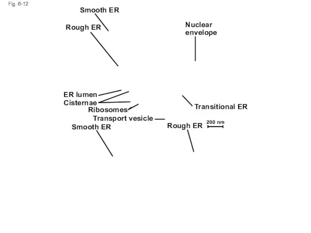
Fig. 6-12
Smooth ER
Rough ER
Nuclear envelope
Transitional ER
Rough ER
Smooth ER
Transport vesicle
Ribosomes
Cisternae
ER lumen
200 nm
Fig. 6-12
Smooth ER
Rough ER
Nuclear envelope
Transitional ER
Rough ER
Smooth ER
Transport vesicle
Ribosomes
Cisternae
ER lumen
200 nm
Functions of Smooth ER
The smooth ER
Synthesizes lipids
Metabolizes carbohydrates
Detoxifies poison
Stores calcium
Copyright ©
Functions of Smooth ER
The smooth ER
Synthesizes lipids
Metabolizes carbohydrates
Detoxifies poison
Stores calcium
Copyright ©
Functions of Rough ER
The rough ER
Has bound ribosomes, which secrete glycoproteins
Functions of Rough ER
The rough ER
Has bound ribosomes, which secrete glycoproteins
The Golgi apparatus consists of flattened membranous sacs called cisternae
Functions of
The Golgi apparatus consists of flattened membranous sacs called cisternae
Functions of
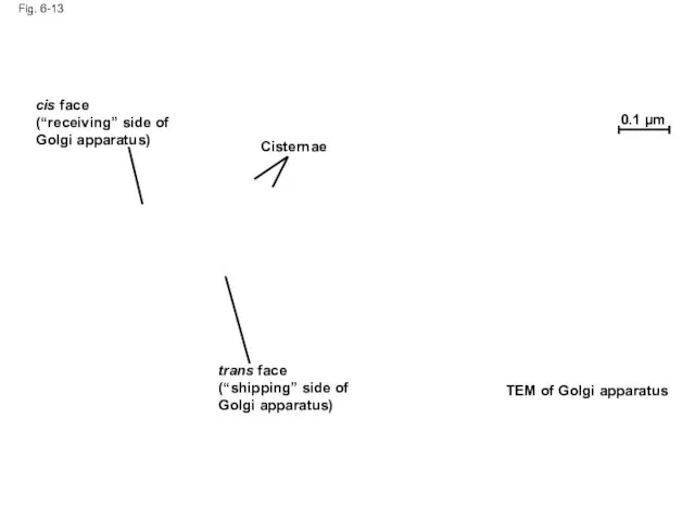
Fig. 6-13
cis face
(“receiving” side of Golgi apparatus)
Cisternae
trans face
(“shipping” side of Golgi
Fig. 6-13
cis face
(“receiving” side of Golgi apparatus)
Cisternae
trans face
(“shipping” side of Golgi
Lysosomes: Digestive Compartments
A lysosome is a membranous sac of hydrolytic enzymes
Lysosomes: Digestive Compartments
A lysosome is a membranous sac of hydrolytic enzymes
Some types of cell can engulf another cell by phagocytosis; this
Some types of cell can engulf another cell by phagocytosis; this
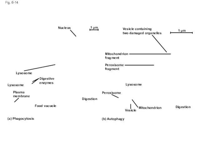
Fig. 6-14
Nucleus
1 µm
Lysosome
Digestive
enzymes
Lysosome
Plasma
membrane
Food vacuole
(a) Phagocytosis
Digestion
(b) Autophagy
Peroxisome
Vesicle
Lysosome
Mitochondrion
Peroxisome
fragment
Mitochondrion
fragment
Vesicle containing
two damaged organelles
1 µm
Digestion
Fig. 6-14
Nucleus
1 µm
Lysosome
Digestive
enzymes
Lysosome
Plasma
membrane
Food vacuole
(a) Phagocytosis
Digestion
(b) Autophagy
Peroxisome
Vesicle
Lysosome
Mitochondrion
Peroxisome
fragment
Mitochondrion
fragment
Vesicle containing
two damaged organelles
1 µm
Digestion
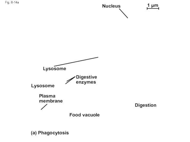
Fig. 6-14a
Nucleus
1 µm
Lysosome
Lysosome
Digestive enzymes
Plasma membrane
Food vacuole
Digestion
(a) Phagocytosis
Fig. 6-14a
Nucleus
1 µm
Lysosome
Lysosome
Digestive enzymes
Plasma membrane
Food vacuole
Digestion
(a) Phagocytosis
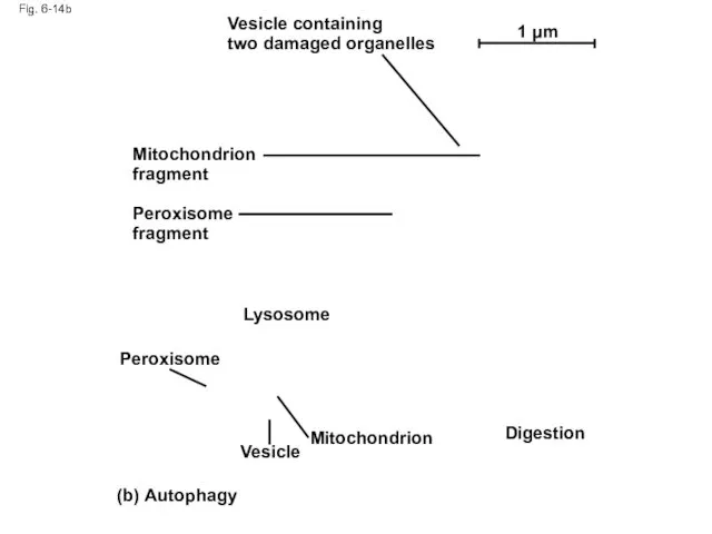
Fig. 6-14b
Vesicle containing
two damaged organelles
Mitochondrion fragment
Peroxisome fragment
Peroxisome
Lysosome
Digestion
Mitochondrion
Vesicle
(b) Autophagy
1 µm
Fig. 6-14b
Vesicle containing
two damaged organelles
Mitochondrion fragment
Peroxisome fragment
Peroxisome
Lysosome
Digestion
Mitochondrion
Vesicle
(b) Autophagy
1 µm
Vacuoles: Diverse Maintenance Compartments
A plant cell or fungal cell may have
Vacuoles: Diverse Maintenance Compartments
A plant cell or fungal cell may have
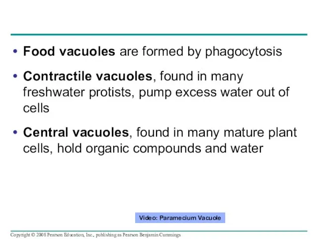
Food vacuoles are formed by phagocytosis
Contractile vacuoles, found in many freshwater
Food vacuoles are formed by phagocytosis
Contractile vacuoles, found in many freshwater
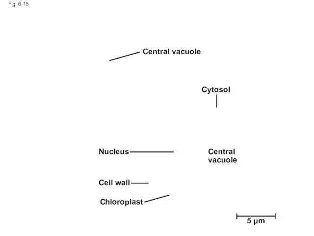
Fig. 6-15
Central vacuole
Cytosol
Central vacuole
Nucleus
Cell wall
Chloroplast
5 µm
Fig. 6-15
Central vacuole
Cytosol
Central vacuole
Nucleus
Cell wall
Chloroplast
5 µm
The Endomembrane System: A Review
The endomembrane system is a complex and
The Endomembrane System: A Review
The endomembrane system is a complex and
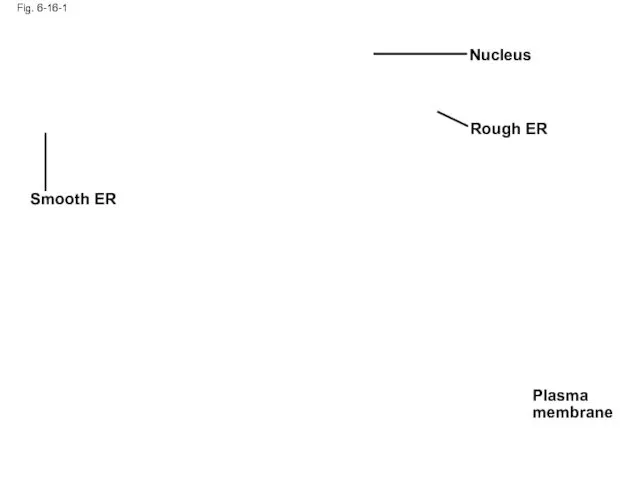
Fig. 6-16-1
Smooth ER
Nucleus
Rough ER
Plasma membrane
Fig. 6-16-1
Smooth ER
Nucleus
Rough ER
Plasma membrane

Fig. 6-16-2
Smooth ER
Nucleus
Rough ER
Plasma membrane
cis Golgi
trans Golgi
Fig. 6-16-2
Smooth ER
Nucleus
Rough ER
Plasma membrane
cis Golgi
trans Golgi
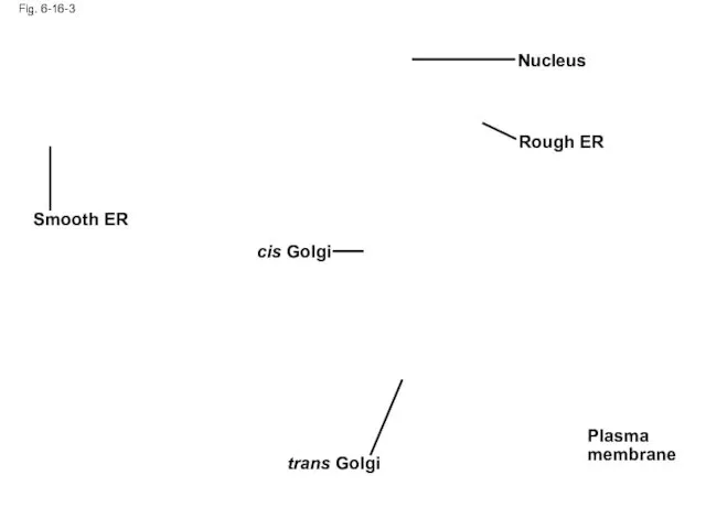
Fig. 6-16-3
Smooth ER
Nucleus
Rough ER
Plasma membrane
cis Golgi
trans Golgi
Fig. 6-16-3
Smooth ER
Nucleus
Rough ER
Plasma membrane
cis Golgi
trans Golgi
Concept 6.5: Mitochondria and chloroplasts change energy from one form to
Concept 6.5: Mitochondria and chloroplasts change energy from one form to
Mitochondria and chloroplasts
Are not part of the endomembrane system
Have a
Mitochondria and chloroplasts
Are not part of the endomembrane system
Have a

Mitochondria: Chemical Energy Conversion
Mitochondria are in nearly all eukaryotic cells
They have
Mitochondria: Chemical Energy Conversion
Mitochondria are in nearly all eukaryotic cells
They have
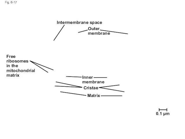
Fig. 6-17
Free ribosomes
in the mitochondrial matrix
Intermembrane space
Outer membrane
Inner membrane
Cristae
Matrix
0.1 µm
Fig. 6-17
Free ribosomes
in the mitochondrial matrix
Intermembrane space
Outer membrane
Inner membrane
Cristae
Matrix
0.1 µm
Chloroplasts: Capture of Light Energy
The chloroplast is a member of a
Chloroplasts: Capture of Light Energy
The chloroplast is a member of a
Chloroplast structure includes:
Thylakoids, membranous sacs, stacked to form a granum
Stroma, the
Chloroplast structure includes:
Thylakoids, membranous sacs, stacked to form a granum
Stroma, the
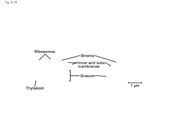
Fig. 6-18
Ribosomes
Thylakoid
Stroma
Granum
Inner and outer membranes
1 µm
Fig. 6-18
Ribosomes
Thylakoid
Stroma
Granum
Inner and outer membranes
1 µm
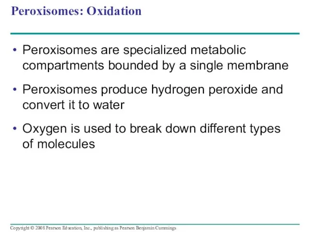
Peroxisomes: Oxidation
Peroxisomes are specialized metabolic compartments bounded by a single membrane
Peroxisomes
Peroxisomes: Oxidation
Peroxisomes are specialized metabolic compartments bounded by a single membrane
Peroxisomes
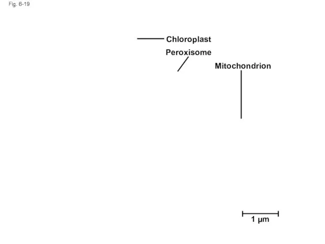
Fig. 6-19
1 µm
Chloroplast
Peroxisome
Mitochondrion
Fig. 6-19
1 µm
Chloroplast
Peroxisome
Mitochondrion
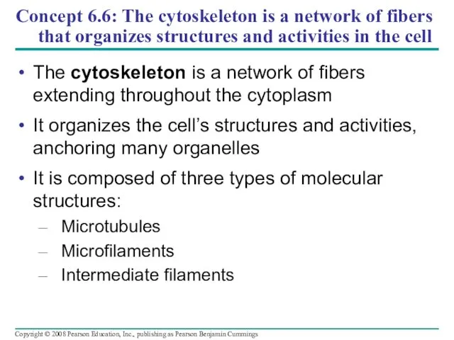
Concept 6.6: The cytoskeleton is a network of fibers that organizes
Concept 6.6: The cytoskeleton is a network of fibers that organizes
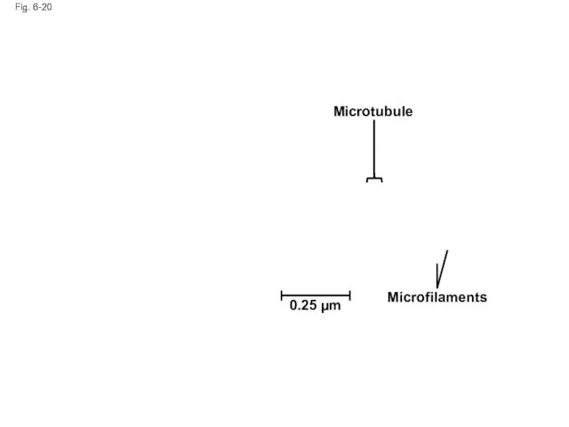
Fig. 6-20
Microtubule
Microfilaments
0.25 µm
Fig. 6-20
Microtubule
Microfilaments
0.25 µm
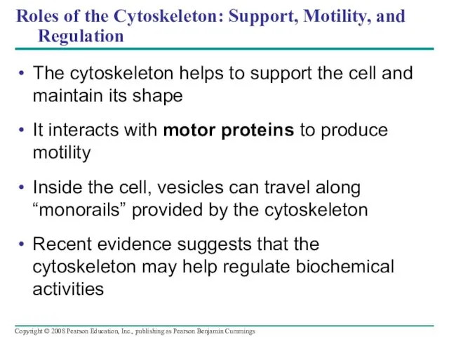
Roles of the Cytoskeleton: Support, Motility, and Regulation
The cytoskeleton helps to
Roles of the Cytoskeleton: Support, Motility, and Regulation
The cytoskeleton helps to
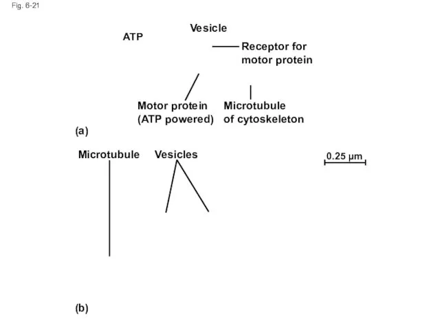
Fig. 6-21
Vesicle
ATP
Receptor for motor protein
Microtubule
of cytoskeleton
Motor protein (ATP powered)
(a)
Microtubule
Vesicles
(b)
0.25 µm
Fig. 6-21
Vesicle
ATP
Receptor for motor protein
Microtubule
of cytoskeleton
Motor protein (ATP powered)
(a)
Microtubule
Vesicles
(b)
0.25 µm
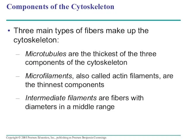
Components of the Cytoskeleton
Three main types of fibers make up the
Components of the Cytoskeleton
Three main types of fibers make up the
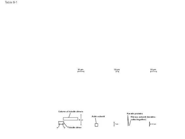
Table 6-1
10 µm
10 µm
10 µm
Column of tubulin dimers
Tubulin dimer
Actin subunit
Table 6-1
10 µm
10 µm
10 µm
Column of tubulin dimers
Tubulin dimer
Actin subunit
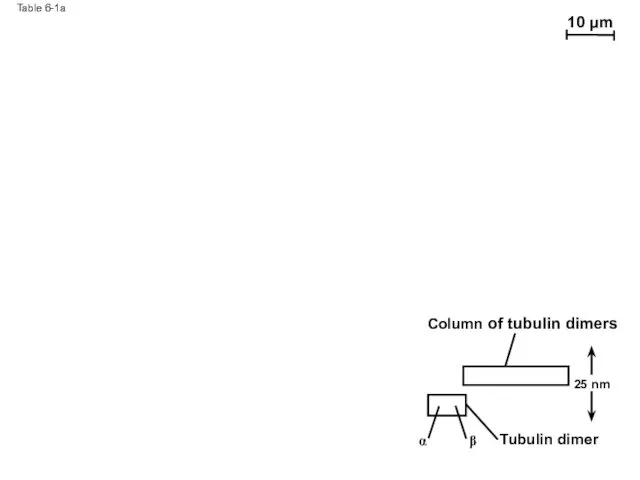
Table 6-1a
10 µm
Column of tubulin dimers
Tubulin dimer
α
β
25 nm
Table 6-1a
10 µm
Column of tubulin dimers
Tubulin dimer
α
β
25 nm
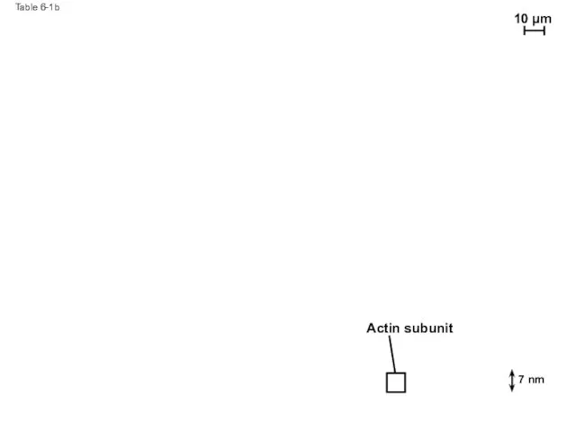
Table 6-1b
Actin subunit
10 µm
7 nm
Table 6-1b
Actin subunit
10 µm
7 nm
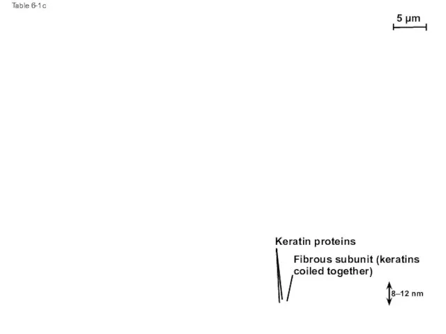
Table 6-1c
5 µm
Keratin proteins
Fibrous subunit (keratins
coiled together)
8–12 nm
Table 6-1c
5 µm
Keratin proteins
Fibrous subunit (keratins
coiled together)
8–12 nm
Microtubules
Microtubules are hollow rods about 25 nm in diameter and about
Microtubules
Microtubules are hollow rods about 25 nm in diameter and about
Centrosomes and Centrioles
In many cells, microtubules grow out from a
Centrosomes and Centrioles
In many cells, microtubules grow out from a
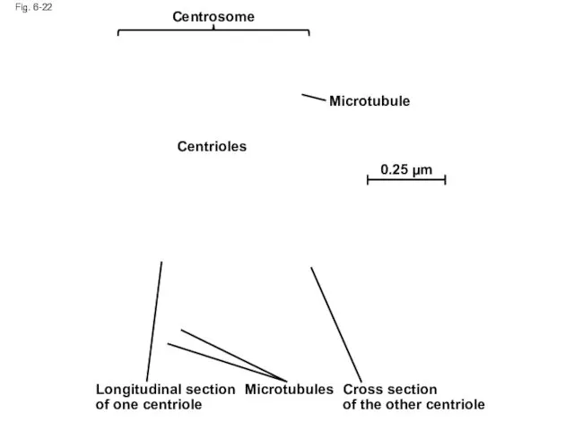
Fig. 6-22
Centrosome
Microtubule
Centrioles
0.25 µm
Longitudinal section of one centriole
Microtubules
Cross section
of the other centriole
Fig. 6-22
Centrosome
Microtubule
Centrioles
0.25 µm
Longitudinal section of one centriole
Microtubules
Cross section
of the other centriole
Cilia and Flagella
Microtubules control the beating of cilia and flagella,
Cilia and Flagella
Microtubules control the beating of cilia and flagella,
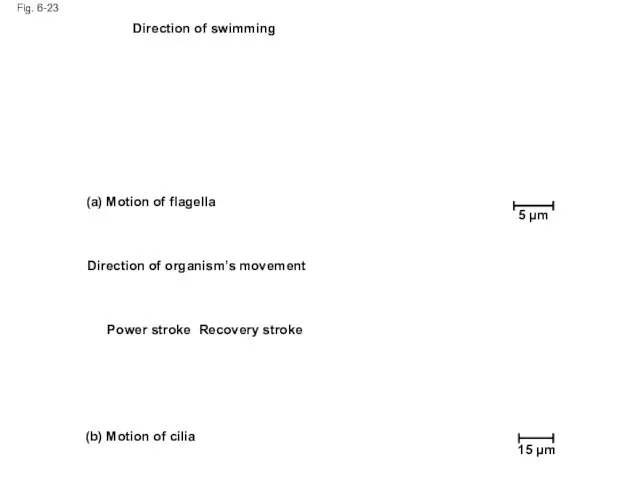
Fig. 6-23
5 µm
Direction of swimming
(a) Motion of flagella
Direction of organism’s movement
Power
Fig. 6-23
5 µm
Direction of swimming
(a) Motion of flagella
Direction of organism’s movement
Power
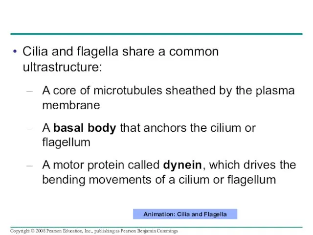
Cilia and flagella share a common ultrastructure:
A core of microtubules sheathed
Cilia and flagella share a common ultrastructure:
A core of microtubules sheathed
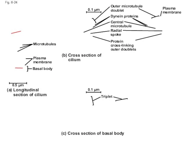
Fig. 6-24
0.1 µm
Triplet
(c) Cross section of basal body
(a)
Longitudinal section of cilium
0.5
Fig. 6-24
0.1 µm
Triplet
(c) Cross section of basal body
(a)
Longitudinal section of cilium
0.5
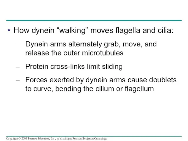
How dynein “walking” moves flagella and cilia:
Dynein arms alternately grab, move,
How dynein “walking” moves flagella and cilia:
Dynein arms alternately grab, move,
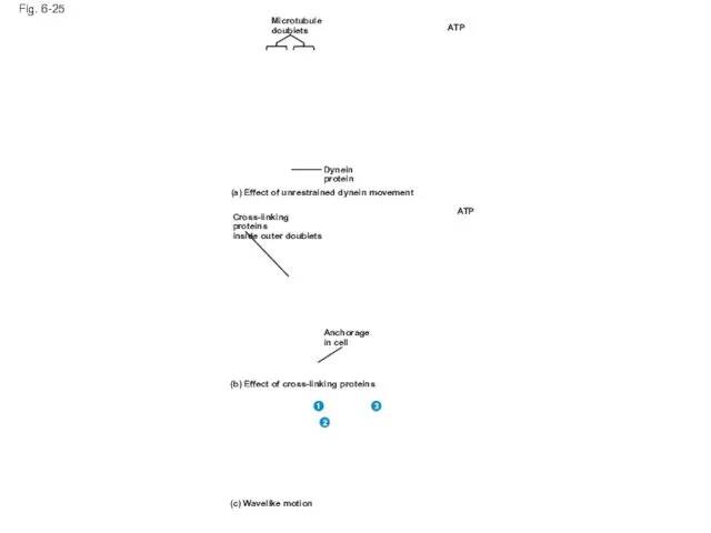
Fig. 6-25
Microtubule
doublets
Dynein
protein
ATP
ATP
(a) Effect of unrestrained dynein movement
Cross-linking proteins
inside outer doublets
Anchorage
in cell
(b)
Fig. 6-25
Microtubule
doublets
Dynein
protein
ATP
ATP
(a) Effect of unrestrained dynein movement
Cross-linking proteins
inside outer doublets
Anchorage
in cell
(b)
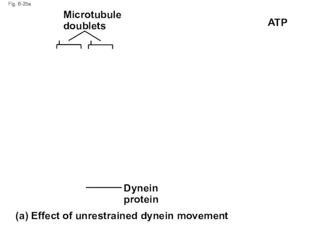
Fig. 6-25a
Microtubule doublets
Dynein protein
(a) Effect of unrestrained dynein movement
ATP
Fig. 6-25a
Microtubule doublets
Dynein protein
(a) Effect of unrestrained dynein movement
ATP
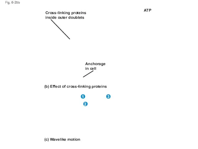
Fig. 6-25b
Cross-linking proteins inside outer doublets
Anchorage in cell
ATP
(b) Effect of cross-linking
Fig. 6-25b
Cross-linking proteins inside outer doublets
Anchorage in cell
ATP
(b) Effect of cross-linking
Microfilaments (Actin Filaments)
Microfilaments are solid rods about 7 nm in diameter,
Microfilaments (Actin Filaments)
Microfilaments are solid rods about 7 nm in diameter,
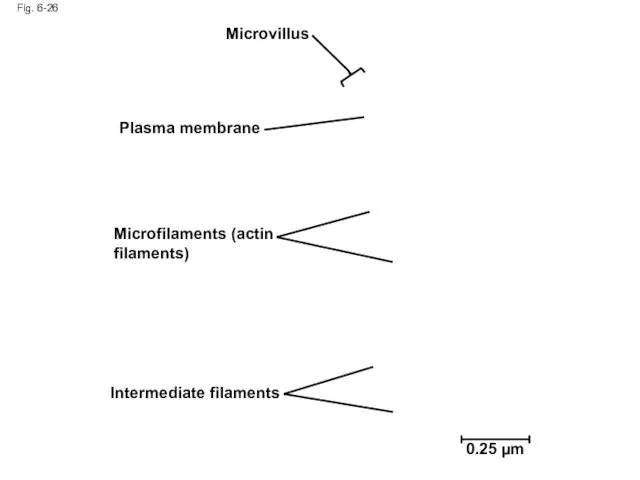
Fig. 6-26
Microvillus
Plasma membrane
Microfilaments (actin filaments)
Intermediate filaments
0.25 µm
Fig. 6-26
Microvillus
Plasma membrane
Microfilaments (actin filaments)
Intermediate filaments
0.25 µm
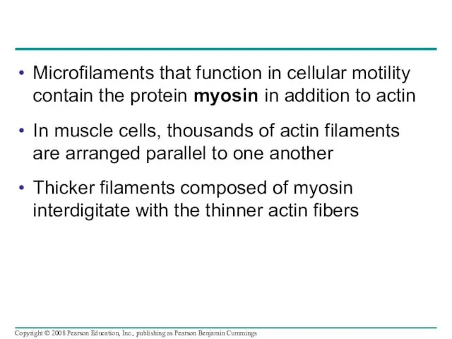
Microfilaments that function in cellular motility contain the protein myosin in
Microfilaments that function in cellular motility contain the protein myosin in
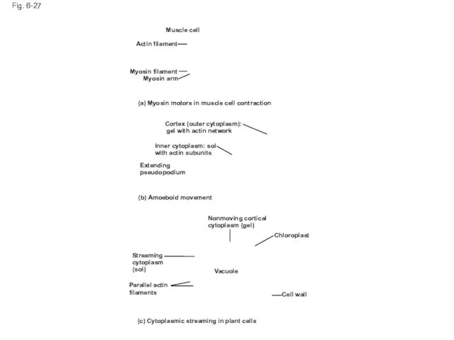
Fig. 6-27
Muscle cell
Actin filament
Myosin filament
Myosin arm
(a) Myosin motors in muscle cell
Fig. 6-27
Muscle cell
Actin filament
Myosin filament
Myosin arm
(a) Myosin motors in muscle cell
Fig, 6-27a
Muscle cell
Actin filament
Myosin filament
Myosin arm
(a) Myosin motors in muscle cell
Fig, 6-27a
Muscle cell
Actin filament
Myosin filament
Myosin arm
(a) Myosin motors in muscle cell
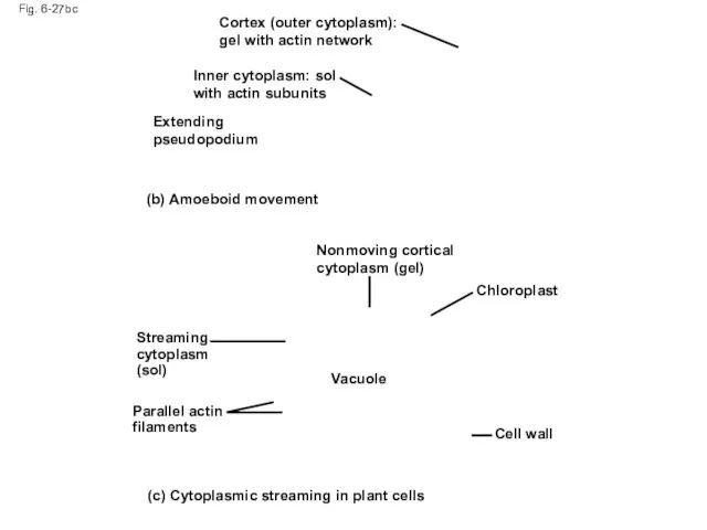
Fig. 6-27bc
Cortex (outer cytoplasm): gel with actin network
Inner cytoplasm: sol with
Fig. 6-27bc
Cortex (outer cytoplasm): gel with actin network
Inner cytoplasm: sol with
Localized contraction brought about by actin and myosin also drives amoeboid
Localized contraction brought about by actin and myosin also drives amoeboid
Cytoplasmic streaming is a circular flow of cytoplasm within cells
This streaming
Cytoplasmic streaming is a circular flow of cytoplasm within cells
This streaming
Intermediate Filaments
Intermediate filaments range in diameter from 8–12 nanometers, larger than
Intermediate Filaments
Intermediate filaments range in diameter from 8–12 nanometers, larger than
Concept 6.7: Extracellular components and connections between cells help coordinate cellular
Concept 6.7: Extracellular components and connections between cells help coordinate cellular
Cell Walls of Plants
The cell wall is an extracellular structure that
Cell Walls of Plants
The cell wall is an extracellular structure that
Plant cell walls may have multiple layers:
Primary cell wall: relatively thin
Plant cell walls may have multiple layers:
Primary cell wall: relatively thin

Fig. 6-28
Secondary cell wall
Primary cell wall
Middle lamella
Central vacuole
Cytosol
Plasma membrane
Plant cell walls
Plasmodesmata
1
Fig. 6-28
Secondary cell wall
Primary cell wall
Middle lamella
Central vacuole
Cytosol
Plasma membrane
Plant cell walls
Plasmodesmata
1
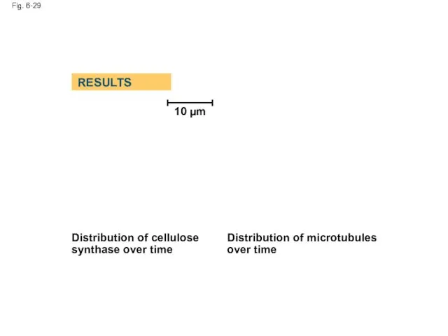
Fig. 6-29
10 µm
Distribution of cellulose synthase over time
Distribution of microtubules over
Fig. 6-29
10 µm
Distribution of cellulose synthase over time
Distribution of microtubules over
The Extracellular Matrix (ECM) of Animal Cells
Animal cells lack cell walls
The Extracellular Matrix (ECM) of Animal Cells
Animal cells lack cell walls
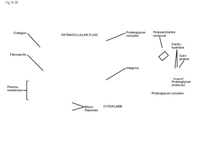
Fig. 6-30
EXTRACELLULAR FLUID
Collagen
Fibronectin
Plasma
membrane
Micro-
filaments
CYTOPLASM
Integrins
Proteoglycan
complex
Polysaccharide
molecule
Carbo-
hydrates
Core
protein
Proteoglycan
molecule
Proteoglycan complex
Fig. 6-30
EXTRACELLULAR FLUID
Collagen
Fibronectin
Plasma
membrane
Micro-
filaments
CYTOPLASM
Integrins
Proteoglycan
complex
Polysaccharide
molecule
Carbo-
hydrates
Core
protein
Proteoglycan
molecule
Proteoglycan complex
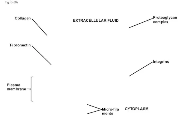
Fig. 6-30a
Collagen
Fibronectin
Plasma membrane
Proteoglycan complex
Integrins
CYTOPLASM
Micro-filaments
EXTRACELLULAR FLUID
Fig. 6-30a
Collagen
Fibronectin
Plasma membrane
Proteoglycan complex
Integrins
CYTOPLASM
Micro-filaments
EXTRACELLULAR FLUID
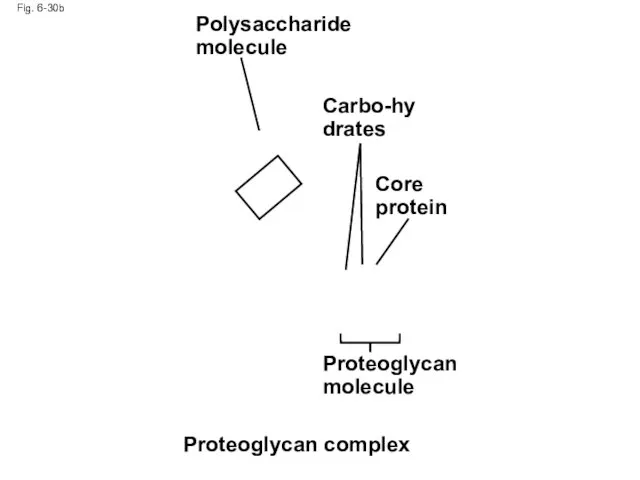
Fig. 6-30b
Polysaccharide molecule
Carbo-hydrates
Core protein
Proteoglycan molecule
Proteoglycan complex
Fig. 6-30b
Polysaccharide molecule
Carbo-hydrates
Core protein
Proteoglycan molecule
Proteoglycan complex
Functions of the ECM:
Support
Adhesion
Movement
Regulation
Copyright © 2008 Pearson Education, Inc., publishing as
Functions of the ECM:
Support
Adhesion
Movement
Regulation
Copyright © 2008 Pearson Education, Inc., publishing as
Intercellular Junctions
Neighboring cells in tissues, organs, or organ systems often adhere,
Intercellular Junctions
Neighboring cells in tissues, organs, or organ systems often adhere,
Plasmodesmata in Plant Cells
Plasmodesmata are channels that perforate plant cell walls
Through
Plasmodesmata in Plant Cells
Plasmodesmata are channels that perforate plant cell walls
Through

Fig. 6-31
Interior of cell
Interior of cell
0.5 µm
Plasmodesmata
Plasma membranes
Cell walls
Fig. 6-31
Interior of cell
Interior of cell
0.5 µm
Plasmodesmata
Plasma membranes
Cell walls
Tight Junctions, Desmosomes, and Gap Junctions in Animal Cells
At tight junctions,
Tight Junctions, Desmosomes, and Gap Junctions in Animal Cells
At tight junctions,
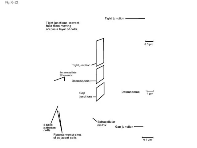
Fig. 6-32
Tight junction
0.5 µm
1 µm
Desmosome
Gap junction
Extracellular
matrix
0.1 µm
Plasma membranes
of adjacent cells
Space
between
cells
Gap
junctions
Desmosome
Intermediate
filaments
Tight junction
Tight
Fig. 6-32
Tight junction
0.5 µm
1 µm
Desmosome
Gap junction
Extracellular
matrix
0.1 µm
Plasma membranes
of adjacent cells
Space
between
cells
Gap
junctions
Desmosome
Intermediate
filaments
Tight junction
Tight
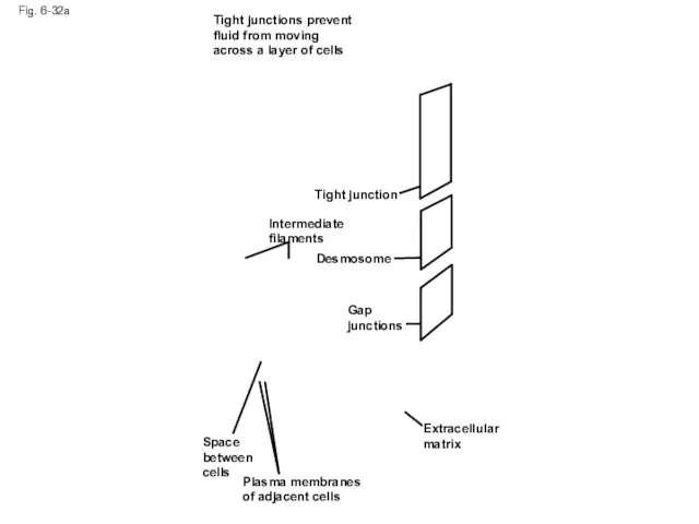
Fig. 6-32a
Tight junctions prevent fluid from moving across a layer of
Fig. 6-32a
Tight junctions prevent fluid from moving across a layer of
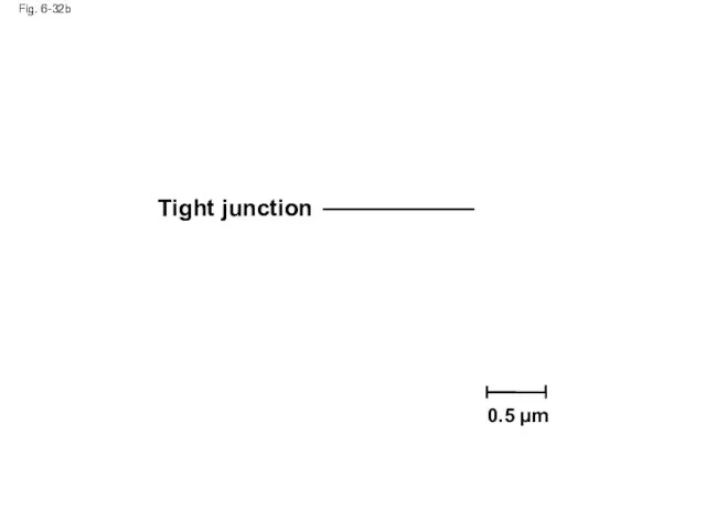
Fig. 6-32b
Tight junction
0.5 µm
Fig. 6-32b
Tight junction
0.5 µm
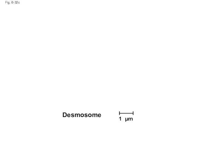
Fig. 6-32c
Desmosome
1 µm
Fig. 6-32c
Desmosome
1 µm
Fig. 6-32d
Gap junction
0.1 µm
Fig. 6-32d
Gap junction
0.1 µm
The Cell: A Living Unit Greater Than the Sum of Its
The Cell: A Living Unit Greater Than the Sum of Its
Fig. 6-33
5 µm
Fig. 6-33
5 µm
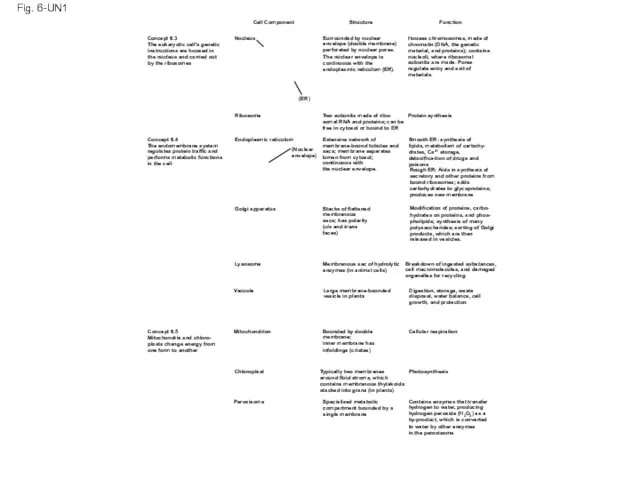
Fig. 6-UN1
Cell Component
Structure
Function
Houses chromosomes, made of
chromatin (DNA, the
Fig. 6-UN1
Cell Component
Structure
Function
Houses chromosomes, made of
chromatin (DNA, the
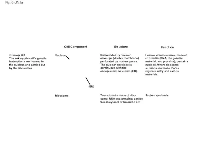
Fig. 6-UN1a
Cell Component
Structure
Function
Concept 6.3
The eukaryotic cell’s genetic
instructions
Fig. 6-UN1a
Cell Component
Structure
Function
Concept 6.3
The eukaryotic cell’s genetic
instructions
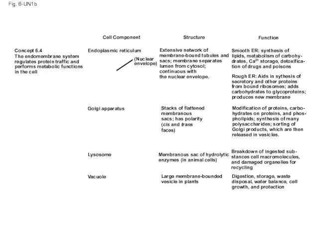
Fig. 6-UN1b
Cell Component
Structure
Function
Concept 6.4
The endomembrane system
regulates protein
Fig. 6-UN1b
Cell Component
Structure
Function
Concept 6.4
The endomembrane system
regulates protein
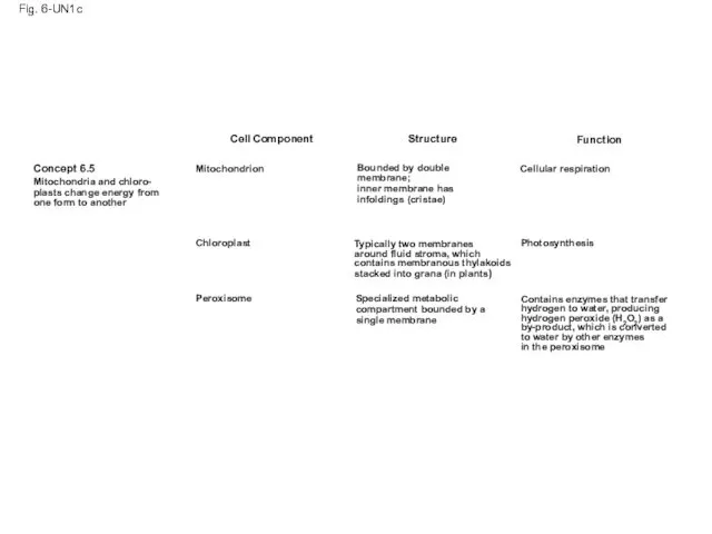
Fig. 6-UN1c
Cell Component
Concept 6.5
Mitochondria and chloro-
plasts change energy from
one form
Fig. 6-UN1c
Cell Component
Concept 6.5
Mitochondria and chloro-
plasts change energy from
one form

Fig. 6-UN2
Fig. 6-UN2

Fig. 6-UN3
Fig. 6-UN3
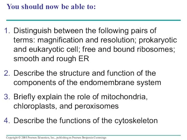
You should now be able to:
Distinguish between the following pairs of
You should now be able to:
Distinguish between the following pairs of
 Моллюски. Членистоногие
 Скелет. Строение скелета
 Систематический обзор беспозвоночных (Черви)
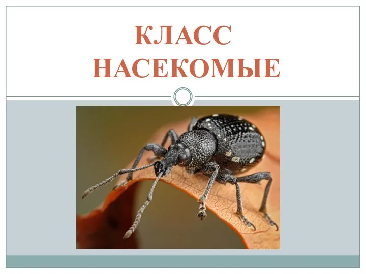 Класс Насекомые. 7 Класс
 Развитие устойчивого экотуризма в национальном парке Красноярские столбы
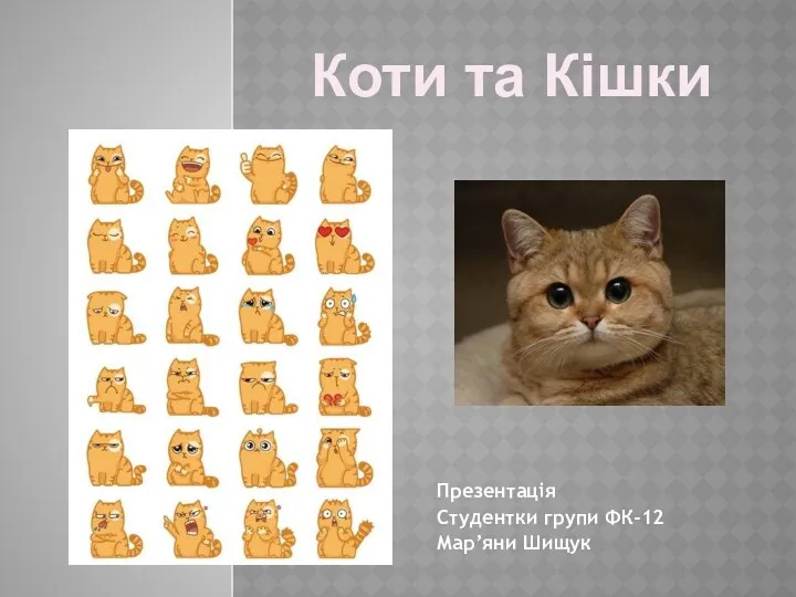 Коти та кішки
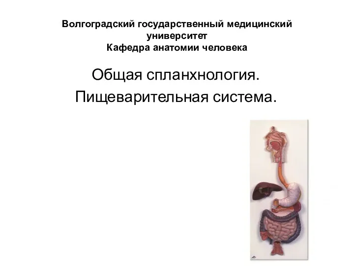 Общая спланхнология. Пищеварительная система
 Санитарно-гигиеническая оценка биологического объекта и готовых продуктов, включающих живые клетки продуцента
 Вкусы и запахи
 Наш живой уголок
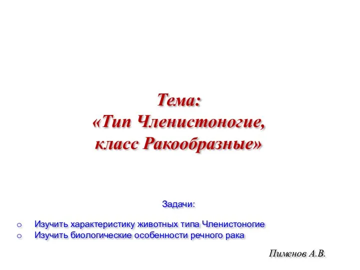 Тип Членистоногие, класс Ракообразные
 Транскрипция. Биосинтез белка
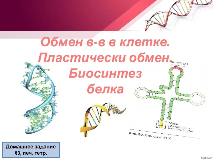 Обмен веществ в клетке. Пластический обмен. Биосинтез белка
 Викторина Материки
 Салонные процедуры
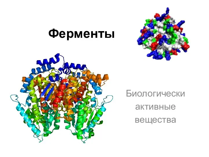 Ферменты. Биологически активные вещества
 Женская половая система
 Пищеварительная система
 Знакомство детей с насекомыми
 Вода и здоровье. Вода в природе и жизни человека
 Строение эукариотической клетки
 Интересное из жизни бабочек
 Половое поведение животных
 Скопа - птица 2018 года
 Презентация Плоды к открытому уроку
 Интересное о грибах
 Корсак
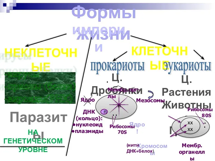 Строение эукариотической клетки