- Anatomical Basis of Breathing

Содержание
- 2. Objectives Describe thoracic wall: bones and muscles Define the muscles of respiration Define the mediastinum and
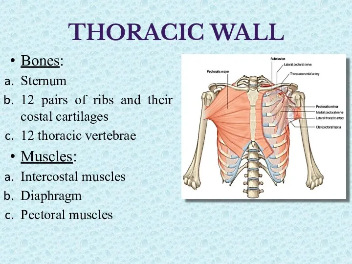
- 3. THORACIC WALL Bones: Sternum 12 pairs of ribs and their costal cartilages 12 thoracic vertebrae Muscles:
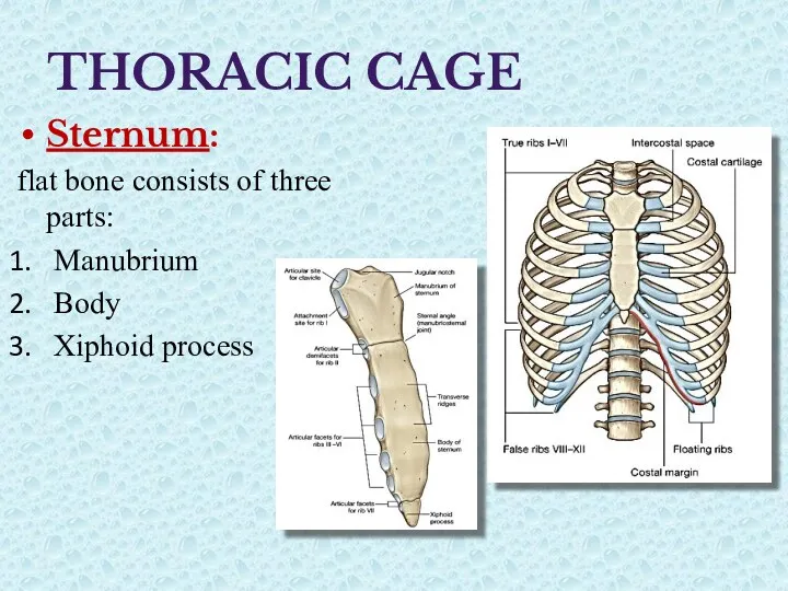
- 4. THORACIC CAGE Sternum: flat bone consists of three parts: Manubrium Body Xiphoid process
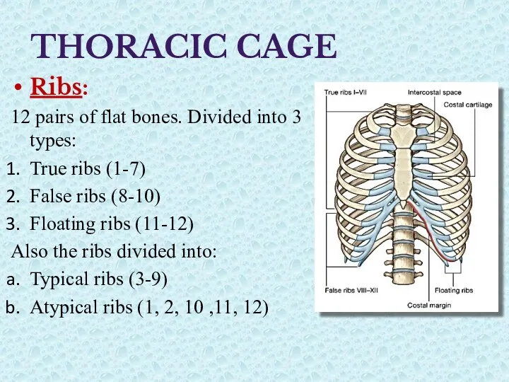
- 5. THORACIC CAGE Ribs: 12 pairs of flat bones. Divided into 3 types: True ribs (1-7) False
- 6. Typical rib Head with two articular surfaces Neck Tubercle with two parts Shaft with an angle
- 7. Atypical ribs Head with one articular surface Neck not present in 11 & 12 No tubercle
- 8. Thoracic Vertebrae Typical thoracic vertebra Body: heart shape, with two articular demi facets Long spinous process
- 9. Thoracic Vertebrae Atypical thoracic vertebra Vertebra 1, 10, 11 and 12 Body has complete articular facet
- 10. Joints Intervertebral joints: Symphyses: vertebral bodies; synovial joints: articular processes Costovertebral joints: synovial Sterno-costal joints: First
- 11. Muscles of thorax Muscles of pectoral region Intercostal muscles Diaphragm
- 12. Intercostal muscles Three flat muscles in each intercostal space. External I.C.M. extend from the inferior edges
- 13. Transversus thoracis from the posterior aspect of the xiphoid process, the inferior part of the body
- 15. Intercostal neurovascular bundle Each intercostal space has its own intercostal blood vessels and nerve. Protected by
- 16. Diaphragm It is a thin musculotendinous structure that fills the inferior thoracic aperture and separates the
- 17. Diaphragm Structures passing through it: Inferior vena cava: T8. Esophagus: T10 Vagus nerves pass through the
- 18. Diaphragm Blood supply: From above, Pericardiacophrenic and Musculophrenic arteries; branches of the internal thoracic artery. From
- 19. Thoracic Cavity The cavity of thorax extends from superior to inferior thoracic apertures. Superior thoracic aperture
- 20. Pleura Each pleural cavity is lined by a single layer of flat mesothelial cells, and an
- 21. Pleural Reflections Superiorly: 3-4 cm above the first costal cartilage. Anteriorly: meet at sternal angle. R.
- 22. Pleural Recesses Spaces where the two layers of pleura become opposed as the lung do not
- 23. Mechanism of Breathing One of the principal functions of the thoracic wall and the diaphragm is
- 24. Breathing Movements Pump handle movement: change the A.P. diameter due to elevation of ribs and sternum
- 25. Breathing Movements Bucket handle movement Increases the lateral dimensions of the thorax, because the middles of
- 26. So, In Inspiration: Diaphragm contracts and depressed that increases vertical diameter of thoracic cavity. Elevation of
- 28. Скачать презентацию
Objectives
Describe thoracic wall: bones and muscles
Define the muscles of respiration
Define the
Objectives
Describe thoracic wall: bones and muscles
Define the muscles of respiration
Define the
THORACIC WALL
Bones:
Sternum
12 pairs of ribs and their costal cartilages
12
THORACIC WALL
Bones:
Sternum
12 pairs of ribs and their costal cartilages
12
THORACIC CAGE
Sternum:
flat bone consists of three parts:
Manubrium
Body
Xiphoid process
THORACIC CAGE
Sternum:
flat bone consists of three parts:
Manubrium
Body
Xiphoid process
THORACIC CAGE
Ribs:
12 pairs of flat bones. Divided into 3 types:
True ribs
THORACIC CAGE
Ribs:
12 pairs of flat bones. Divided into 3 types:
True ribs
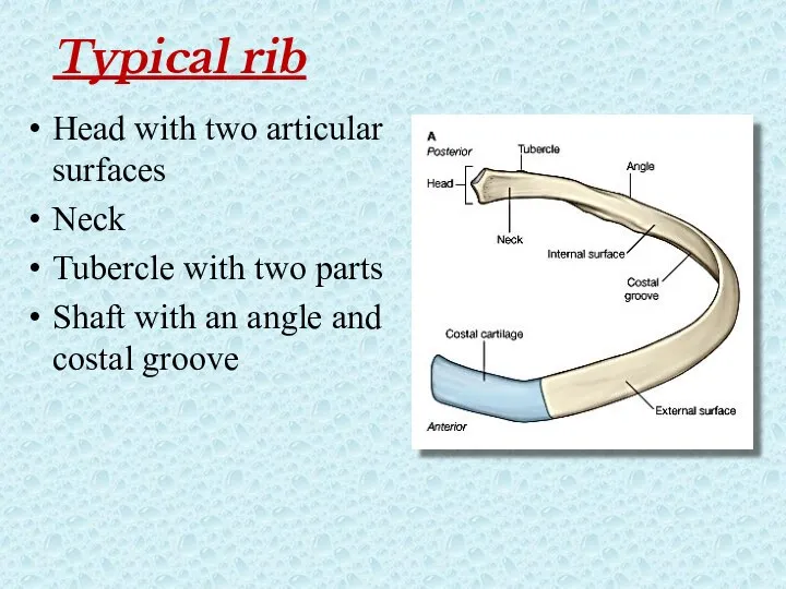
Typical rib
Head with two articular surfaces
Neck
Tubercle with two parts
Shaft with an
Typical rib
Head with two articular surfaces
Neck
Tubercle with two parts
Shaft with an
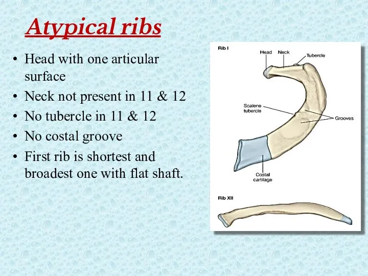
Atypical ribs
Head with one articular surface
Neck not present in 11 &
Atypical ribs
Head with one articular surface
Neck not present in 11 &
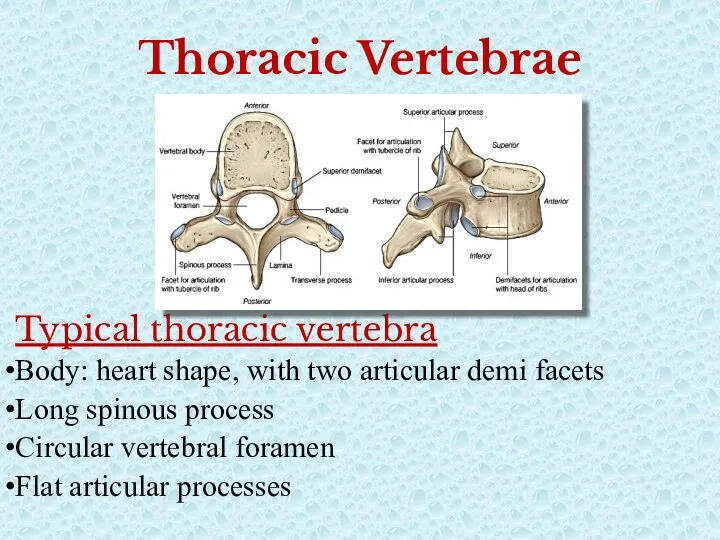
Thoracic Vertebrae
Typical thoracic vertebra
Body: heart shape, with two articular demi facets
Long
Thoracic Vertebrae
Typical thoracic vertebra
Body: heart shape, with two articular demi facets
Long
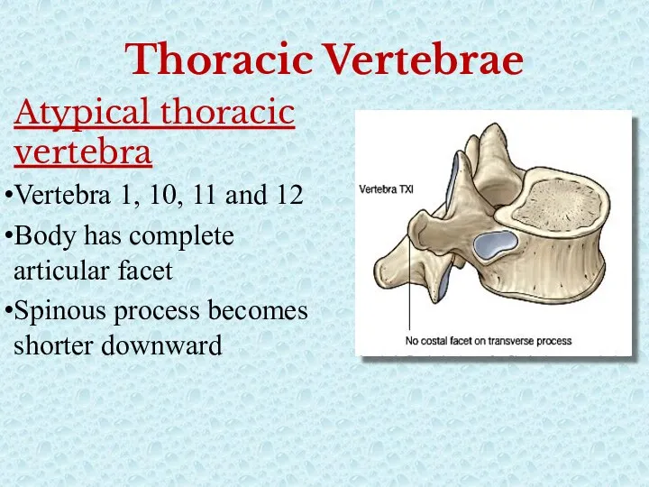
Thoracic Vertebrae
Atypical thoracic vertebra
Vertebra 1, 10, 11 and 12
Body has complete
Thoracic Vertebrae
Atypical thoracic vertebra
Vertebra 1, 10, 11 and 12
Body has complete

Joints
Intervertebral joints:
Symphyses: vertebral bodies;
synovial joints: articular processes
Costovertebral joints:
Joints
Intervertebral joints:
Symphyses: vertebral bodies;
synovial joints: articular processes
Costovertebral joints:
Muscles of thorax
Muscles of pectoral region
Intercostal muscles
Diaphragm
Muscles of thorax
Muscles of pectoral region
Intercostal muscles
Diaphragm
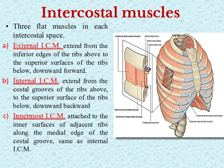
Intercostal muscles
Three flat muscles in each intercostal space.
External I.C.M. extend from
Intercostal muscles
Three flat muscles in each intercostal space.
External I.C.M. extend from
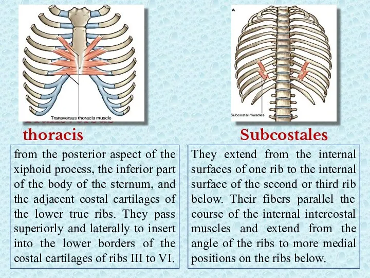
Transversus thoracis
from the posterior aspect of the xiphoid process, the inferior
Transversus thoracis
from the posterior aspect of the xiphoid process, the inferior
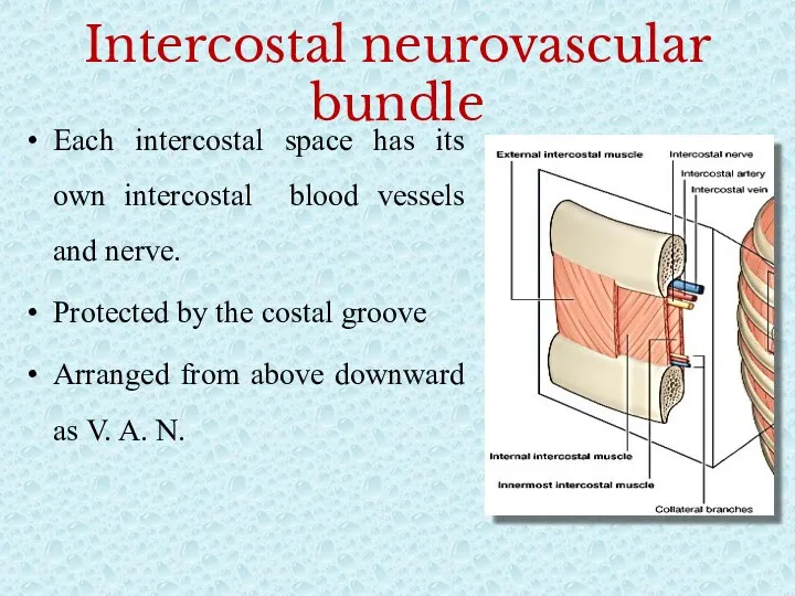
Intercostal neurovascular bundle
Each intercostal space has its own intercostal blood vessels
Intercostal neurovascular bundle
Each intercostal space has its own intercostal blood vessels
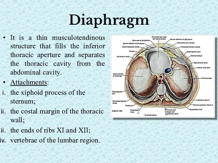
Diaphragm
It is a thin musculotendinous structure that fills the inferior thoracic
Diaphragm
It is a thin musculotendinous structure that fills the inferior thoracic
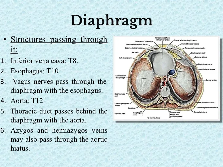
Diaphragm
Structures passing through it:
Inferior vena cava: T8.
Esophagus: T10
Vagus nerves
Diaphragm
Structures passing through it:
Inferior vena cava: T8.
Esophagus: T10
Vagus nerves
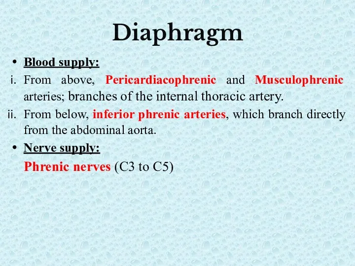
Diaphragm
Blood supply:
From above, Pericardiacophrenic and Musculophrenic arteries; branches of the
Diaphragm
Blood supply:
From above, Pericardiacophrenic and Musculophrenic arteries; branches of the
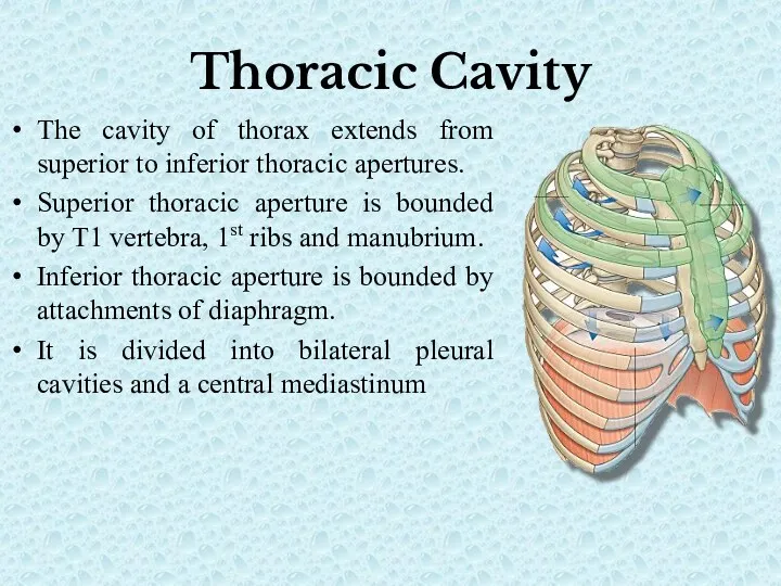
Thoracic Cavity
The cavity of thorax extends from superior to inferior thoracic
Thoracic Cavity
The cavity of thorax extends from superior to inferior thoracic
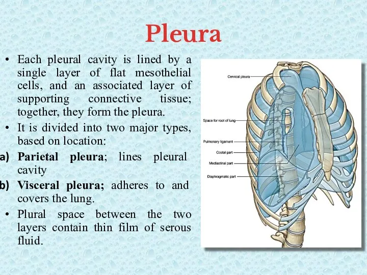
Pleura
Each pleural cavity is lined by a single layer of
Pleura
Each pleural cavity is lined by a single layer of
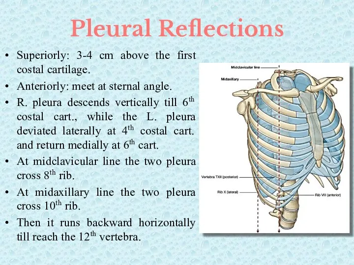
Pleural Reflections
Superiorly: 3-4 cm above the first costal cartilage.
Anteriorly: meet at
Pleural Reflections
Superiorly: 3-4 cm above the first costal cartilage.
Anteriorly: meet at
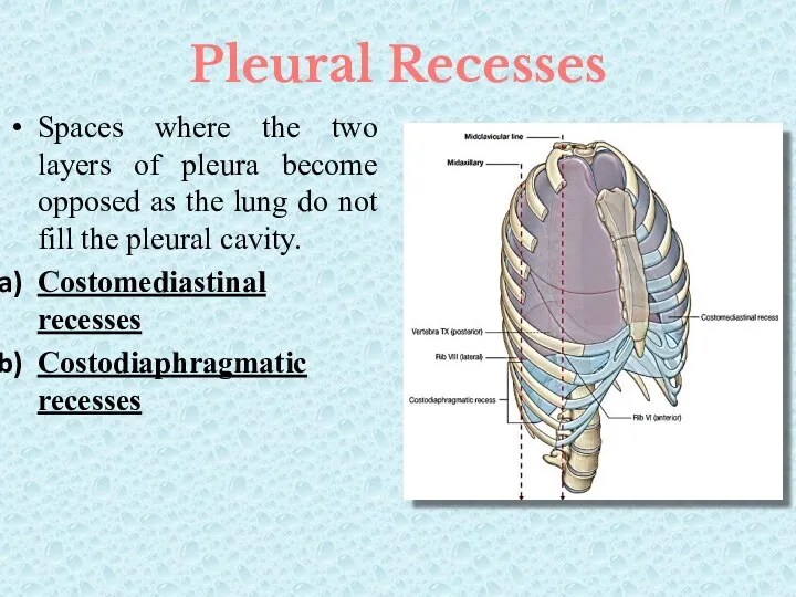
Pleural Recesses
Spaces where the two layers of pleura become opposed as
Pleural Recesses
Spaces where the two layers of pleura become opposed as
Mechanism of Breathing
One of the principal functions of the thoracic wall
Mechanism of Breathing
One of the principal functions of the thoracic wall
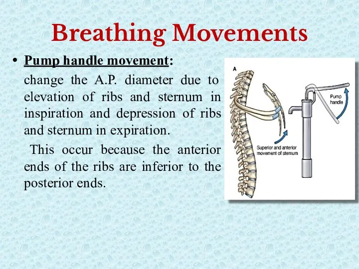
Breathing Movements
Pump handle movement:
change the A.P. diameter due to elevation of
Breathing Movements
Pump handle movement:
change the A.P. diameter due to elevation of
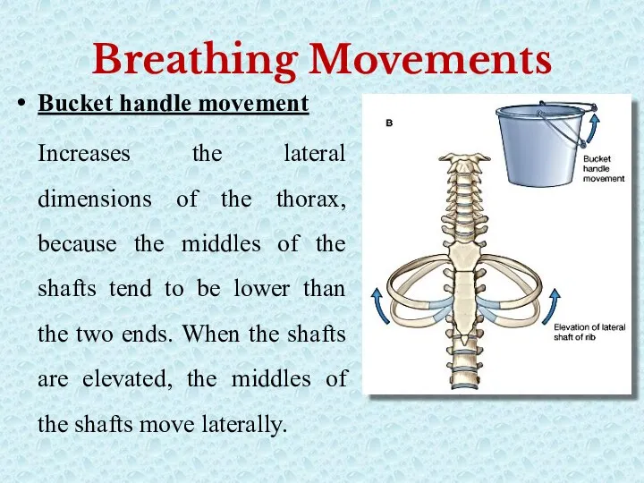
Breathing Movements
Bucket handle movement
Increases the lateral dimensions of the thorax,
Breathing Movements
Bucket handle movement
Increases the lateral dimensions of the thorax,
So,
In Inspiration:
Diaphragm contracts and depressed that increases vertical diameter of thoracic
So,
In Inspiration:
Diaphragm contracts and depressed that increases vertical diameter of thoracic
 Рекомбинантные антитела для диагностики и терапии
 Структурно-функциональная организация клетки
 Причины разнообразия жизни на Земле
 Митохондрия
 Утворення перлин
 Организация микробиологической лабораторной службы
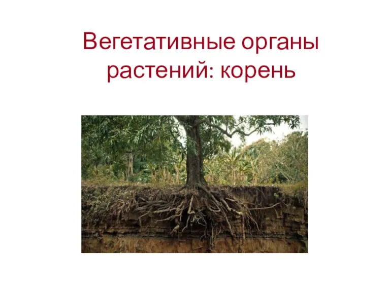 Вегетативные органы растений: корень
 Формы естественного отбора
 Биологическая викторина Птицы Калужской области
 Пищеварительная система (наддиафрагмальный отдел пищеварительной трубки)
 5 популярных характеристик при сравнении организмов
 Муравьи
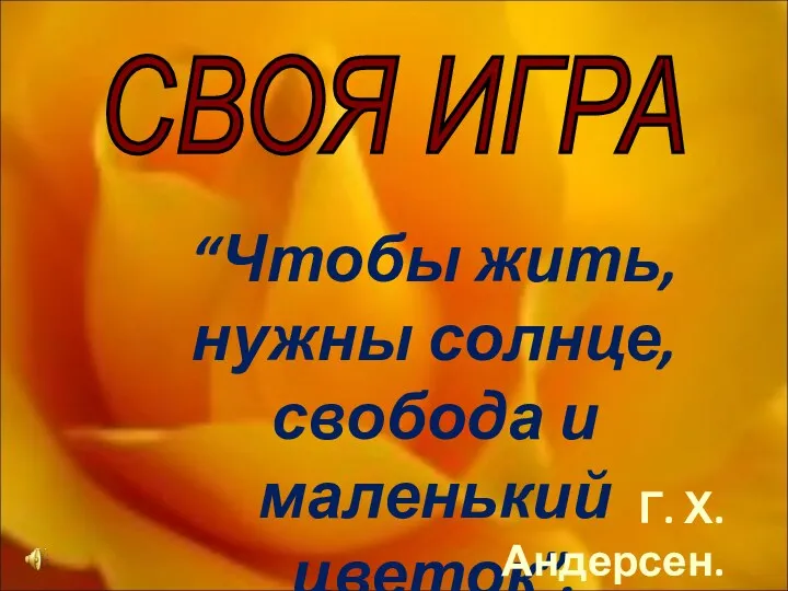 Своя игра. Мир растений
 Водная среда обитания организмов
 Шляпочные грибы
 Общая характеристика типа Моллюски
 Животные России
 Эволюция и видообразование. Современные представления
 Растительные сообщества городской системы
 Биотехнология в селекции растений. Часть 7. Селекция на качество продукции
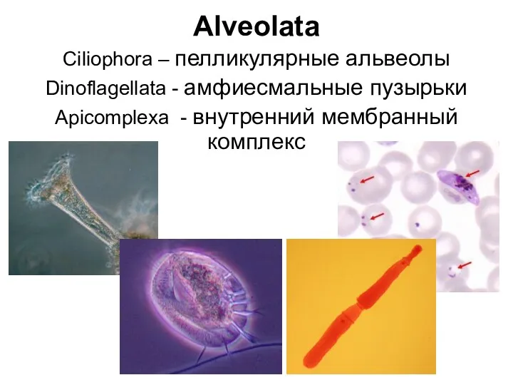 Альвеолата. Инфузории
 Ядро клетки. Хромосомный набор клетки
 Животные и птицы (фотографии)
 Функция желез внутренней секреции
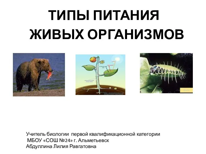 Типы питания живых организмов
 Хвойный лес. Экосистема хвойного леса
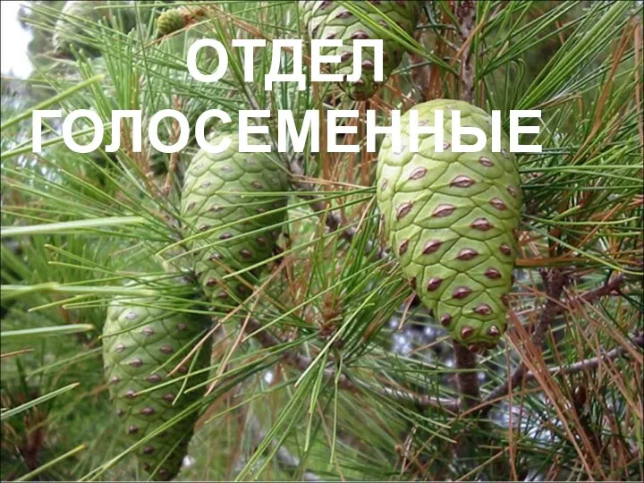 Отдел голосеменные
 Профессия агроном