- Antigen-antibody reactions and selected tests

Содержание
- 2. NATURE OF ANTIGEN-ANTIBODY REACTIONS Lock and Key Concept The combining site of an antibody is located
- 3. AFFINITY AND AVIDITY Affinity Antibody affinity is the strength of the reaction between a single antigenic
- 5. Avidity Avidity is a measure of the overall strength of binding of an antigen with many
- 7. Possible effects on soluble protein of immobilization Protein is shown as having three antigenic sites (epitopes).
- 8. SPECIFICITY AND CROSS REACTIVITY Specificity Specificity refers to the ability of an individual antibody combining site
- 9. Diagnosis of infectious and parasitic diseases and the establishment of detection antibody titers (serodiagnosis); Diagnosis of
- 10. Cross reactivity Cross reactivity refers to the ability of an individual antibody combining site to react
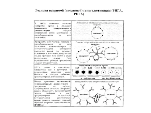
- 11. Agglutination test Agglutination test (agglutinacio - склеивание) - gluing and precipitation of the bacteria under the
- 12. STATEMENT OF MICROAGGLUTINATION TEST
- 13. THE RESULTS OF MICROAGGLUTINATION TEST
- 15. In positive cases precipitate has the form of a thin film of the red blood cells
- 16. PRECIPITATION TEST Principle: When interacting of soluble antigen with antibody in the presence of electrolyte (NaCl)
- 17. THE PRINCIPLE OF PRECIPITATION TEST
- 18. Ring-precipitation test Formation of the Ag-Ab complex - + The test is carried out by layering
- 19. THE PRINCIPLE OF RADIAL IMMUNODIFFUSION TEST The test is carried out in agar gel plates or
- 20. Radial Immunodiffusion (RID)
- 21. Complement fixation test
- 22. Materials and Reagents 1. Sheep erythrocytes suspension (5% suspension of washed sheep RBCs) 2. Hemolysin (rabbit
- 23. The principle of the complement fixation test is illustrated in Figure. Antigen is mixed with the
- 24. Procedure of Complement Fixation Test Complement Fixation Test (CFT) consists of two stage: First step (Complement
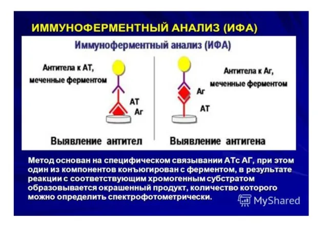
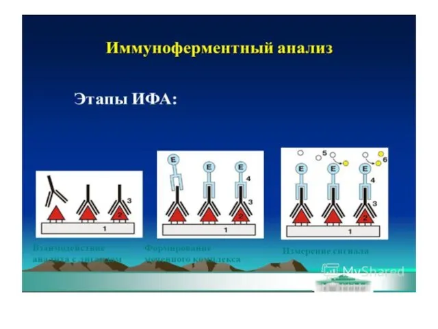
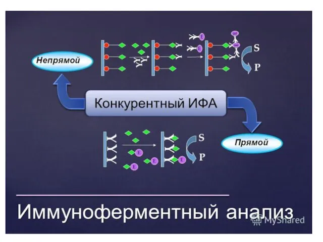
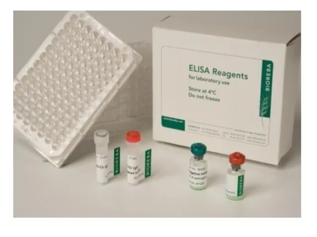
- 25. Enzyme linked immunosorbent assay ELISA - it is serological test in which for the visualization of
- 26. IgG IgM Серологические реакции, основанные на использовании меток МЕТКИ Ферментные Флюоресцирующие радиоизотопные
- 29. 96-луночный планшет для ИФА
- 31. Спектрофотометр для ИФА
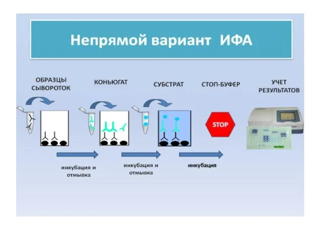
- 32. ПРИНЦИП НЕПРЯМОГО ИФА
- 34. This variant of ELISA is extremely common for the determination of antigens possessing more than one
- 35. Принцип сэндвич-ИФА
- 37. Конкурентный ИФА для определения антигена (антибиотиков) Конъюгат АБ-носитель Субстрат Антибиотик Специфические АТ Антивидовой конъюгат
- 39. Конкурентный ИФА для определения специфических антител
- 40. ИСПОЛЬЗОВАНИЕ АВИДИН-БИОТИНОВОЙ СИСТЕМЫ В ИФА
- 42. Dot-ИФА
- 43. Сравнительная характеристика dot-ИФА и ИФА.
- 44. ПРИНЦИП ИММУНОХРОМАТОГРАФИЧЕСКОГО АНАЛИЗА
- 45. Оборудование для производства иммунохроматографической тест - системы Презиционный диспенсер автоматический Автоматический гильотинный резак Вакуумный сушильный шкаф
- 46. Иммунохроматографическое определение антигена (I – аналитическая зона, II – контрольная зона)
- 48. Скачать презентацию
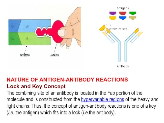
NATURE OF ANTIGEN-ANTIBODY REACTIONS
Lock and Key Concept
The combining site of an
NATURE OF ANTIGEN-ANTIBODY REACTIONS
Lock and Key Concept
The combining site of an
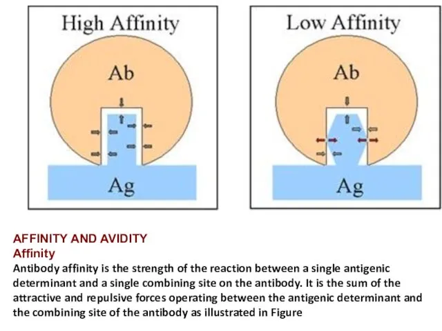
AFFINITY AND AVIDITY
Affinity
Antibody affinity is the strength of the reaction between
AFFINITY AND AVIDITY
Affinity
Antibody affinity is the strength of the reaction between
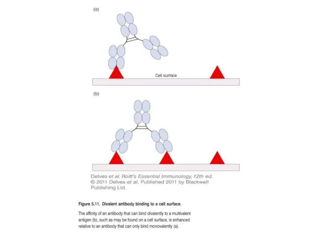
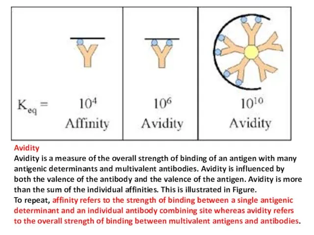
Avidity
Avidity is a measure of the overall strength of binding of
Avidity
Avidity is a measure of the overall strength of binding of
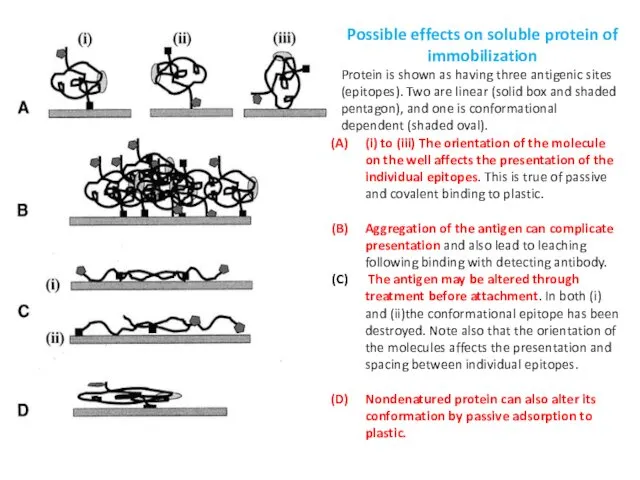
Possible effects on soluble protein of immobilization
Protein is shown as
Possible effects on soluble protein of immobilization
Protein is shown as
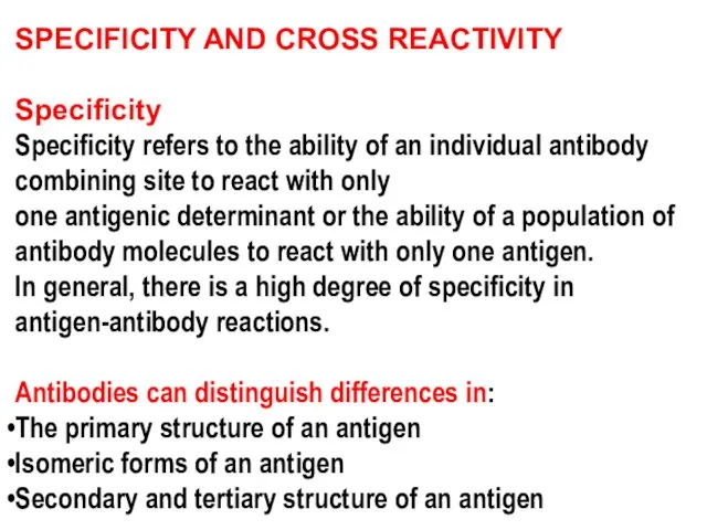
SPECIFICITY AND CROSS REACTIVITY
Specificity
Specificity refers to the ability of an individual
SPECIFICITY AND CROSS REACTIVITY
Specificity
Specificity refers to the ability of an individual
Diagnosis of infectious and parasitic diseases and the establishment of detection
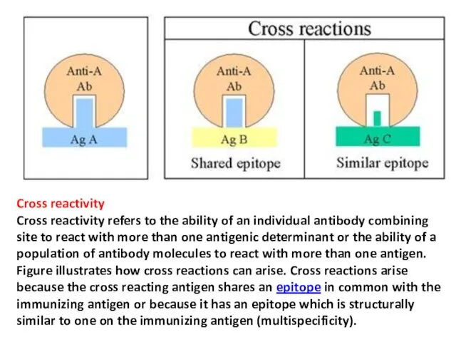
Cross reactivity
Cross reactivity refers to the ability of an individual antibody
Cross reactivity
Cross reactivity refers to the ability of an individual antibody
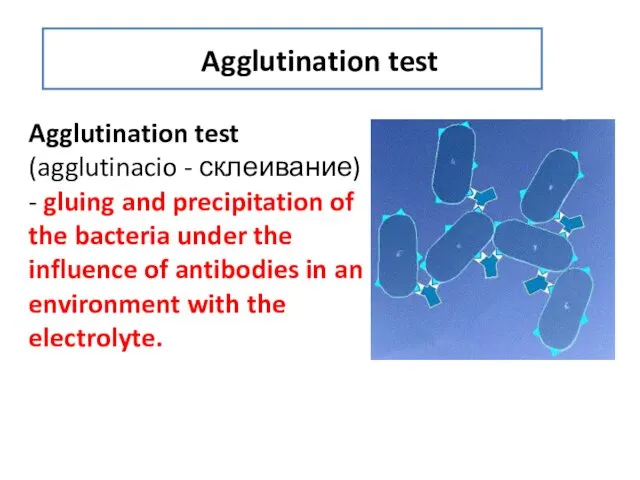
Agglutination test
Agglutination test (agglutinacio - склеивание) - gluing and precipitation of
Agglutination test
Agglutination test (agglutinacio - склеивание) - gluing and precipitation of
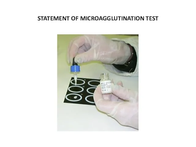
STATEMENT OF MICROAGGLUTINATION TEST
STATEMENT OF MICROAGGLUTINATION TEST
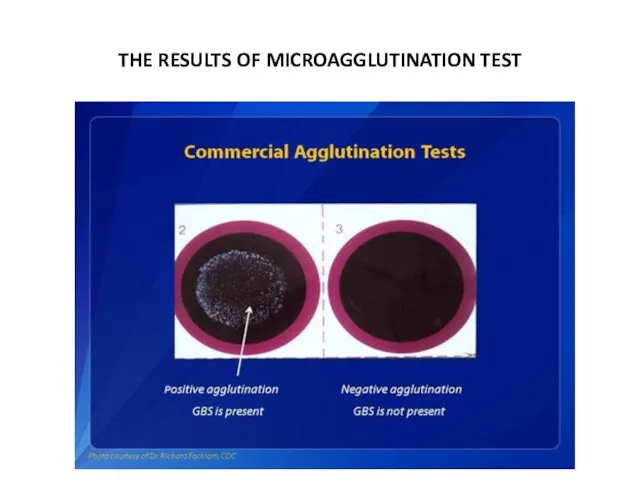
THE RESULTS OF MICROAGGLUTINATION TEST
THE RESULTS OF MICROAGGLUTINATION TEST
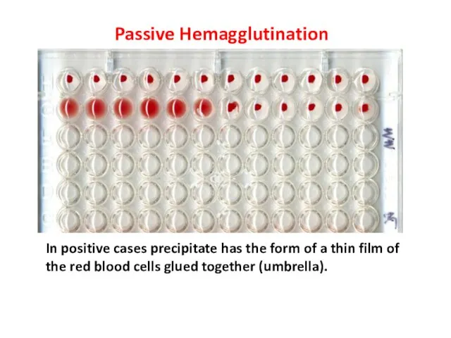
In positive cases precipitate has the form of a thin film
In positive cases precipitate has the form of a thin film
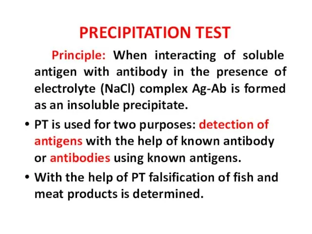
PRECIPITATION TEST
Principle: When interacting of soluble antigen with antibody in
PRECIPITATION TEST
Principle: When interacting of soluble antigen with antibody in
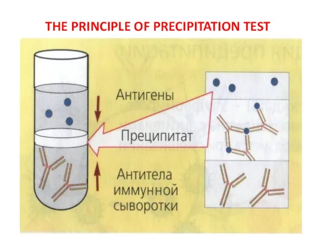
THE PRINCIPLE OF PRECIPITATION TEST
THE PRINCIPLE OF PRECIPITATION TEST
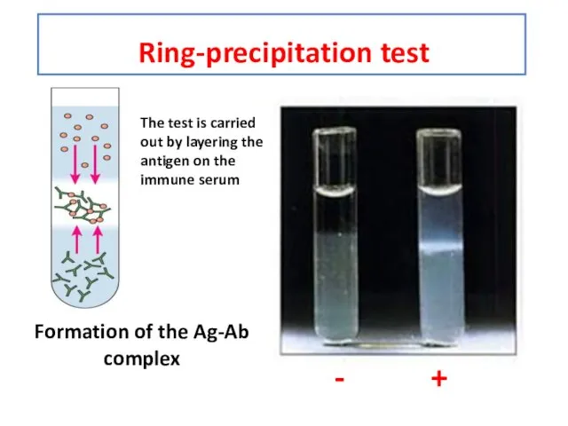
Ring-precipitation test
Formation of the Ag-Ab complex
- +
The test is carried
Ring-precipitation test
Formation of the Ag-Ab complex
- +
The test is carried
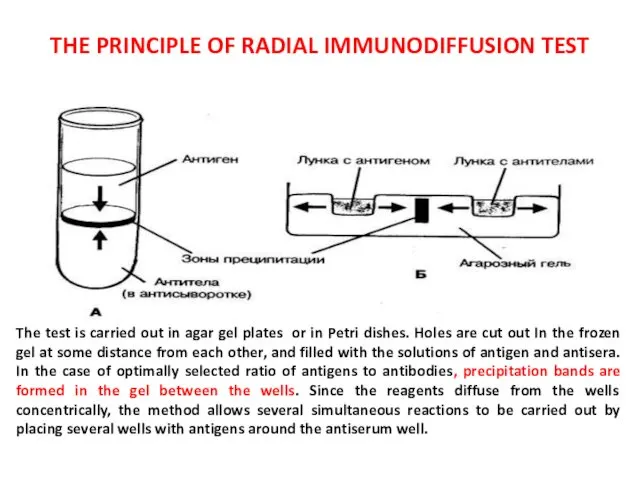
THE PRINCIPLE OF RADIAL IMMUNODIFFUSION TEST
The test is carried out in
THE PRINCIPLE OF RADIAL IMMUNODIFFUSION TEST
The test is carried out in
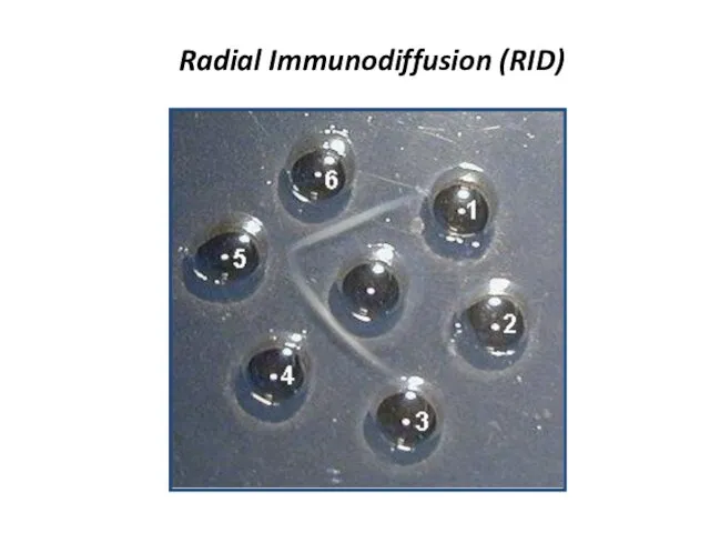
Radial Immunodiffusion (RID)
Radial Immunodiffusion (RID)
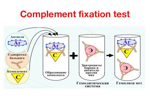
Complement fixation test
Complement fixation test
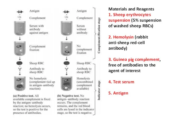
Materials and Reagents
1. Sheep erythrocytes suspension (5% suspension of washed sheep RBCs)
2.
Materials and Reagents
1. Sheep erythrocytes suspension (5% suspension of washed sheep RBCs)
2.
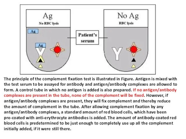
The principle of the complement fixation test is illustrated in Figure.
The principle of the complement fixation test is illustrated in Figure.
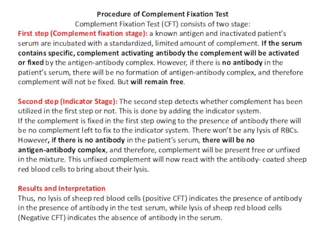
Procedure of Complement Fixation Test
Complement Fixation Test (CFT) consists of two
Procedure of Complement Fixation Test
Complement Fixation Test (CFT) consists of two
Enzyme linked immunosorbent assay
ELISA - it is serological test in which
Enzyme linked immunosorbent assay
ELISA - it is serological test in which
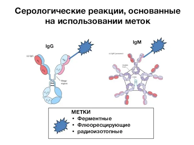
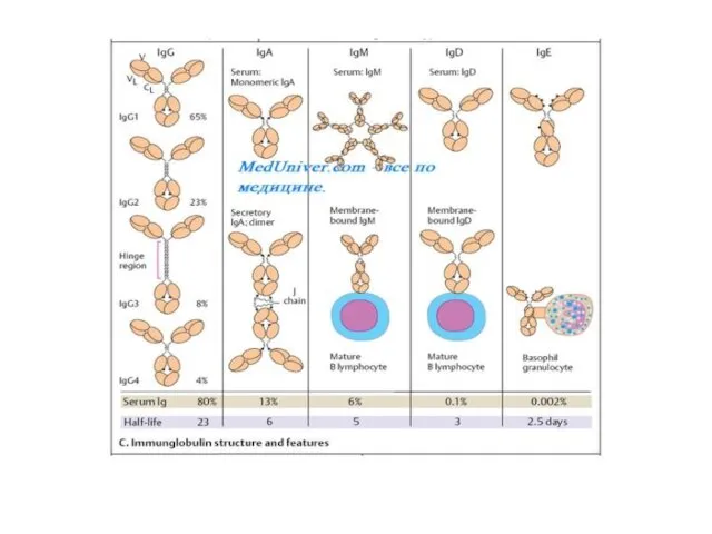
IgG
IgM
Серологические реакции, основанные на использовании меток
МЕТКИ
Ферментные
Флюоресцирующие
радиоизотопные
IgG
IgM
Серологические реакции, основанные на использовании меток
МЕТКИ
Ферментные
Флюоресцирующие
радиоизотопные
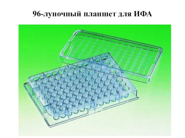
96-луночный планшет для ИФА
96-луночный планшет для ИФА


Спектрофотометр для ИФА
Спектрофотометр для ИФА
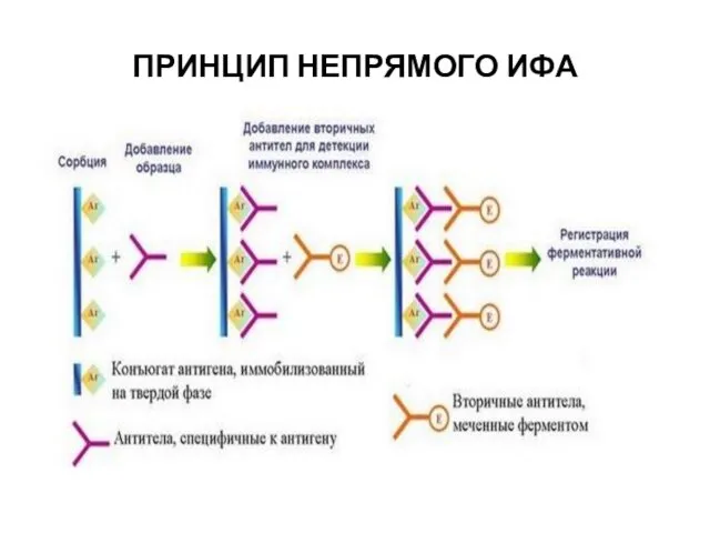
ПРИНЦИП НЕПРЯМОГО ИФА
ПРИНЦИП НЕПРЯМОГО ИФА
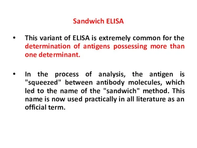
This variant of ELISA is extremely common for the determination of
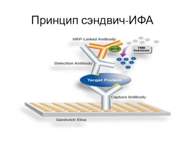
Принцип сэндвич-ИФА
Принцип сэндвич-ИФА

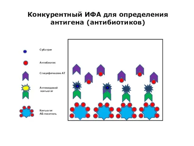
Конкурентный ИФА для определения антигена (антибиотиков)
Конъюгат
АБ-носитель
Субстрат
Антибиотик
Специфические АТ
Антивидовой
конъюгат
Конкурентный ИФА для определения антигена (антибиотиков)
Конъюгат
АБ-носитель
Субстрат
Антибиотик
Специфические АТ
Антивидовой
конъюгат
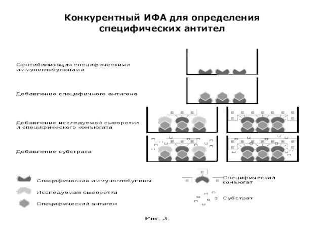
Конкурентный ИФА для определения специфических антител
Конкурентный ИФА для определения специфических антител
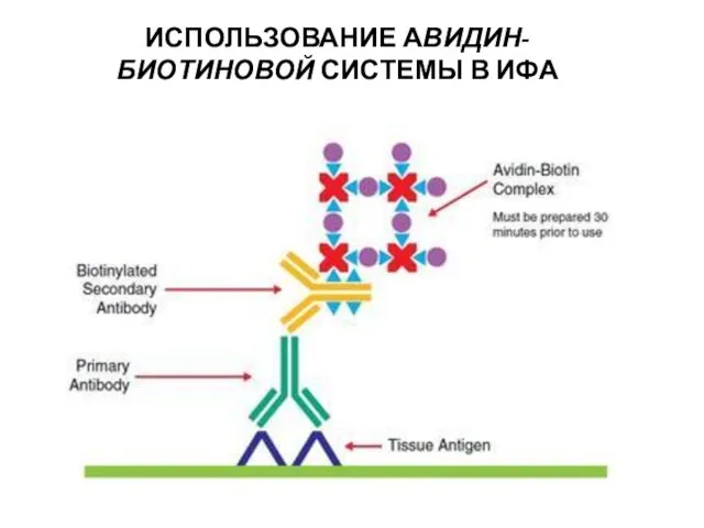
ИСПОЛЬЗОВАНИЕ АВИДИН-БИОТИНОВОЙ СИСТЕМЫ В ИФА
ИСПОЛЬЗОВАНИЕ АВИДИН-БИОТИНОВОЙ СИСТЕМЫ В ИФА
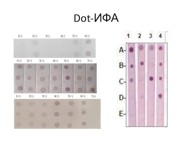
Dot-ИФА
Dot-ИФА
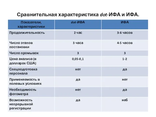
Сравнительная характеристика dot-ИФА и ИФА.
Сравнительная характеристика dot-ИФА и ИФА.
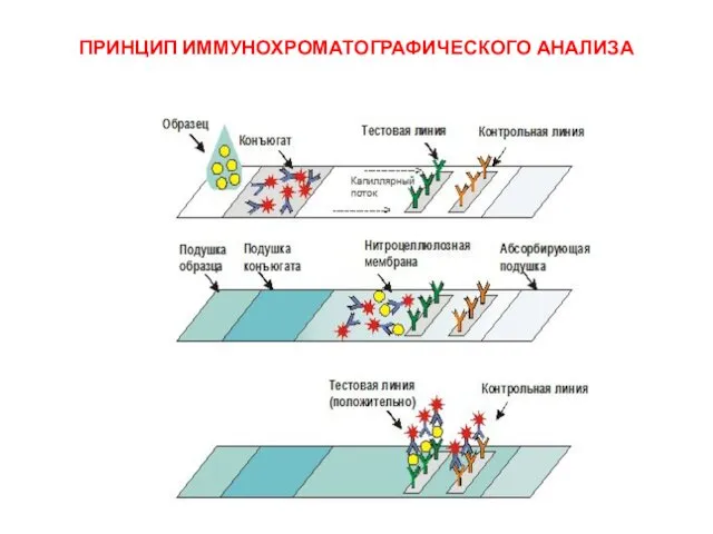
ПРИНЦИП ИММУНОХРОМАТОГРАФИЧЕСКОГО АНАЛИЗА
ПРИНЦИП ИММУНОХРОМАТОГРАФИЧЕСКОГО АНАЛИЗА
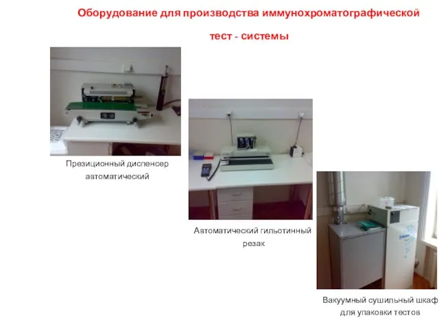
Оборудование для производства иммунохроматографической тест - системы
Презиционный диспенсер
автоматический
Автоматический
Оборудование для производства иммунохроматографической тест - системы
Презиционный диспенсер
автоматический
Автоматический
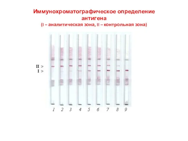
Иммунохроматографическое определение
антигена
(I – аналитическая зона, II – контрольная зона)
Иммунохроматографическое определение
антигена
(I – аналитическая зона, II – контрольная зона)
 Клеткалық биотехнология
 Биотехнология түсініктері, даму тарихы, негізгі әдістері
 Изменчивость: наследственная и ненаследственная
 Головний мозок людини
 Система работы учителя биологии по формированию потребности к здоровому образу жизни.ЗДОРОВЬЕ ШКОЛЬНИКОВ-ЗАЛОГ УСПЕШНОГО ОБУЧЕНИЯ.
 Киты. Внешний вид, габариты, строение тела
 Размножение и развитие растений
 Биохимия мышечной деятельности. Общая характеристика механизма энергообеспечения. Лекция № 6
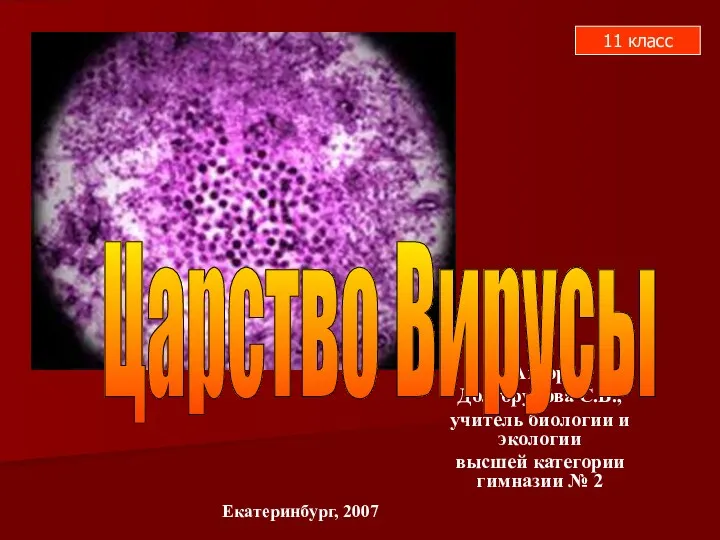 Вирусы. (11 класс)
 Микробиология зерна, крупы, муки и хлеба
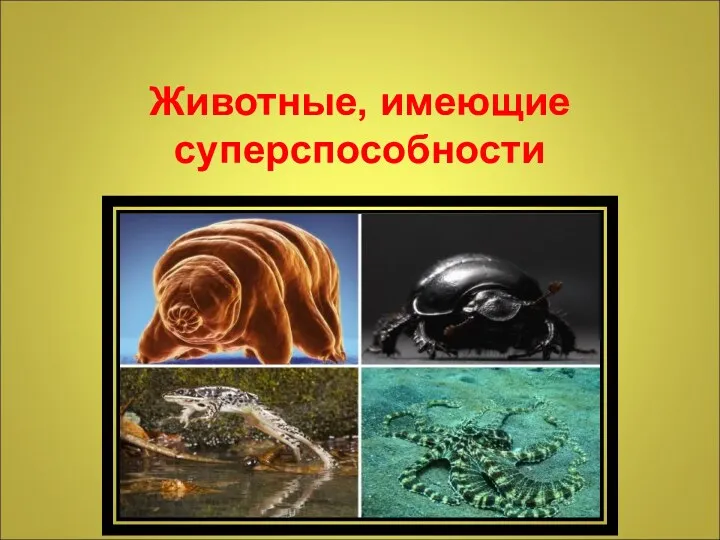 Животные, имеющие суперспособности
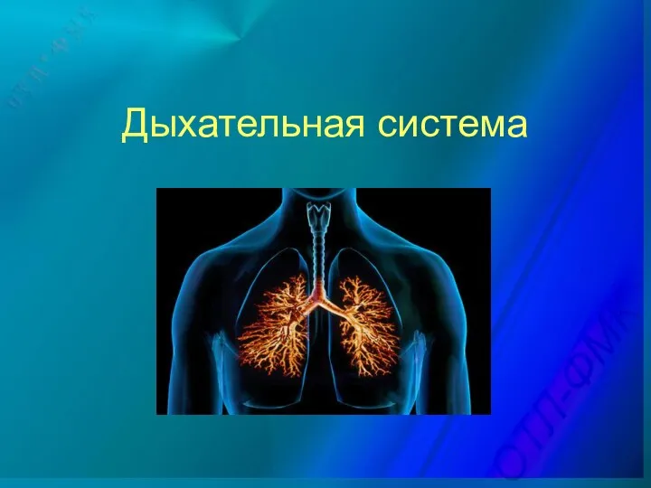 Дыхательная система
 Биотехнологии. Генная инженерия
 Биологическое окисление. Этапы катаболизма. Цикл Кребса
 Сибирский кедр
 Гибель клетки и ее роль в патологических процессах
 Пищевая микробиология. Брожение
 Адаптация - приспособления организмов к среде обитания
 Витамины группы B
 Съедобные и ядовитые грибы
 Будова та функції скелетних м'язів
 Генетика микроорганизмов
 Игровой урок Знатоки млекопитающих
 Классификация животных.Основные систематические группы.
 Почему осенью листья опадают?
 Строение и функции органов дыхания
 Свойства живых организмов
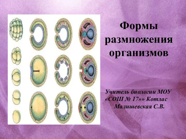 Формы размножения организмов