- Blood smear. DLC 2

Содержание
- 3. WBCs are classified into two groups:
- 4. Cell diameter: 10-15 µm. Nucleus: multi-lobed (2-5 lobes), dark purple-blue in color. Cytoplasm: Pink with fine
- 5. Cell diameter: 12-17 µm. Nucleus: Bi-lobed, spectacle shape, purple in color. Cytoplasm: has orange -red granules.
- 6. Cell diameter: 10-15 µm. Nucleus: Bi-lobed, purple in color. Cytoplasm: dark blue or purple granules. Normal
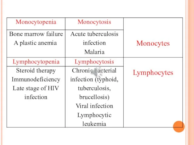
- 7. Cell diameter: small 7-9 /large 12-16 µm. Nucleus: large, round to indented fills the cell. Cytoplasm:
- 8. Cell diameter: 12-20 µm. Nucleus: large and curved, like kidney shape. Cytoplasm: large amount of pale
- 9. Automated hematology autoanalyzer… as part of CBC (however not totally accurate need microscopic assessment). Manual method
- 10. Blood smear: is a blood test that gives information about the number and shape of blood
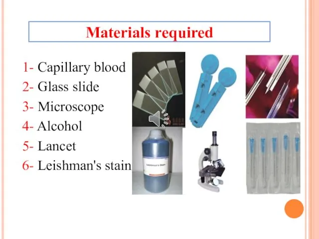
- 11. 1- Capillary blood 2- Glass slide 3- Microscope 4- Alcohol 5- Lancet 6- Leishman's stain Materials
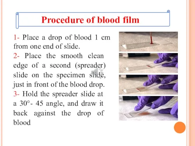
- 12. 1- Place a drop of blood 1 cm from one end of slide. 2- Place the
- 13. 4- Allow the blood to spread almost to the edges of the slide 5- Push the
- 15. 1- Cover the slide with concentrated Leishman stain for about 2-3 min. 2- Diluted the slide
- 16. Examination of the stained blood film: 1- Place the slide on the microscope stage. 2- Examine
- 17. Normal adult differential WBC counts are:
- 21. Скачать презентацию

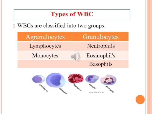
WBCs are classified into two groups:
WBCs are classified into two groups:
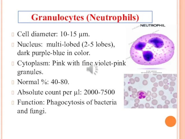
Cell diameter: 10-15 µm.
Nucleus: multi-lobed (2-5 lobes), dark purple-blue in color.
Cell diameter: 10-15 µm.
Nucleus: multi-lobed (2-5 lobes), dark purple-blue in color.
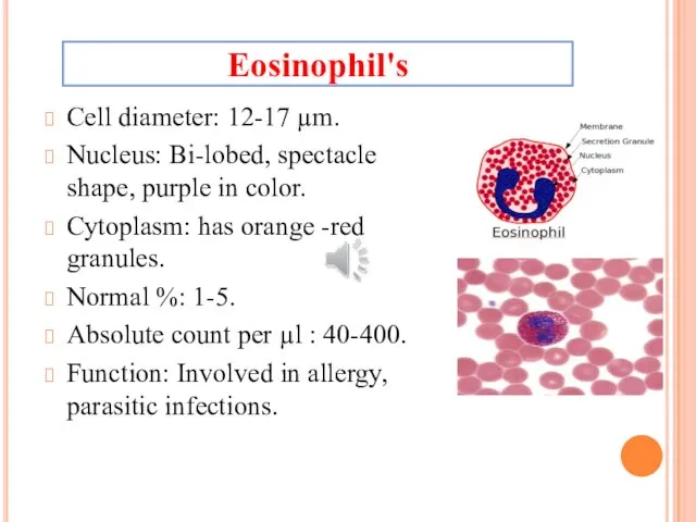
Cell diameter: 12-17 µm.
Nucleus: Bi-lobed, spectacle shape, purple in color.
Cytoplasm: has
Cell diameter: 12-17 µm.
Nucleus: Bi-lobed, spectacle shape, purple in color.
Cytoplasm: has
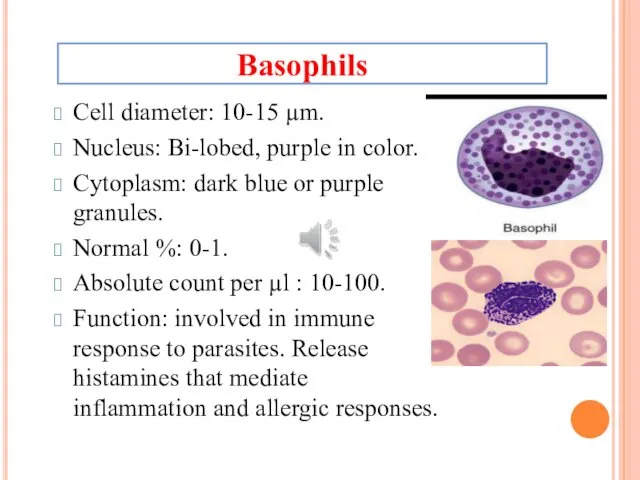
Cell diameter: 10-15 µm.
Nucleus: Bi-lobed, purple in color.
Cytoplasm: dark blue or
Cell diameter: 10-15 µm.
Nucleus: Bi-lobed, purple in color.
Cytoplasm: dark blue or
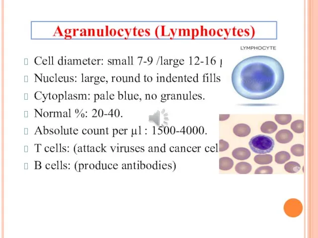
Cell diameter: small 7-9 /large 12-16 µm.
Nucleus: large, round to indented
Cell diameter: small 7-9 /large 12-16 µm.
Nucleus: large, round to indented
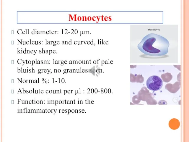
Cell diameter: 12-20 µm.
Nucleus: large and curved, like kidney shape.
Cytoplasm: large
Cell diameter: 12-20 µm.
Nucleus: large and curved, like kidney shape.
Cytoplasm: large
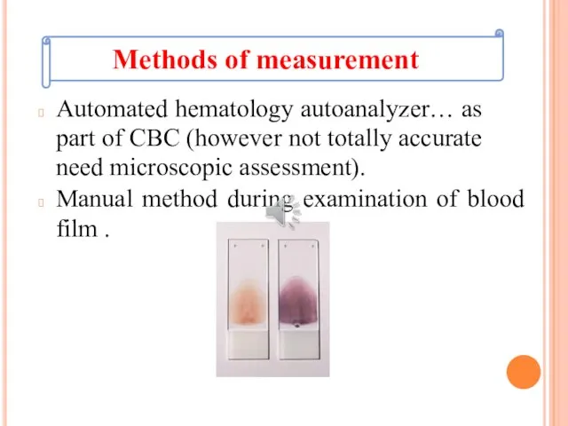
Automated hematology autoanalyzer… as part of CBC (however not totally accurate
Automated hematology autoanalyzer… as part of CBC (however not totally accurate

Blood smear: is a blood test that gives information about the
Blood smear: is a blood test that gives information about the
1- Capillary blood
2- Glass slide
3- Microscope
4- Alcohol
5- Lancet
6- Leishman's stain
Materials required
1- Capillary blood
2- Glass slide
3- Microscope
4- Alcohol
5- Lancet
6- Leishman's stain
Materials required
1- Place a drop of blood 1 cm from one end
1- Place a drop of blood 1 cm from one end
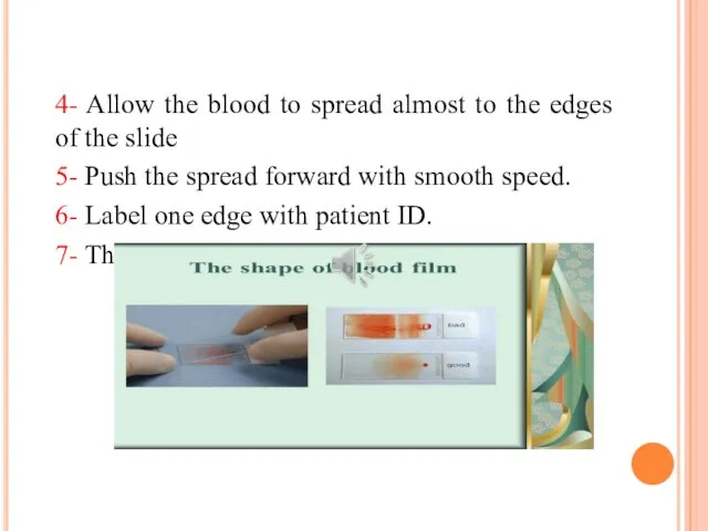
4- Allow the blood to spread almost to the edges of

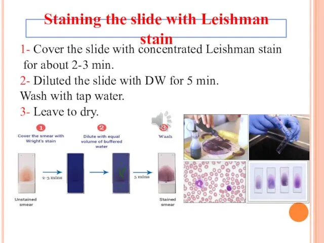
1- Cover the slide with concentrated Leishman stain
for about 2-3
1- Cover the slide with concentrated Leishman stain
for about 2-3
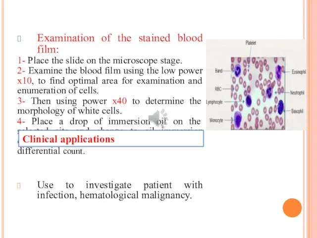
Examination of the stained blood film:
1- Place the slide on
Examination of the stained blood film:
1- Place the slide on
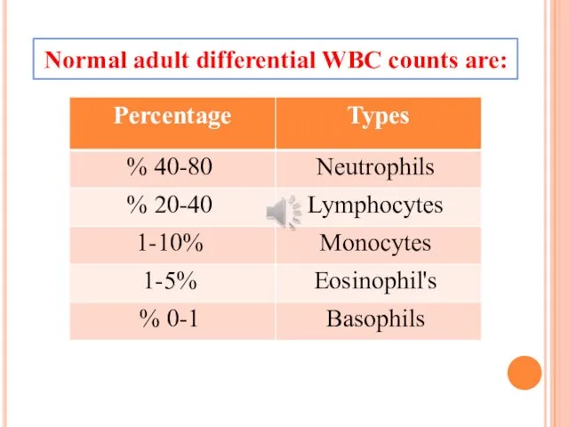
Normal adult differential WBC counts are:
Normal adult differential WBC counts are:

 Биологическое действие радиации
 Индивидуальное развитие организмов
 Waterflood Design and Operational Best Practices
 Нейроны. Функции, строение, классификация
 20. Біохімія мязів і сполучної тканини
 Красная книга Воронежской области
 Прецептрон. Практична реалізація
 Генетическая инженерия растений
 Салонные процедуры
 Плеснивые грибы и грибы паразиты
 Эволюция. Теория Ламарка
 Музей московского зоопарка
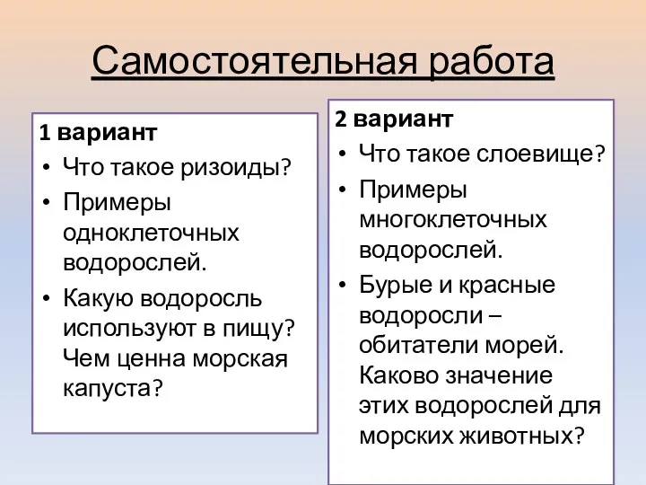 Презентация по теме Лишайники
 Анатомия ЦНС. Анатомия и физиология спинного мозга
 Сердце и кровообращение.
 Грибы
Грибы Интерактивный кроссворд Насекомые
 презентация Пушистый доктор
 Дикие и домашние животные
 Семя. Внешнее и внутреннее строение семени
 Генетический контроль метаболизма азота, фосфора и ретроградная регуляция
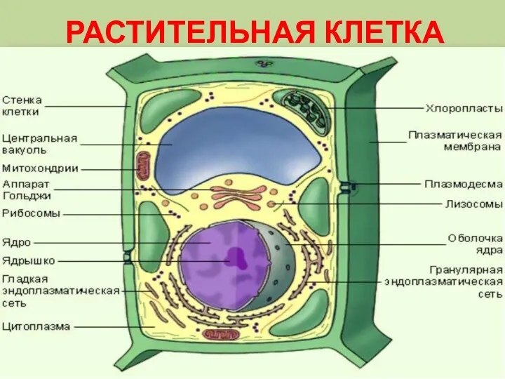 Растительная клетка
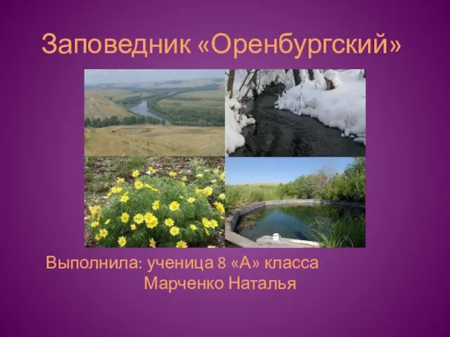 Заповедник Оренбургский
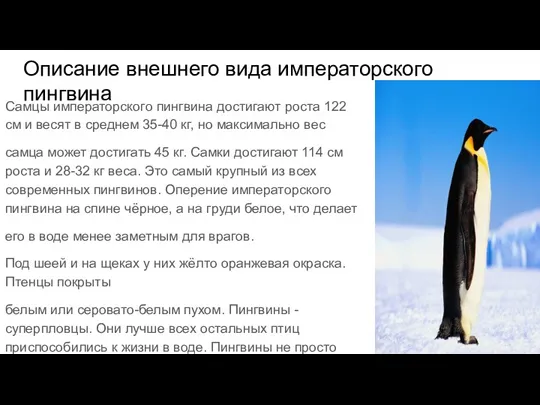 Пингвины (1)
 Сторінка дослідника. Твій город на підвіконні. Урок №76. Я досліджую світ
 Механическая обработка молока
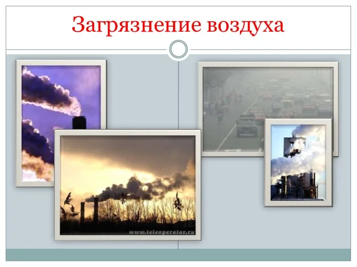 Презентация Загрязнение воздуха
 Звери-млекопитающие (урок окружающего мира 1 класс)