- Eye lecture

Содержание
- 2. THE SENSORY ORGANS RECIEVE INFORMATION ABOUT THE STATE OF THE EXTERNAL ENVIRONMENT AND THE ACTIVITY OF
- 3. Eyeball (eye) - is the peripheral part of the visual analyzer. Through the organ of vision
- 4. Three tunics are distinguished in the eyeball: Outer fibrous (sclera and its transparent part- the cornea)
- 6. Functionally several apparatuses are distinguished: receptor (retina), dioptric or light-refracting (cornea, crystalline lens, vitreous body, fluid
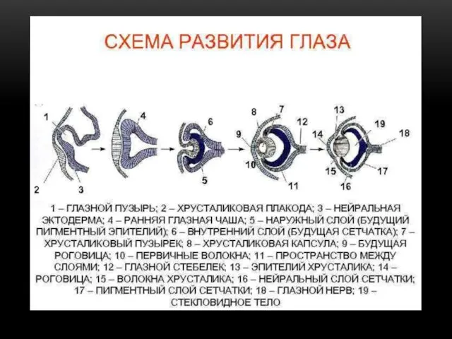
- 7. THE ORGAN OF VISION DEVELOPS FROM 3 EMBRYONIC SOURCES: ECTODERM, NEURAL TUBE AND MESENCHYME DEVELOPMENT begins
- 8. DEVELOPMENT OF EYE At the same time, the ectoderm opposite the eye vesicles, while puffing, forms
- 10. STAGES OF EYE DEVELOPMENT
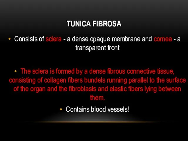
- 12. TUNICA FIBROSA Consists of sclera - a dense opaque membrane and cornea - a transparent front
- 13. LIMBUS The sclera passes into the cornea in the limb region, on the inner surface of
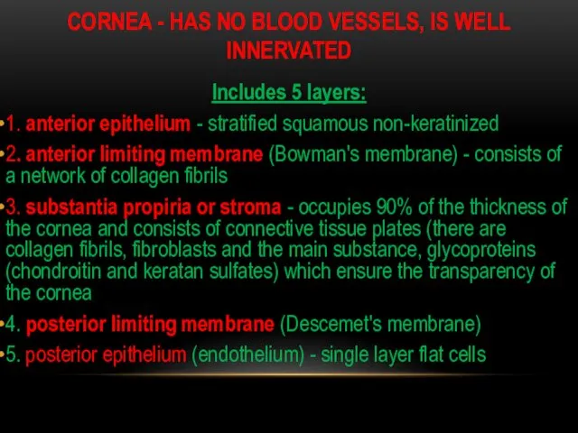
- 14. CORNEA - HAS NO BLOOD VESSELS, IS WELL INNERVATED Includes 5 layers: 1. anterior epithelium -
- 15. CORNEA- TRANSPARENT PART OF TUNICA FIBROSA
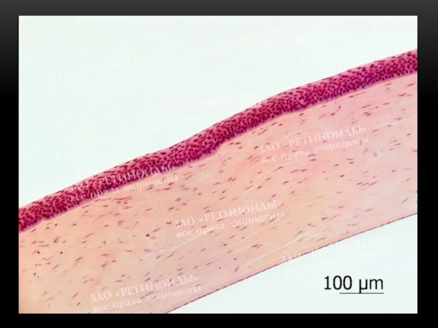
- 17. HOROID Includes: choroid ciliary body iris Actually, the choroid consists of Loose irregular connective tissue and
- 18. CILIARY BODY It is formed by the ciliary muscle (smooth muscle) and ciliary processes that fix
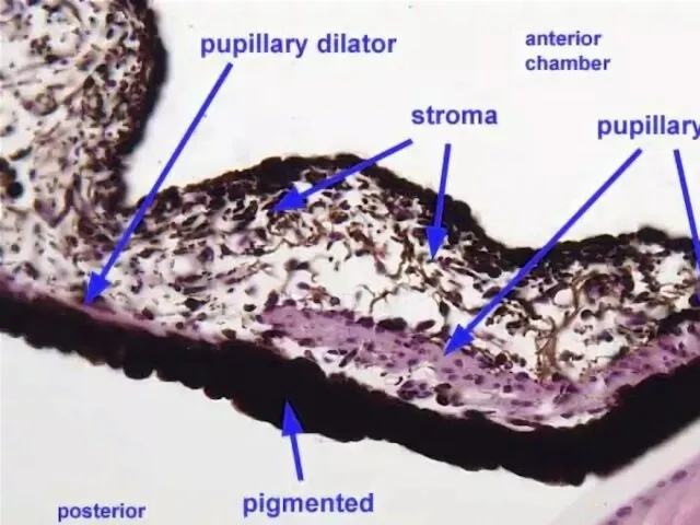
- 19. IRIS the anterior part of the choroid, separates the anterior and posterior chambers of the eye,
- 20. РАДУЖКА И ХРУСТАЛИК
- 21. THE LENS - DOES NOT HAVE BLOOD VESSELS Consists of: 1. Capsule - a thin transparent
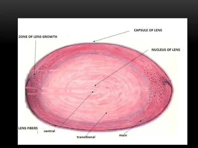
- 22. LENS FIBERS Elongated hexagonal epithelial cells lying parallel to the lens surface in concentric layers and
- 25. VITREOUS BODY A transparent jelly-like mass filling the space between the lens and the retina. It
- 26. RETINA Photosensitive tunica of the eye. It is divided into the visual part, lining the inside
- 27. The retina consists of nerve tissue + pigment epithelium. Nerve tissue includes neurons and neuroglia
- 28. NEURONS They form a three-membered chain of radially located neurons connected to each other by synapses:
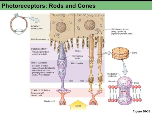
- 29. PHOTORECEPTORS - are bipolar neurons - rods and cones. They consist of three parts: perikarion +
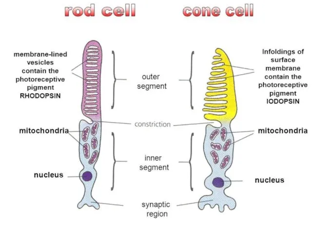
- 30. ROD - With narrow elongated outer segments The outer segment of the process has a cylindrical
- 31. RODOPSIN Rodopsin decomposes under the influence of light with the appearance of an electrical signal, and
- 32. INNER SEGMENT It contains mitochondria, centriole, rER, sER, Golgi complex and provides the outer segment with
- 33. CONES The structure is similar to rods But The outer segments of them are conical in
- 34. CONS The inner segment has a drop of fat inside, surrounded by mitochondria (ellipsoid) Contains larger
- 35. RETINA
- 37. BIPOLAR NEURONS Dendrites are associated with axons of photoreceptor cells, and their axons transmit impulses to
- 38. MULTIPOLAR (GANGLIONIC) NEURON Large multipolar cells with an eccentrically located nucleus. Contain well-developed organelles. Their dendrites
- 39. HORIZONTAL NEURON Associative multipolar neurons, their dendrites and axon are synaptically connected with the axons of
- 40. AMACRINE NEURONS Unipolar associative neurons whose dendrites form bonds with axons of bipolar cells and ganglionic
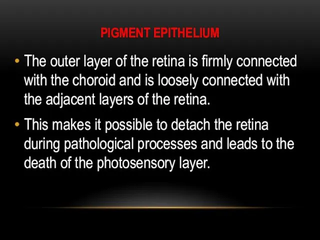
- 41. PIGMENT EPITHELIUM The outer layer of the retina is firmly connected with the choroid and is
- 42. FUNCTIONS OF THE PIGMENT EPITHELIUM Accumulation and transport of vitamin A to photoreceptors Phagocytosis and digestion
- 43. NEUROGLIA OF RETINA It is represented by radial gliocytes (Muller cells), astrocytes and microglia Astrocytes are
- 44. MULLER CELLS Large star shaped cells stretch across the entire thickness of the retina perpendicular to
- 45. LAYERS OF RETINA 8 layers without limiting membraines Pigment epithelium A layer of rods and cones
- 46. LAYERS OF RETINA 10 LAYERS 1. pigment epithelium 2. layer of rods and cones - represented
- 48. Скачать презентацию
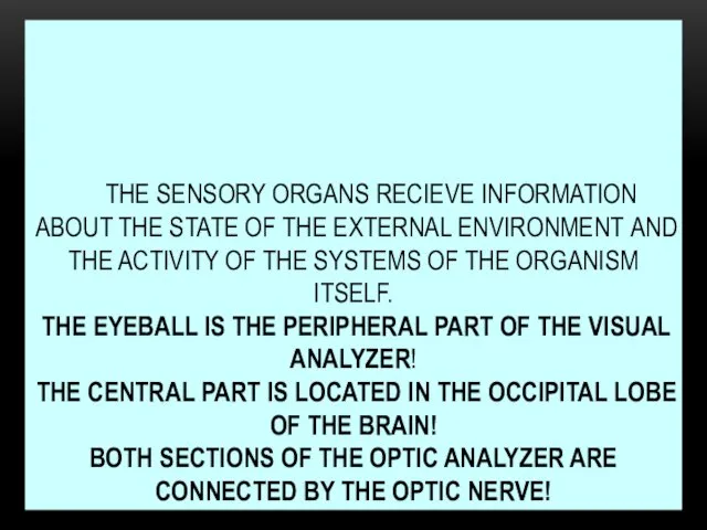
THE SENSORY ORGANS RECIEVE INFORMATION ABOUT THE STATE OF THE EXTERNAL
THE SENSORY ORGANS RECIEVE INFORMATION ABOUT THE STATE OF THE EXTERNAL

Eyeball (eye) - is the peripheral part of the visual analyzer.
Eyeball (eye) - is the peripheral part of the visual analyzer.
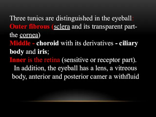
Three tunics are distinguished in the eyeball: Outer fibrous (sclera and
Three tunics are distinguished in the eyeball: Outer fibrous (sclera and
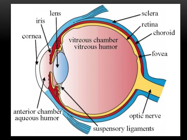
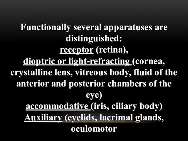
Functionally several apparatuses are distinguished:
receptor (retina),
dioptric or light-refracting (cornea, crystalline
Functionally several apparatuses are distinguished:
receptor (retina),
dioptric or light-refracting (cornea, crystalline
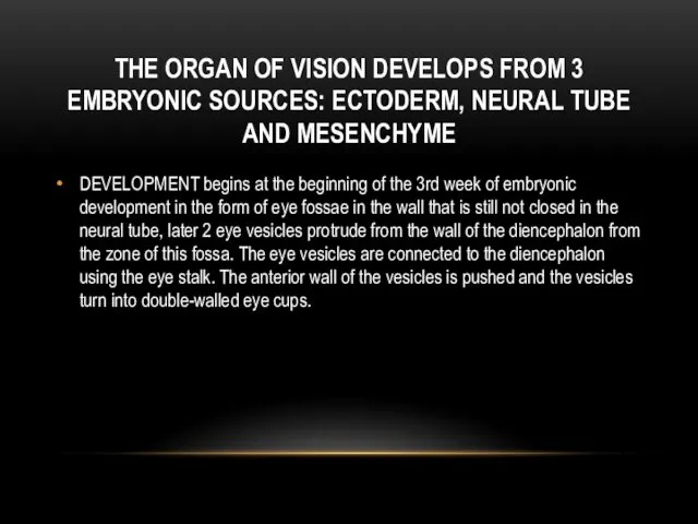
THE ORGAN OF VISION DEVELOPS FROM 3 EMBRYONIC SOURCES: ECTODERM, NEURAL
THE ORGAN OF VISION DEVELOPS FROM 3 EMBRYONIC SOURCES: ECTODERM, NEURAL
DEVELOPMENT OF EYE
At the same time, the ectoderm opposite the eye
DEVELOPMENT OF EYE
At the same time, the ectoderm opposite the eye
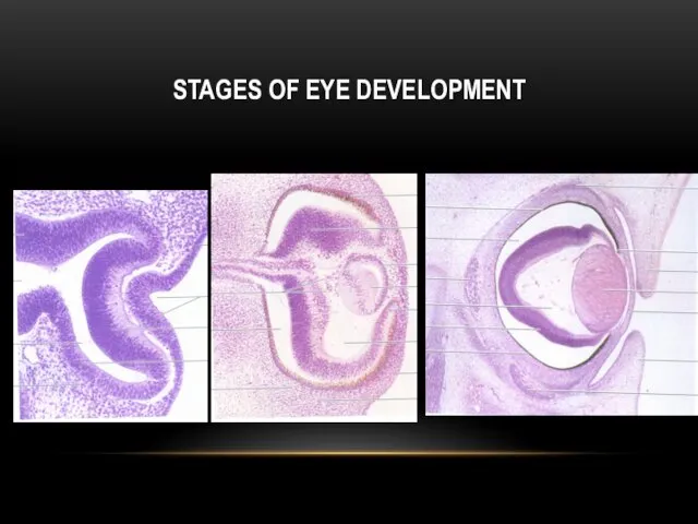
STAGES OF EYE DEVELOPMENT
STAGES OF EYE DEVELOPMENT

TUNICA FIBROSA
Consists of sclera - a dense opaque membrane and cornea
TUNICA FIBROSA
Consists of sclera - a dense opaque membrane and cornea
LIMBUS
The sclera passes into the cornea in the limb region, on
LIMBUS
The sclera passes into the cornea in the limb region, on
CORNEA - HAS NO BLOOD VESSELS, IS WELL INNERVATED
Includes 5 layers:
1.
CORNEA - HAS NO BLOOD VESSELS, IS WELL INNERVATED
Includes 5 layers:
1.
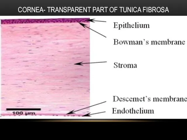
CORNEA- TRANSPARENT PART OF TUNICA FIBROSA
CORNEA- TRANSPARENT PART OF TUNICA FIBROSA
HOROID
Includes:
choroid
ciliary body
iris
Actually, the choroid consists of Loose irregular connective tissue
HOROID
Includes:
choroid
ciliary body
iris
Actually, the choroid consists of Loose irregular connective tissue
CILIARY BODY
It is formed by the ciliary muscle (smooth muscle) and
CILIARY BODY
It is formed by the ciliary muscle (smooth muscle) and
IRIS
the anterior part of the choroid, separates the anterior and posterior
IRIS
the anterior part of the choroid, separates the anterior and posterior
РАДУЖКА И ХРУСТАЛИК
РАДУЖКА И ХРУСТАЛИК
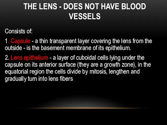
THE LENS - DOES NOT HAVE BLOOD VESSELS
Consists of:
1. Capsule -
THE LENS - DOES NOT HAVE BLOOD VESSELS
Consists of:
1. Capsule -
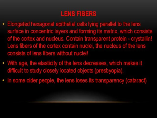
LENS FIBERS
Elongated hexagonal epithelial cells lying parallel to the lens surface
LENS FIBERS
Elongated hexagonal epithelial cells lying parallel to the lens surface

VITREOUS BODY
A transparent jelly-like mass filling the space between the lens
VITREOUS BODY
A transparent jelly-like mass filling the space between the lens
RETINA
Photosensitive tunica of the eye.
It is divided into the visual part,
RETINA
Photosensitive tunica of the eye.
It is divided into the visual part,
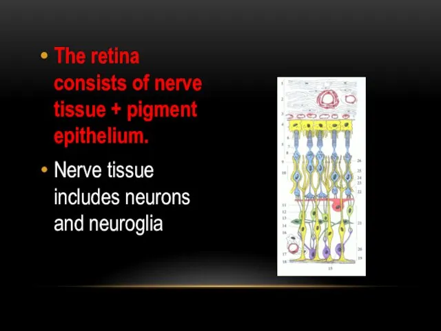
The retina consists of nerve tissue + pigment epithelium.
Nerve tissue includes
The retina consists of nerve tissue + pigment epithelium.
Nerve tissue includes
NEURONS
They form a three-membered chain of radially located neurons connected to
NEURONS
They form a three-membered chain of radially located neurons connected to
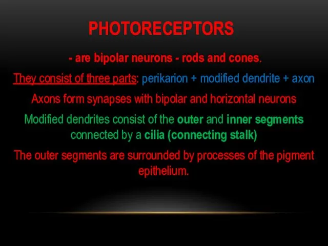
PHOTORECEPTORS
- are bipolar neurons - rods and cones.
They consist of
PHOTORECEPTORS
- are bipolar neurons - rods and cones.
They consist of
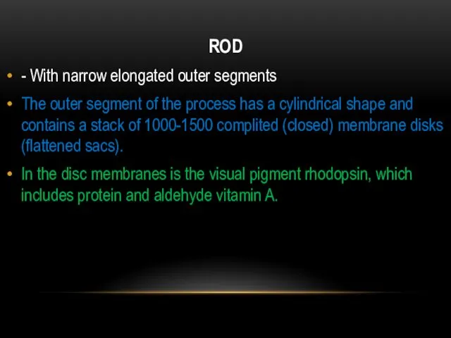
ROD
- With narrow elongated outer segments
The outer segment of the process
ROD
- With narrow elongated outer segments
The outer segment of the process
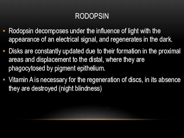
RODOPSIN
Rodopsin decomposes under the influence of light with the appearance of
RODOPSIN
Rodopsin decomposes under the influence of light with the appearance of
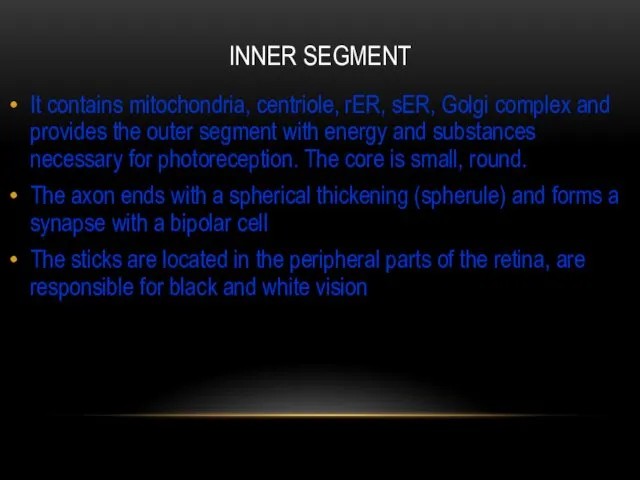
INNER SEGMENT
It contains mitochondria, centriole, rER, sER, Golgi complex and provides
INNER SEGMENT
It contains mitochondria, centriole, rER, sER, Golgi complex and provides
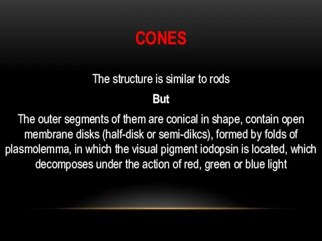
CONES
The structure is similar to rods
But
The outer segments of them are
CONES
The structure is similar to rods
But
The outer segments of them are
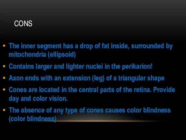
CONS
The inner segment has a drop of fat inside, surrounded by
CONS
The inner segment has a drop of fat inside, surrounded by
RETINA
RETINA
BIPOLAR NEURONS
Dendrites are associated with axons of photoreceptor cells, and their
BIPOLAR NEURONS
Dendrites are associated with axons of photoreceptor cells, and their
MULTIPOLAR (GANGLIONIC) NEURON
Large multipolar cells with an eccentrically located nucleus. Contain
MULTIPOLAR (GANGLIONIC) NEURON
Large multipolar cells with an eccentrically located nucleus. Contain
HORIZONTAL NEURON
Associative multipolar neurons, their dendrites and axon are synaptically connected
HORIZONTAL NEURON
Associative multipolar neurons, their dendrites and axon are synaptically connected
AMACRINE NEURONS
Unipolar associative neurons whose dendrites form bonds with axons of
AMACRINE NEURONS
Unipolar associative neurons whose dendrites form bonds with axons of
PIGMENT EPITHELIUM
The outer layer of the retina is firmly connected with
PIGMENT EPITHELIUM
The outer layer of the retina is firmly connected with
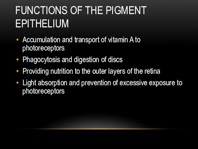
FUNCTIONS OF THE PIGMENT EPITHELIUM
Accumulation and transport of vitamin A to
FUNCTIONS OF THE PIGMENT EPITHELIUM
Accumulation and transport of vitamin A to
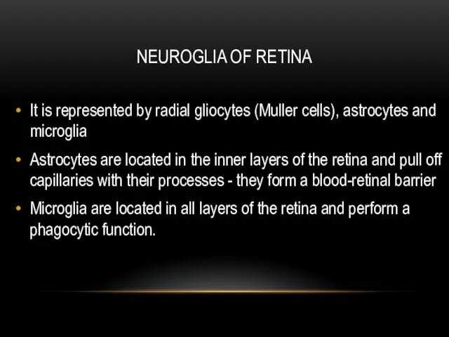
NEUROGLIA OF RETINA
It is represented by radial gliocytes (Muller cells), astrocytes
NEUROGLIA OF RETINA
It is represented by radial gliocytes (Muller cells), astrocytes
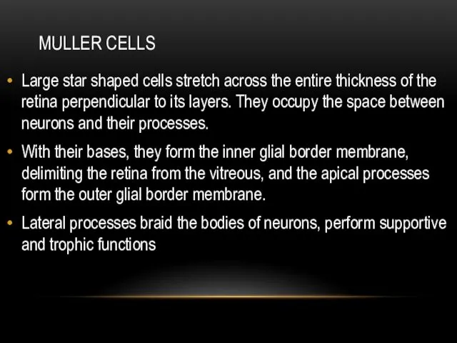
MULLER CELLS
Large star shaped cells stretch across the entire thickness of
MULLER CELLS
Large star shaped cells stretch across the entire thickness of
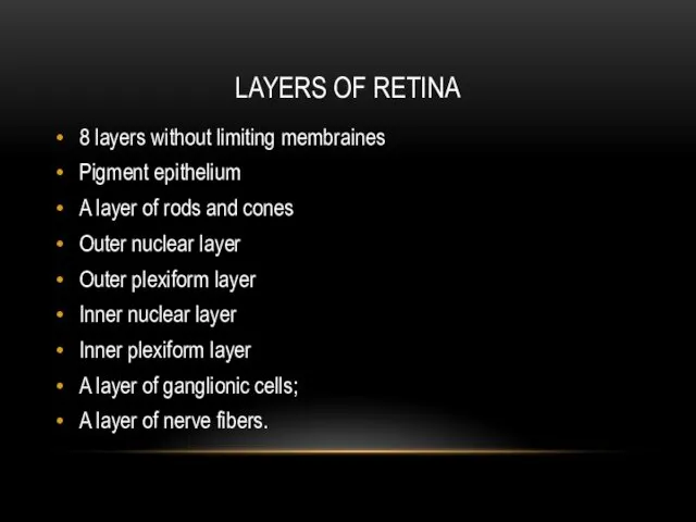
LAYERS OF RETINA
8 layers without limiting membraines
Pigment epithelium
A layer of rods
LAYERS OF RETINA
8 layers without limiting membraines
Pigment epithelium
A layer of rods
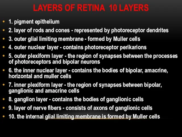
LAYERS OF RETINA 10 LAYERS
1. pigment epithelium
2. layer of rods and
LAYERS OF RETINA 10 LAYERS
1. pigment epithelium
2. layer of rods and
 Каркаралинский государственный национальный природный парк
 Презентация Систематика растений.
 Задания № 28. Соотнесение морфологических признаков организма. Породы лошадей. ОГЭ биология
 Загадки о животных (1 класс)
 блок_3_Ботаника спор-семенн
 Обмен белков
 Молекулярно-генетические методы диагностики
 Эволюция микроорганизмов
 Комнатные растения
 Плісеневі гриби
 Обучающая презентация Система живой природы
 Мышечная система
 Итог производственной практики
 Моногибридное скрещивание. Задача
 Дыхательная система человека
 Наследственность. Генетика человека. Часть 1
 Продолговатый мозг. Черепно-мозговые нервы (IX - XII)
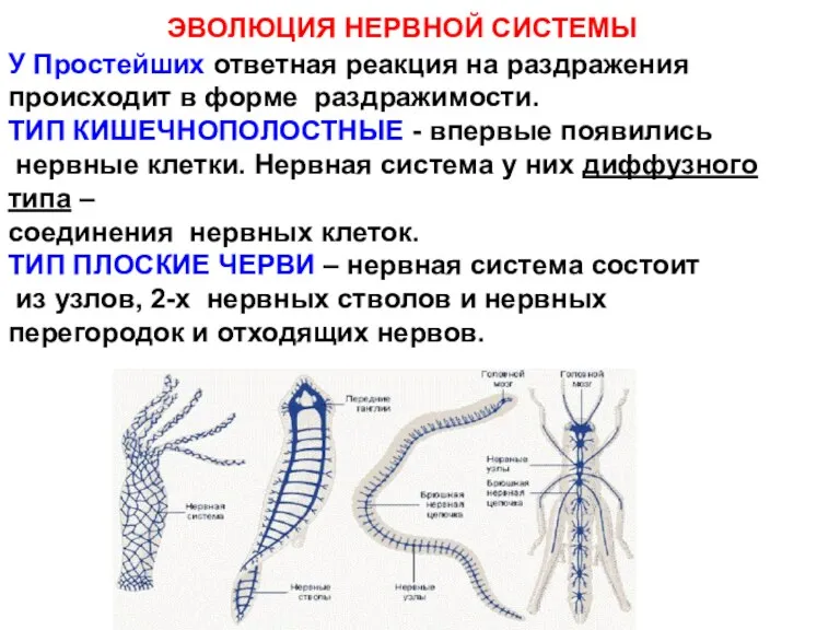 Эволюция нервной системы
 Что такое хвоинки (для дошкольников)
 Насекомые. Отгадай загадки
 Орхидеи. Классификация
 Уровни организацииживой материи
 Тип Кольчатые черви
 Пресмыкающиеся
 Розв`язування елементарних вправ з реплікації, транскрипції, трансляції. Практична робота №2
 водоросли 7 класс Диск
 Анатомия и физиология женской половой системы. Лекция № 46
 Презентация к уроку биологии в 8 классе транспортные системы организма