- General Musculoskeletal Screening: Upper Extremities

Содержание
- 2. 8/27/02 Gregory Crovetti, M.D. General Approach History Inspection Range of Motion (ROM) Palpation Muscular and neurological
- 3. 8/27/02 Gregory Crovetti, M.D. History An accurate history is essential Will give you diagnosis 80-90% of
- 4. 8/27/02 Gregory Crovetti, M.D. General Inspection Observe how the patient moves as they go into the
- 5. 8/27/02 Gregory Crovetti, M.D. Inspection of Specific Area Look for asymmetry between sides Swelling Deformities Atrophy
- 6. 8/27/02 Gregory Crovetti, M.D. Range of Motion (Active) Have patient range the joints Watch for decreased
- 7. 8/27/02 Gregory Crovetti, M.D. Range of Motion (Passive) Next range the joints passively, comparing the end
- 8. 8/27/02 Gregory Crovetti, M.D. Palpation When palpating a structure, you need to know the anatomy of
- 9. 8/27/02 Gregory Crovetti, M.D. Muscular and Neurological Check the following comparing one side to the other:
- 10. 8/27/02 Gregory Crovetti, M.D. Generalized Screening Exam If any abnormalities, a more thorough exam of the
- 11. 8/27/02 Gregory Crovetti, M.D. Neck: Active Range of Motion Chin to chest (flexion) “look at ceiling”
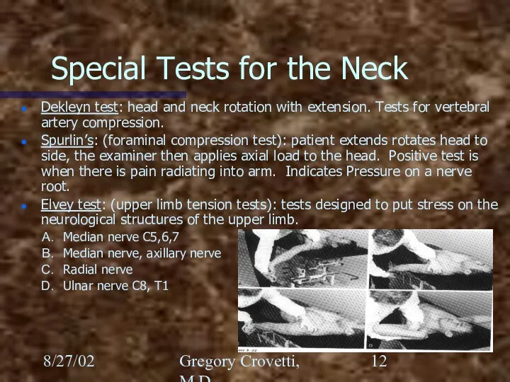
- 12. 8/27/02 Gregory Crovetti, M.D. Special Tests for the Neck Dekleyn test: head and neck rotation with
- 13. 8/27/02 Gregory Crovetti, M.D. Shoulder Exam Inspection Palpation Passive Range of Motion Active Range of Motion
- 14. 8/27/02 Gregory Crovetti, M.D. The Shoulder Joints of the shoulder Glenohumeral Sternoclavicular Acromioclavicular Scapular thoracic (not
- 15. 8/27/02 Gregory Crovetti, M.D. Glenohumeral Joint
- 16. 8/27/02 Gregory Crovetti, M.D. Glenohumeral Ligaments Folds in the anterior capsule produce the superior, middle and
- 17. 8/27/02 Gregory Crovetti, M.D. Glenoid Labrum Glenoid labrum: a fibrocartilaginous rim to increase the contact area
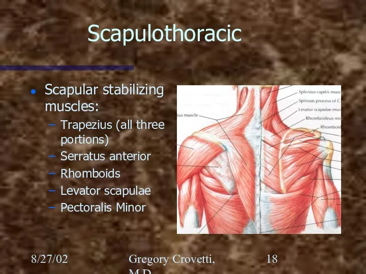
- 18. 8/27/02 Gregory Crovetti, M.D. Scapulothoracic Scapular stabilizing muscles: Trapezius (all three portions) Serratus anterior Rhomboids Levator
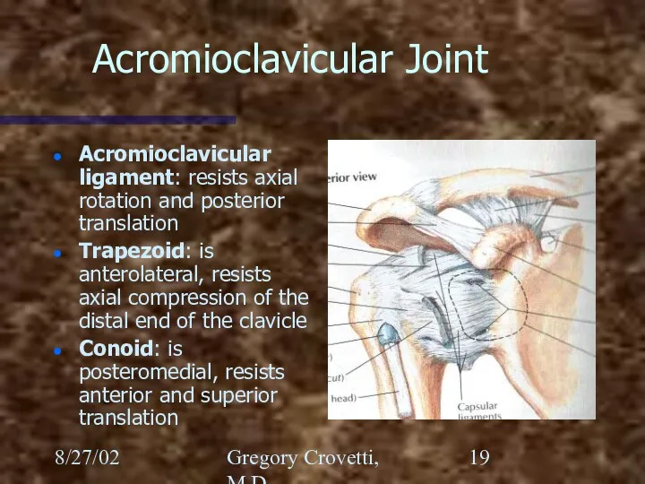
- 19. 8/27/02 Gregory Crovetti, M.D. Acromioclavicular Joint Acromioclavicular ligament: resists axial rotation and posterior translation Trapezoid: is
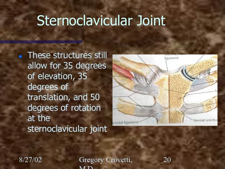
- 20. 8/27/02 Gregory Crovetti, M.D. Sternoclavicular Joint These structures still allow for 35 degrees of elevation, 35
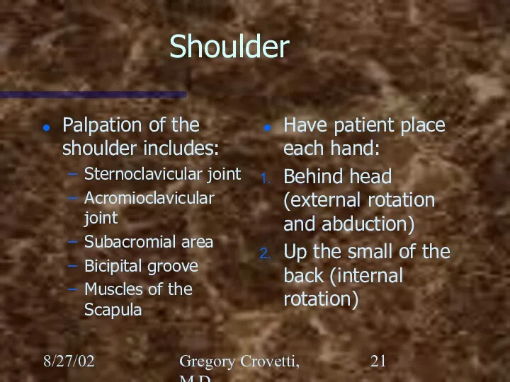
- 21. 8/27/02 Gregory Crovetti, M.D. Shoulder Palpation of the shoulder includes: Sternoclavicular joint Acromioclavicular joint Subacromial area
- 22. 8/27/02 Gregory Crovetti, M.D. Shoulder Rotator cuff: Supraspinatus Infraspinatus Teres Minor Subscapularis
- 23. 8/27/02 Gregory Crovetti, M.D.
- 24. 8/27/02 Gregory Crovetti, M.D. Special Tests for the Shoulder Apprehension (crank) test: The arm is abducted
- 25. 8/27/02 Gregory Crovetti, M.D. Special Tests for the Shoulder Feagin test: arm abducted to 90 elbow
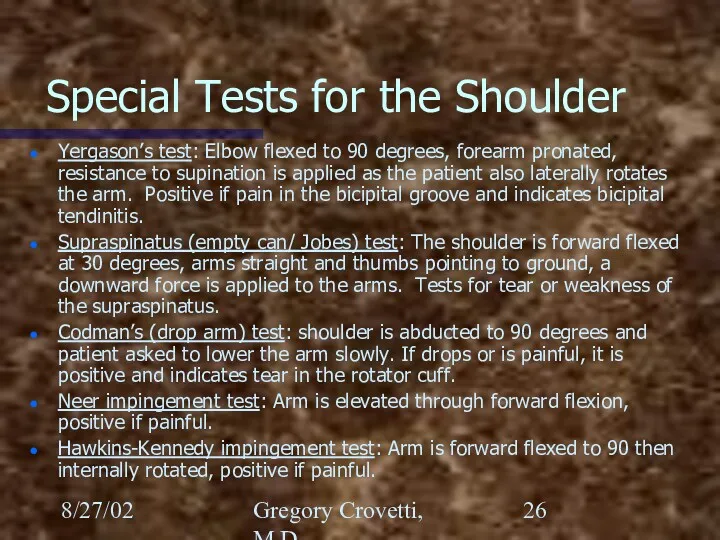
- 26. 8/27/02 Gregory Crovetti, M.D. Special Tests for the Shoulder Yergason’s test: Elbow flexed to 90 degrees,
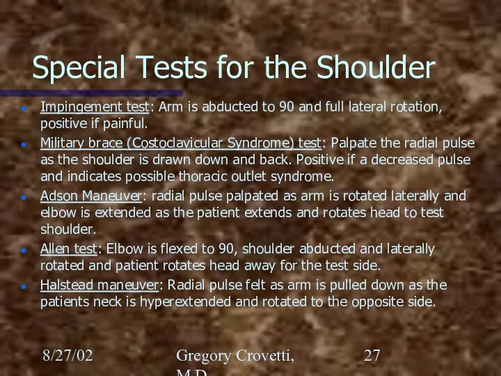
- 27. 8/27/02 Gregory Crovetti, M.D. Special Tests for the Shoulder Impingement test: Arm is abducted to 90
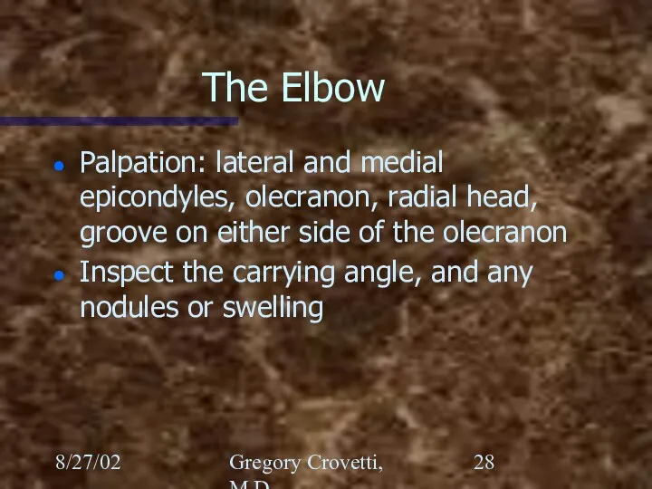
- 28. 8/27/02 Gregory Crovetti, M.D. The Elbow Palpation: lateral and medial epicondyles, olecranon, radial head, groove on
- 29. 8/27/02 Gregory Crovetti, M.D.
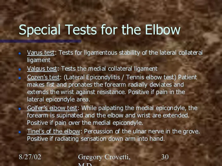
- 30. 8/27/02 Gregory Crovetti, M.D. Special Tests for the Elbow Varus test: Tests for ligamentous stability of
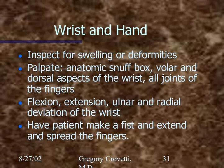
- 31. 8/27/02 Gregory Crovetti, M.D. Wrist and Hand Inspect for swelling or deformities Palpate: anatomic snuff box,
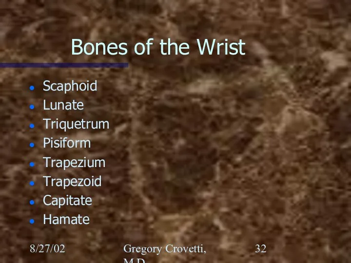
- 32. 8/27/02 Gregory Crovetti, M.D. Bones of the Wrist Scaphoid Lunate Triquetrum Pisiform Trapezium Trapezoid Capitate Hamate
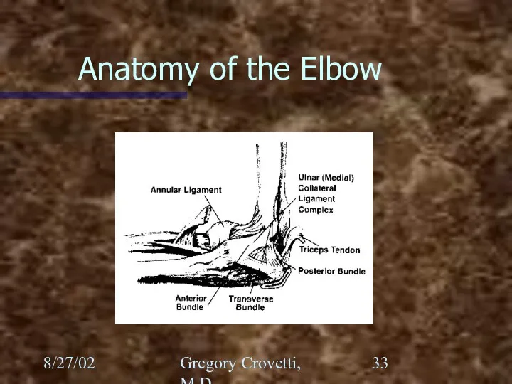
- 33. 8/27/02 Gregory Crovetti, M.D. Anatomy of the Elbow
- 34. 8/27/02 Gregory Crovetti, M.D. Nerves of the Hand Ulnar Radial Median Palmar branch of the median
- 35. 8/27/02 Gregory Crovetti, M.D.
- 36. 8/27/02 Gregory Crovetti, M.D.
- 37. 8/27/02 Gregory Crovetti, M.D.
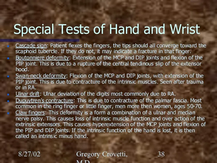
- 38. 8/27/02 Gregory Crovetti, M.D. Special Tests of Hand and Wrist Cascade sign: Patient flexes the fingers,
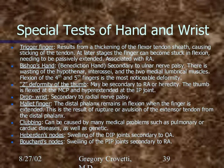
- 39. 8/27/02 Gregory Crovetti, M.D. Special Tests of Hand and Wrist Trigger finger: Results from a thickening
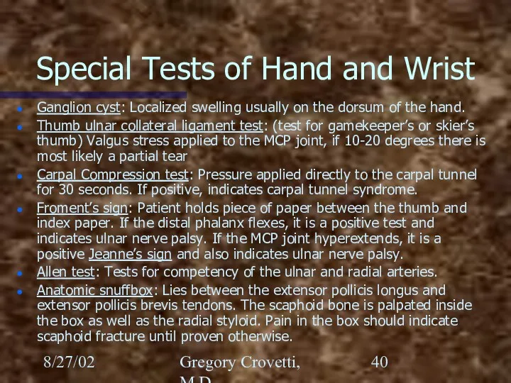
- 40. 8/27/02 Gregory Crovetti, M.D. Special Tests of Hand and Wrist Ganglion cyst: Localized swelling usually on
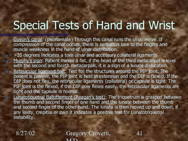
- 41. 8/27/02 Gregory Crovetti, M.D. Special Tests of Hand and Wrist Guyon’s canal: (pisohamate) Through this canal
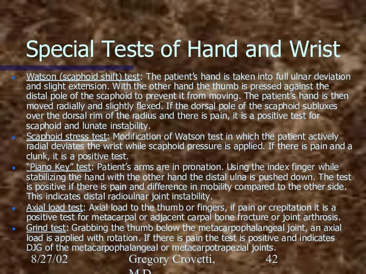
- 42. 8/27/02 Gregory Crovetti, M.D. Special Tests of Hand and Wrist Watson (scaphoid shift) test: The patient’s
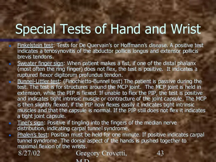
- 43. 8/27/02 Gregory Crovetti, M.D. Special Tests of Hand and Wrist Finkelstein test: Tests for De Quervain’s
- 45. Скачать презентацию
8/27/02
Gregory Crovetti, M.D.
General Approach
History
Inspection
Range of Motion (ROM)
Palpation
Muscular and neurological exams
8/27/02
Gregory Crovetti, M.D.
General Approach
History
Inspection
Range of Motion (ROM)
Palpation
Muscular and neurological exams
8/27/02
Gregory Crovetti, M.D.
History
An accurate history is essential
Will give you diagnosis 80-90%
8/27/02
Gregory Crovetti, M.D.
History
An accurate history is essential
Will give you diagnosis 80-90%
8/27/02
Gregory Crovetti, M.D.
General Inspection
Observe how the patient moves as they go
8/27/02
Gregory Crovetti, M.D.
General Inspection
Observe how the patient moves as they go
8/27/02
Gregory Crovetti, M.D.
Inspection of Specific Area
Look for asymmetry between sides
Swelling
Deformities
Atrophy
Erythema
8/27/02
Gregory Crovetti, M.D.
Inspection of Specific Area
Look for asymmetry between sides
Swelling
Deformities
Atrophy
Erythema
8/27/02
Gregory Crovetti, M.D.
Range of Motion (Active)
Have patient range the joints
Watch for
8/27/02
Gregory Crovetti, M.D.
Range of Motion (Active)
Have patient range the joints
Watch for
8/27/02
Gregory Crovetti, M.D.
Range of Motion (Passive)
Next range the joints passively, comparing
8/27/02
Gregory Crovetti, M.D.
Range of Motion (Passive)
Next range the joints passively, comparing
8/27/02
Gregory Crovetti, M.D.
Palpation
When palpating a structure, you need to know the
8/27/02
Gregory Crovetti, M.D.
Palpation
When palpating a structure, you need to know the
8/27/02
Gregory Crovetti, M.D.
Muscular and Neurological
Check the following comparing one side to
8/27/02
Gregory Crovetti, M.D.
Muscular and Neurological
Check the following comparing one side to
8/27/02
Gregory Crovetti, M.D.
Generalized Screening Exam
If any abnormalities, a more thorough exam
8/27/02
Gregory Crovetti, M.D.
Generalized Screening Exam
If any abnormalities, a more thorough exam
8/27/02
Gregory Crovetti, M.D.
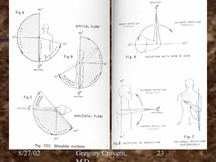
Neck: Active Range of Motion
Chin to chest (flexion)
“look at
8/27/02
Gregory Crovetti, M.D.
Neck: Active Range of Motion
Chin to chest (flexion)
“look at
8/27/02
Gregory Crovetti, M.D.
Special Tests for the Neck
Dekleyn test: head and neck
8/27/02
Gregory Crovetti, M.D.
Special Tests for the Neck
Dekleyn test: head and neck
8/27/02
Gregory Crovetti, M.D.
Shoulder Exam
Inspection
Palpation
Passive Range of Motion
Active Range of Motion
Appley scratch
8/27/02
Gregory Crovetti, M.D.
Shoulder Exam
Inspection
Palpation
Passive Range of Motion
Active Range of Motion
Appley scratch
8/27/02
Gregory Crovetti, M.D.
The Shoulder
Joints of the shoulder
Glenohumeral
Sternoclavicular
Acromioclavicular
Scapular thoracic (not a true
8/27/02
Gregory Crovetti, M.D.
The Shoulder
Joints of the shoulder
Glenohumeral
Sternoclavicular
Acromioclavicular
Scapular thoracic (not a true
8/27/02
Gregory Crovetti, M.D.
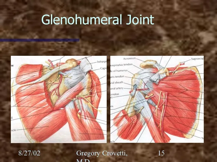
Glenohumeral Joint
8/27/02
Gregory Crovetti, M.D.
Glenohumeral Joint
8/27/02
Gregory Crovetti, M.D.
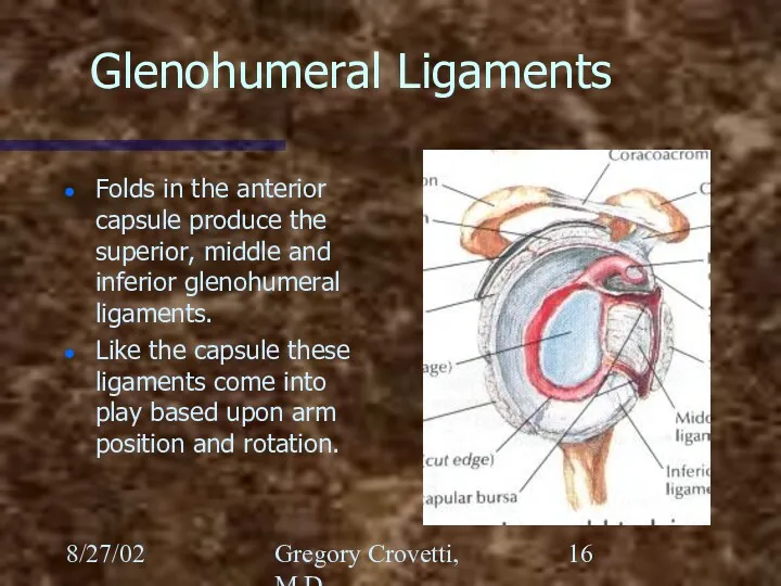
Glenohumeral Ligaments
Folds in the anterior capsule produce the superior,
8/27/02
Gregory Crovetti, M.D.
Glenohumeral Ligaments
Folds in the anterior capsule produce the superior,
8/27/02
Gregory Crovetti, M.D.
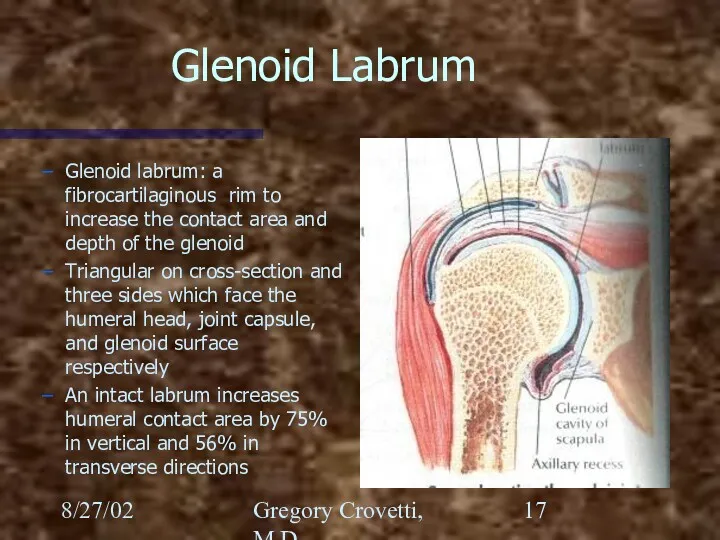
Glenoid Labrum
Glenoid labrum: a fibrocartilaginous rim to increase the
8/27/02
Gregory Crovetti, M.D.
Glenoid Labrum
Glenoid labrum: a fibrocartilaginous rim to increase the
8/27/02
Gregory Crovetti, M.D.
Scapulothoracic
Scapular stabilizing muscles:
Trapezius (all three portions)
Serratus anterior
Rhomboids
Levator scapulae
Pectoralis Minor
8/27/02
Gregory Crovetti, M.D.
Scapulothoracic
Scapular stabilizing muscles:
Trapezius (all three portions)
Serratus anterior
Rhomboids
Levator scapulae
Pectoralis Minor
8/27/02
Gregory Crovetti, M.D.
Acromioclavicular Joint
Acromioclavicular ligament: resists axial rotation and posterior translation
Trapezoid:
8/27/02
Gregory Crovetti, M.D.
Acromioclavicular Joint
Acromioclavicular ligament: resists axial rotation and posterior translation
Trapezoid:
8/27/02
Gregory Crovetti, M.D.
Sternoclavicular Joint
These structures still allow for 35 degrees of
8/27/02
Gregory Crovetti, M.D.
Sternoclavicular Joint
These structures still allow for 35 degrees of
8/27/02
Gregory Crovetti, M.D.
Shoulder
Palpation of the shoulder includes:
Sternoclavicular joint
Acromioclavicular joint
Subacromial area
Bicipital
8/27/02
Gregory Crovetti, M.D.
Shoulder
Palpation of the shoulder includes:
Sternoclavicular joint
Acromioclavicular joint
Subacromial area
Bicipital
8/27/02
Gregory Crovetti, M.D.
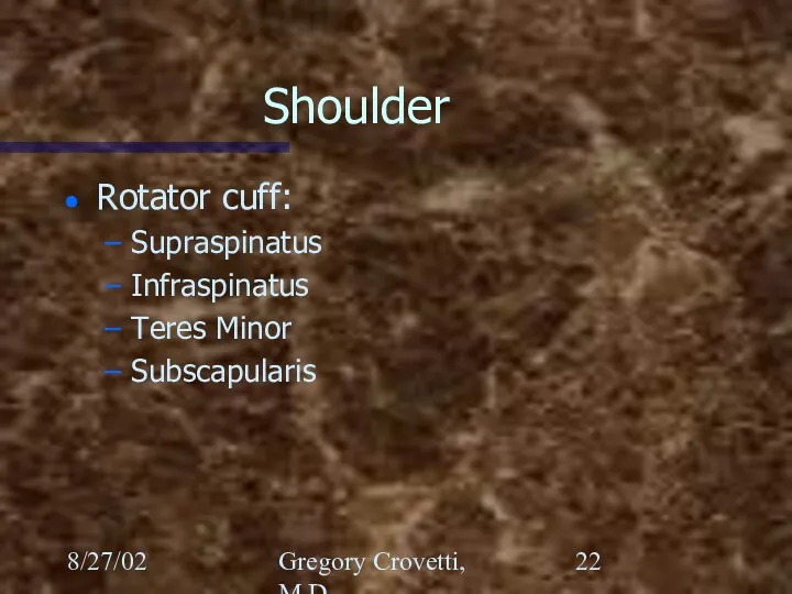
Shoulder
Rotator cuff:
Supraspinatus
Infraspinatus
Teres Minor
Subscapularis
8/27/02
Gregory Crovetti, M.D.
Shoulder
Rotator cuff:
Supraspinatus
Infraspinatus
Teres Minor
Subscapularis
8/27/02
Gregory Crovetti, M.D.
8/27/02
Gregory Crovetti, M.D.
8/27/02
Gregory Crovetti, M.D.
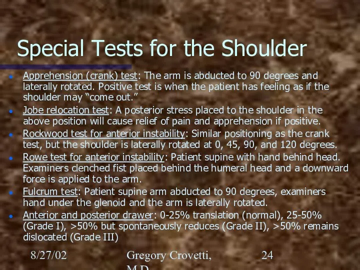
Special Tests for the Shoulder
Apprehension (crank) test: The arm
8/27/02
Gregory Crovetti, M.D.
Special Tests for the Shoulder
Apprehension (crank) test: The arm
8/27/02
Gregory Crovetti, M.D.
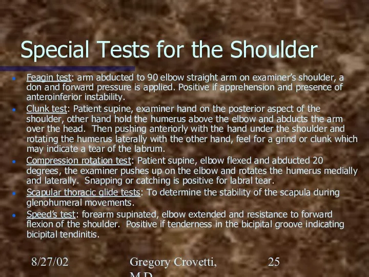
Special Tests for the Shoulder
Feagin test: arm abducted to
8/27/02
Gregory Crovetti, M.D.
Special Tests for the Shoulder
Feagin test: arm abducted to
8/27/02
Gregory Crovetti, M.D.
Special Tests for the Shoulder
Yergason’s test: Elbow flexed to
8/27/02
Gregory Crovetti, M.D.
Special Tests for the Shoulder
Yergason’s test: Elbow flexed to
8/27/02
Gregory Crovetti, M.D.
Special Tests for the Shoulder
Impingement test: Arm is abducted
8/27/02
Gregory Crovetti, M.D.
Special Tests for the Shoulder
Impingement test: Arm is abducted
8/27/02
Gregory Crovetti, M.D.
The Elbow
Palpation: lateral and medial epicondyles, olecranon, radial head,
8/27/02
Gregory Crovetti, M.D.
The Elbow
Palpation: lateral and medial epicondyles, olecranon, radial head,
8/27/02
Gregory Crovetti, M.D.
8/27/02
Gregory Crovetti, M.D.
8/27/02
Gregory Crovetti, M.D.
Special Tests for the Elbow
Varus test: Tests for ligamentous
8/27/02
Gregory Crovetti, M.D.
Special Tests for the Elbow
Varus test: Tests for ligamentous
8/27/02
Gregory Crovetti, M.D.
Wrist and Hand
Inspect for swelling or deformities
Palpate: anatomic snuff
8/27/02
Gregory Crovetti, M.D.
Wrist and Hand
Inspect for swelling or deformities
Palpate: anatomic snuff
8/27/02
Gregory Crovetti, M.D.
Bones of the Wrist
Scaphoid
Lunate
Triquetrum
Pisiform
Trapezium
Trapezoid
Capitate
Hamate
8/27/02
Gregory Crovetti, M.D.
Bones of the Wrist
Scaphoid
Lunate
Triquetrum
Pisiform
Trapezium
Trapezoid
Capitate
Hamate
8/27/02
Gregory Crovetti, M.D.
Anatomy of the Elbow
8/27/02
Gregory Crovetti, M.D.
Anatomy of the Elbow
8/27/02
Gregory Crovetti, M.D.
Nerves of the Hand
Ulnar
Radial
Median
Palmar branch of the median
8/27/02
Gregory Crovetti, M.D.
Nerves of the Hand
Ulnar
Radial
Median
Palmar branch of the median
8/27/02
Gregory Crovetti, M.D.
8/27/02
Gregory Crovetti, M.D.
8/27/02
Gregory Crovetti, M.D.
8/27/02
Gregory Crovetti, M.D.
8/27/02
Gregory Crovetti, M.D.
8/27/02
Gregory Crovetti, M.D.
8/27/02
Gregory Crovetti, M.D.
Special Tests of Hand and Wrist
Cascade sign: Patient flexes
8/27/02
Gregory Crovetti, M.D.
Special Tests of Hand and Wrist
Cascade sign: Patient flexes
8/27/02
Gregory Crovetti, M.D.
Special Tests of Hand and Wrist
Trigger finger: Results from
8/27/02
Gregory Crovetti, M.D.
Special Tests of Hand and Wrist
Trigger finger: Results from
8/27/02
Gregory Crovetti, M.D.
Special Tests of Hand and Wrist
Ganglion cyst: Localized swelling
8/27/02
Gregory Crovetti, M.D.
Special Tests of Hand and Wrist
Ganglion cyst: Localized swelling
8/27/02
Gregory Crovetti, M.D.
Special Tests of Hand and Wrist
Guyon’s canal: (pisohamate) Through
8/27/02
Gregory Crovetti, M.D.
Special Tests of Hand and Wrist
Guyon’s canal: (pisohamate) Through
8/27/02
Gregory Crovetti, M.D.
Special Tests of Hand and Wrist
Watson (scaphoid shift) test:
8/27/02
Gregory Crovetti, M.D.
Special Tests of Hand and Wrist
Watson (scaphoid shift) test:
8/27/02
Gregory Crovetti, M.D.
Special Tests of Hand and Wrist
Finkelstein test: Tests for
8/27/02
Gregory Crovetti, M.D.
Special Tests of Hand and Wrist
Finkelstein test: Tests for
 Класс Птицы
 Виноград. Види винограда
 Урок по биологии в 5 классе Жизнь в мировом океане
 Мутационная изменчивость. Закон гомологических рядов
 Влияние человека на экосистемы
 Животные Австралии
 Органическое вещество почв. (Лекция 8)
 Исследовательская работа
 Взаимосвязи компонентов природы. Природный комплекс. (проект Урок в Москве)
 Местообитание и экологические ниши
 Учение об эволюции
 Основные этапы развития генетики
 Презентация Человек как житель биосферы
 Строение и функции головного мозга
 Хризантема овощная
 Группа праголосеменные растения
 тема Водоросли
 Клетка – элементарная единица жизни на земле
 Мал өсіру әдістері
 Tissue system in plants
 Як змінюється восени життя птахів. Перелітні та осілі птахи
 Строение нейрона. Синапс
 Творческое объединение Друзья природы
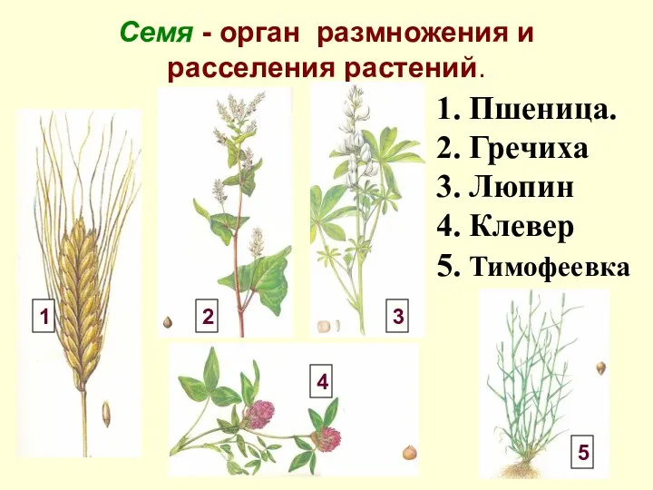 Семя - орган размножения и расселения растений
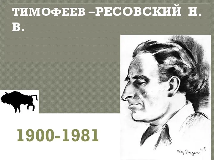 Тимофеев-Ресовский Н.В (1900-1981)
 Значение комнатных растений в жизни человека
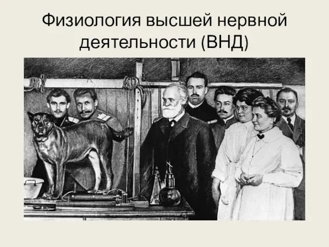 Физиология высшей нервной деятельности
 Выделительная система