- Human circulatory system

Содержание
- 2. The human circulatory system functions like a network of highways. It transports materials around the body.
- 3. The Human Circulatory System It consists of: HEART BLOOD VESSELS BLOOD
- 4. The Heart
- 5. HEART FACTS: About 250-340 grams, In your life time, pumps about 300 million liter of blood,
- 6. Main structure of the heart The heart is made of a special type of muscle called
- 7. External Structure
- 8. Internal Structure
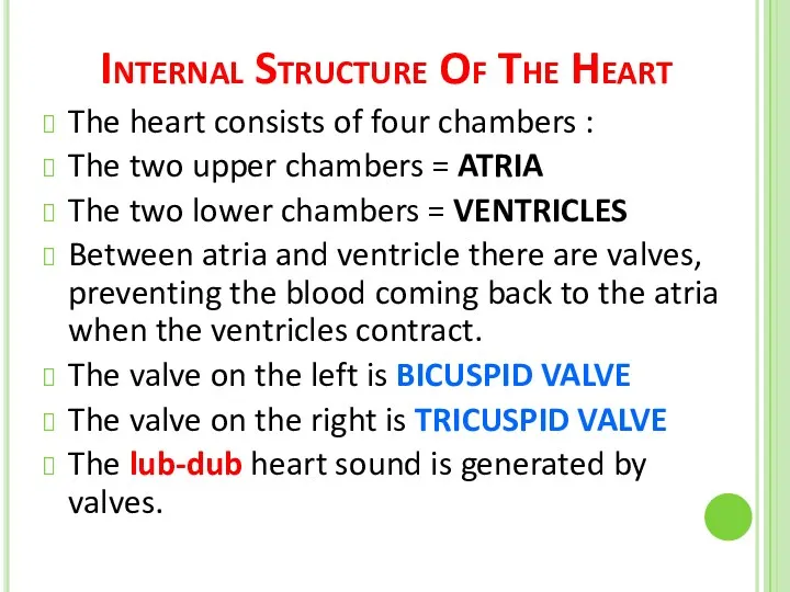
- 9. Internal Structure Of The Heart The heart consists of four chambers : The two upper chambers
- 11. VALVES
- 12. Semilunar Valves Semilunar valves are found between the arteries and the ventricles. They prevent the blood
- 13. VALVES
- 15. C A R D I A C C Y C L E
- 16. The heart pumps blood into the body. Relaxation of heart is known as diastole. Contraction of
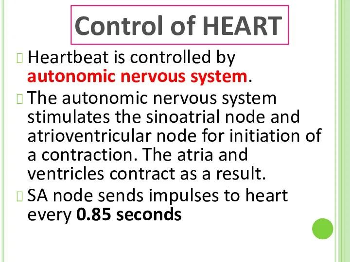
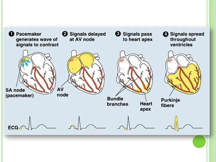
- 20. Heartbeat is controlled by autonomic nervous system. The autonomic nervous system stimulates the sinoatrial node and
- 23. Heart Rate Parasympathetic nerves reduces the heart rate. Sympathetic nervs speed up the heart rate. Acetylcholine
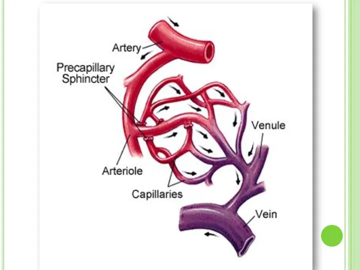
- 24. BLOOD VESSELS There are 3 types of vessels in our body. These are; ARTERIES VEINS CAPILLARIES
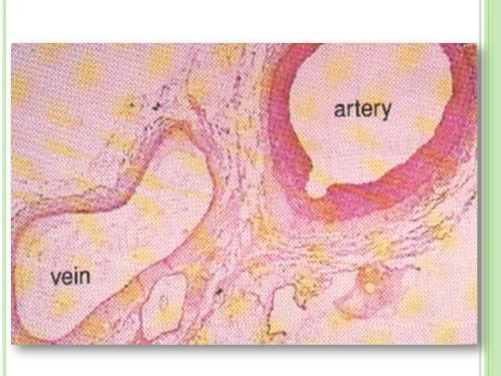
- 27. 1. Arteries Arteries carry blood away from heart to the different tissues of the body. Artery
- 29. 2. Veins Veins carry blood to heart Their walls are much thinner than the walls of
- 32. 3. Capillaries Capillary walls are only one cell thick. Gas and nutrient molecules pass easily through
- 35. artery vein arteriole venule capillary
- 37. Blood Pressure Blood exerts pressure on the walls of vessels during circulation Blood pressure increases when
- 38. Measuring Blood Pressure
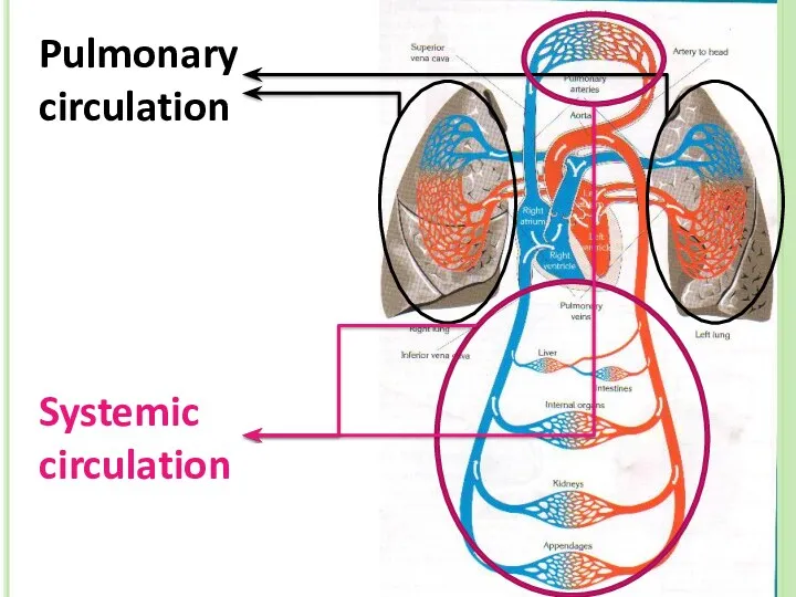
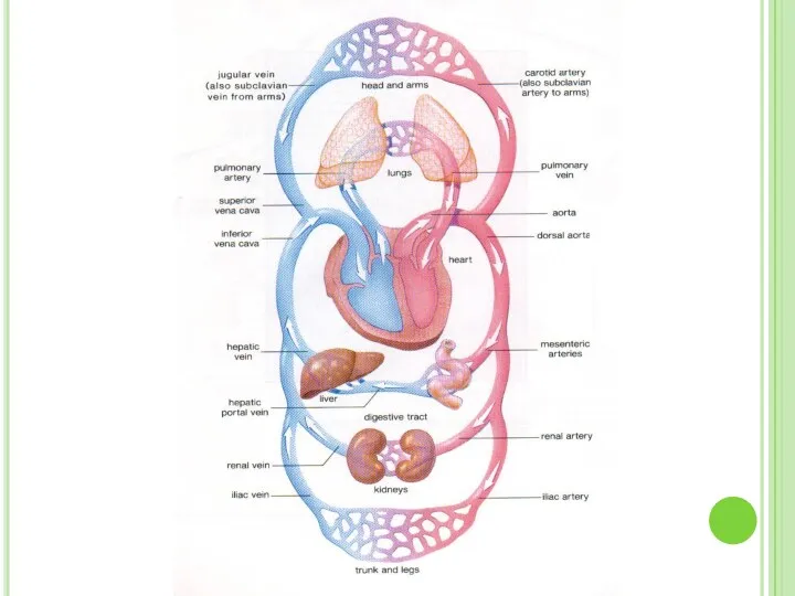
- 41. Blood Circulation There two types of circulation in human body: 1. Pulmonary Circulation: Oxygen poor blood
- 42. Pulmonary circulation Systemic circulation
- 44. Blood Movement Left ventricle pumps oxygenated blood to body, that’s why it’s walls are thicker Right
- 46. Internal Structure
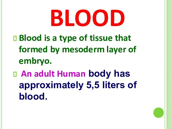
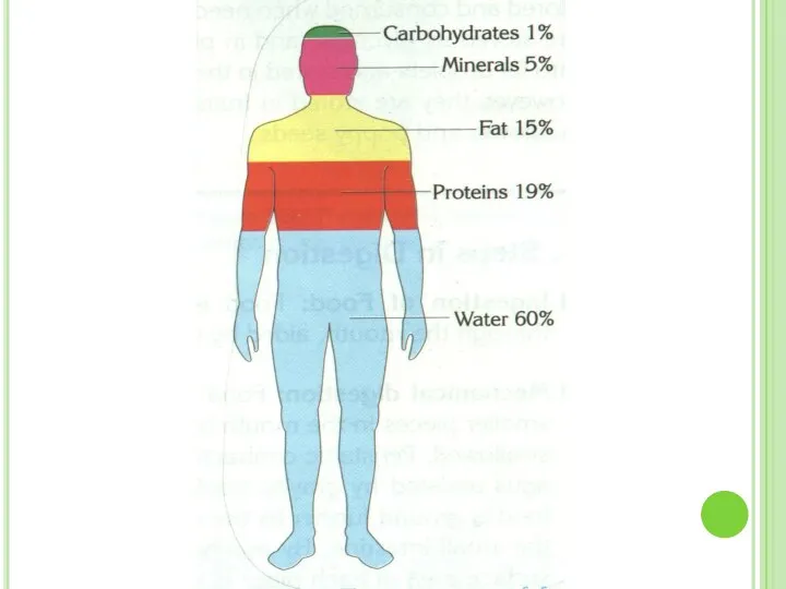
- 47. BLOOD Blood is a type of tissue that formed by mesoderm layer of embryo. An adult
- 48. FUNCTIONS OF BLOOD Transport of materials Hormone transport Homeostasis Immune response Blood Clotting
- 49. BLOOD COMPONENTS Blood contain 2 main parts. These are: Blood Plasma Blood cells
- 50. Blood Plasma Plasma is liquid part of blood. It includes water (90%) and dissolved proteins. It
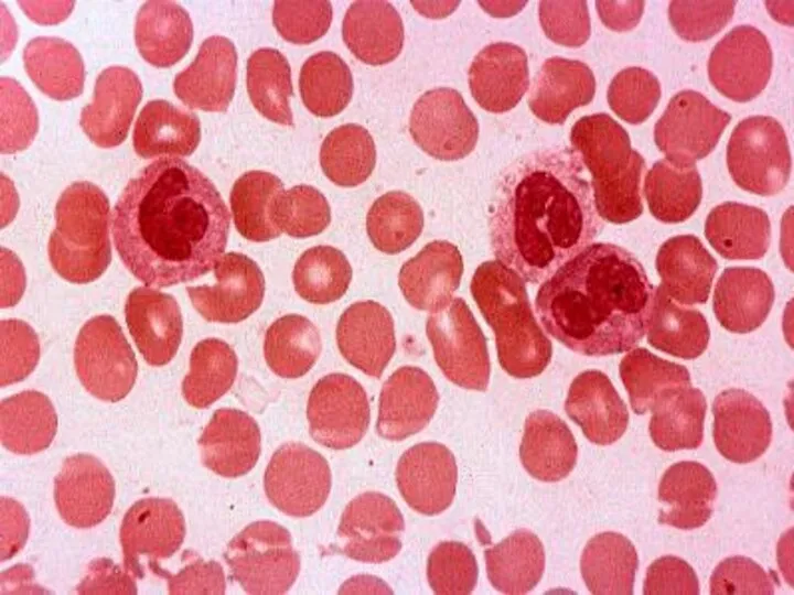
- 52. Blood Cells There are three types of blood cells: Erythrocytes (=Red Blood Cells) Leucocytes (=White Blood
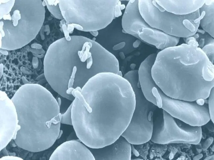
- 54. There are approximately 5 to 5,5 million of erythrocytes per cubic millimeter of blood. The major
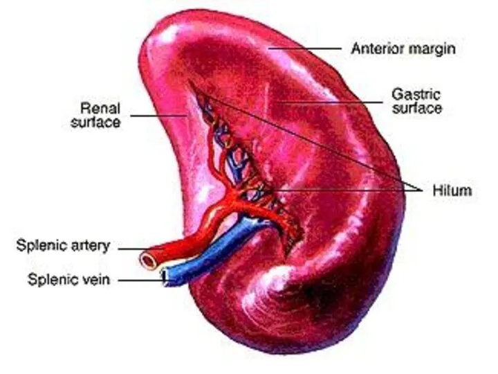
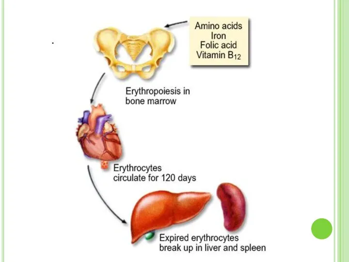
- 55. Mammalian erythrocytes have no nucleus at adult (maturation) stage. They are produced by red bone marrow.
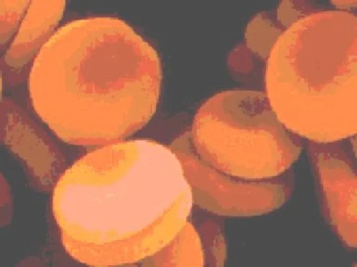
- 59. HEMOGLOBIN Erythrocytes are filled with hemoglobin. Hemoglobin is iron (Fe) containing pigment. It gives red color
- 62. LEUCOCYTES Leucocytes protect the body from infections. They are produced by red bone marrow and lymph
- 63. Normally there are only 6000 to 8000 leucocytes per cubic millimeter of blood. When there is
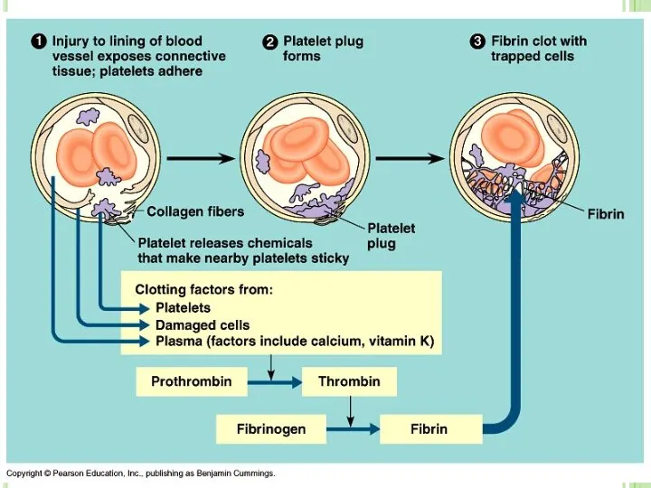
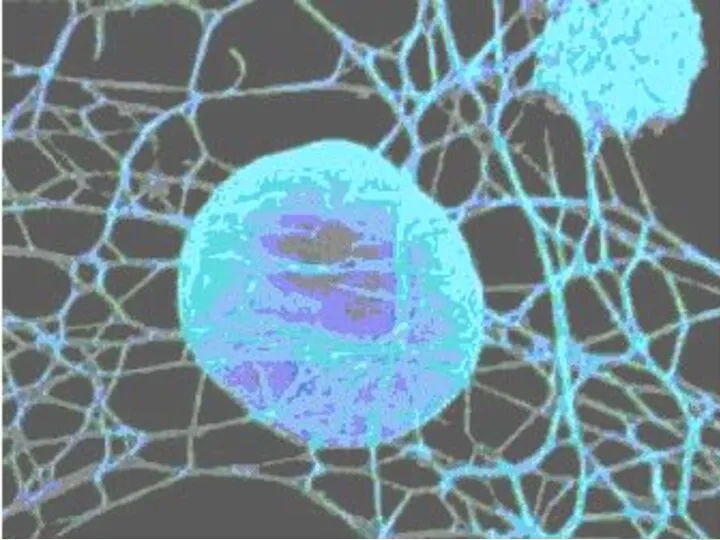
- 66. PLATELETS Platelets are produced by bone marrow. They play major role in blood clotting. Blood clotting
- 67. THE MECHANISM OF BLOOD CLOTTING Prothrombin (In liver) Vitamin K Thrombogen Thrombocytes + O2 Thrombokinase Thrombin
- 70. Diseases related to circulatory system Anemia Leukemia Arteriosclerosis
- 71. Anemia
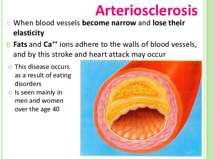
- 72. Arteriosclerosis When blood vessels become narrow and lose their elasticity Fats and Ca++ ions adhere to
- 73. Heart attack, infarcts
- 75. Скачать презентацию
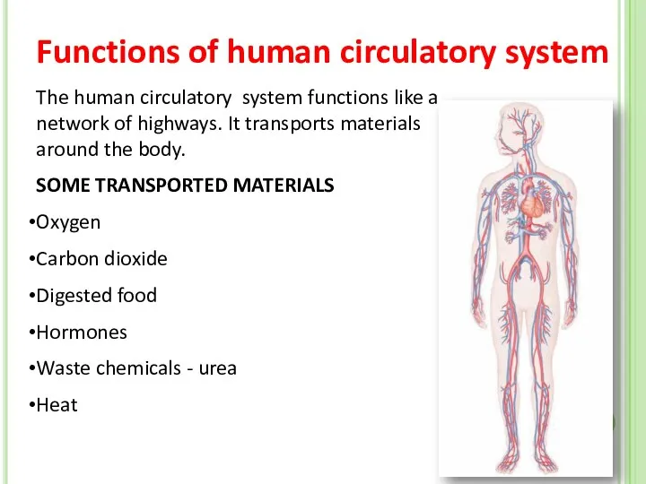
The human circulatory system functions like a network of highways. It
The human circulatory system functions like a network of highways. It
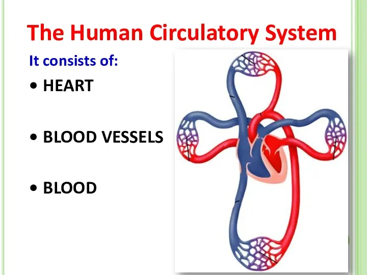
The Human Circulatory System
It consists of:
HEART
BLOOD VESSELS
BLOOD
The Human Circulatory System
It consists of:
HEART
BLOOD VESSELS
BLOOD
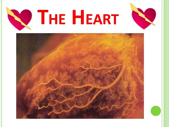
The Heart
The Heart
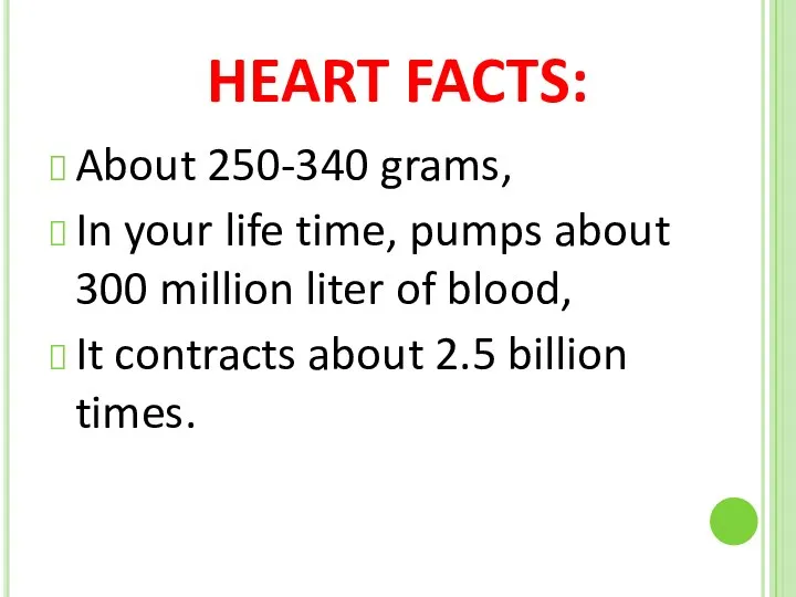
HEART FACTS:
About 250-340 grams,
In your life time, pumps about 300 million
HEART FACTS:
About 250-340 grams,
In your life time, pumps about 300 million
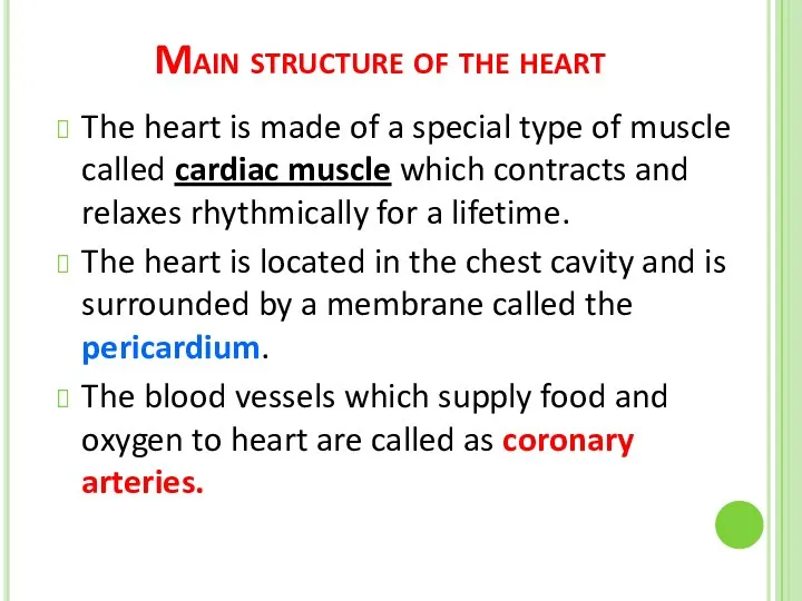
Main structure of the heart
The heart is made of a special
Main structure of the heart
The heart is made of a special
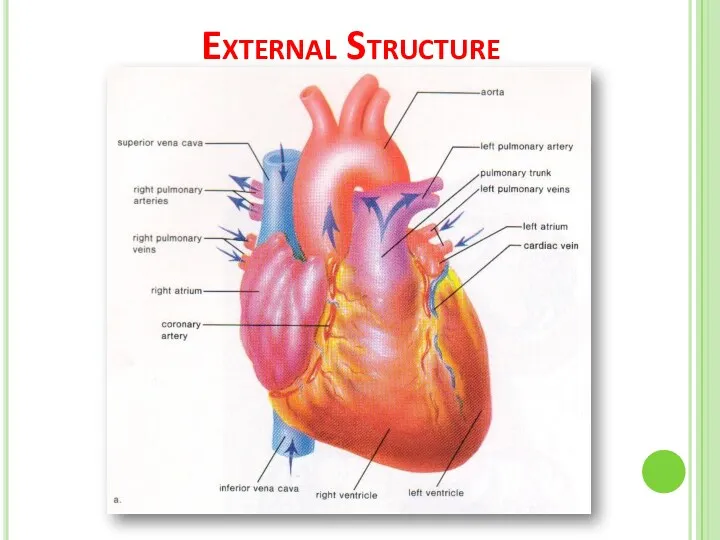
External Structure
External Structure
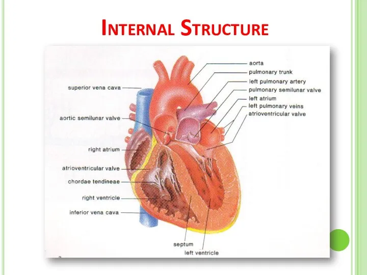
Internal Structure
Internal Structure
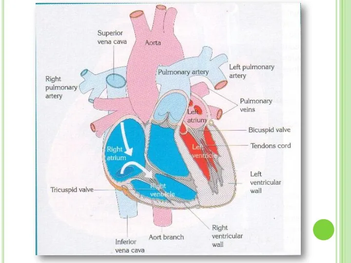
Internal Structure Of The Heart
The heart consists of four chambers :
The
Internal Structure Of The Heart
The heart consists of four chambers :
The

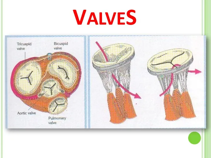
VALVES
VALVES
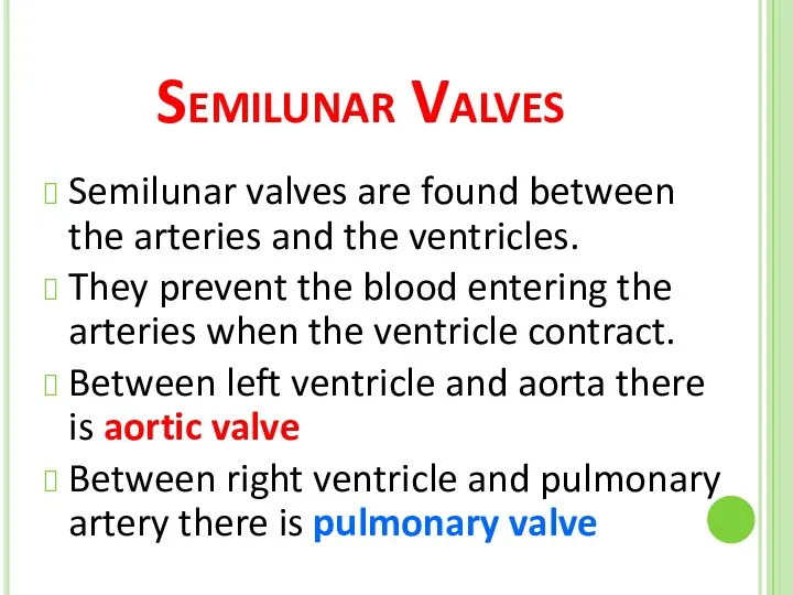
Semilunar Valves
Semilunar valves are found between the arteries and the ventricles.
They
Semilunar Valves
Semilunar valves are found between the arteries and the ventricles.
They
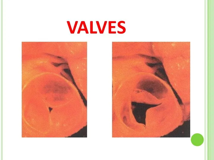
VALVES
VALVES
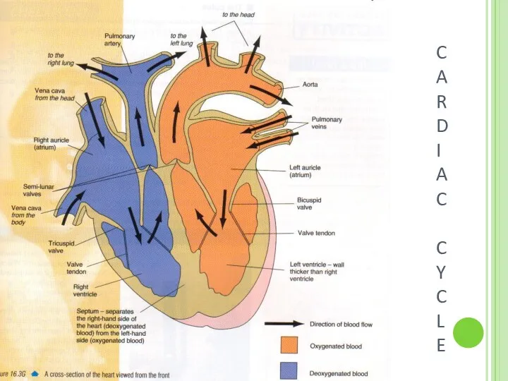
C
A
R
D
I
A
C
C
Y
C
L
E
C
A
R
D
I
A
C
C
Y
C
L
E
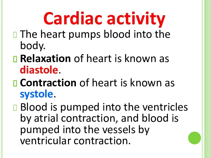
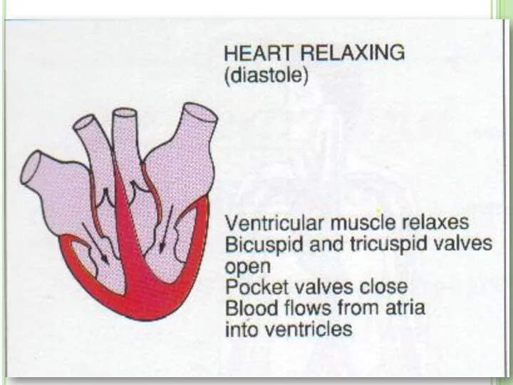
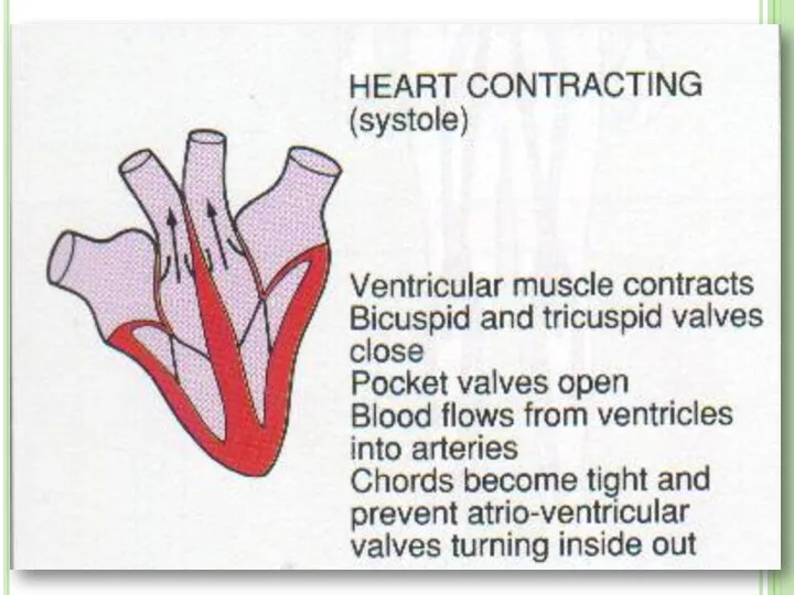
The heart pumps blood into the body.
Relaxation of heart is
The heart pumps blood into the body.
Relaxation of heart is

Heartbeat is controlled by autonomic nervous system.
The autonomic nervous system
Heartbeat is controlled by autonomic nervous system.
The autonomic nervous system

Heart Rate
Parasympathetic nerves reduces the heart rate.
Sympathetic nervs speed up the
Heart Rate
Parasympathetic nerves reduces the heart rate.
Sympathetic nervs speed up the
BLOOD VESSELS
There are 3 types of vessels in our body.
These are;
ARTERIES
VEINS
CAPILLARIES
BLOOD VESSELS
There are 3 types of vessels in our body.
These are;
ARTERIES
VEINS
CAPILLARIES


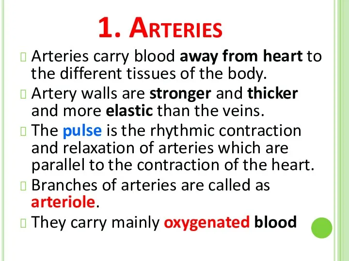
1. Arteries
Arteries carry blood away from heart to the different tissues
1. Arteries
Arteries carry blood away from heart to the different tissues

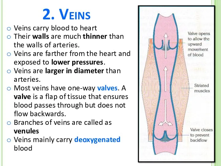
2. Veins
Veins carry blood to heart
Their walls are much thinner than
2. Veins
Veins carry blood to heart
Their walls are much thinner than

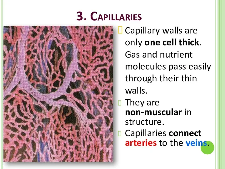
3. Capillaries
Capillary walls are only one cell thick. Gas and nutrient
3. Capillaries
Capillary walls are only one cell thick. Gas and nutrient

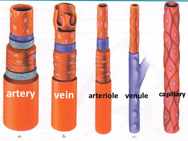
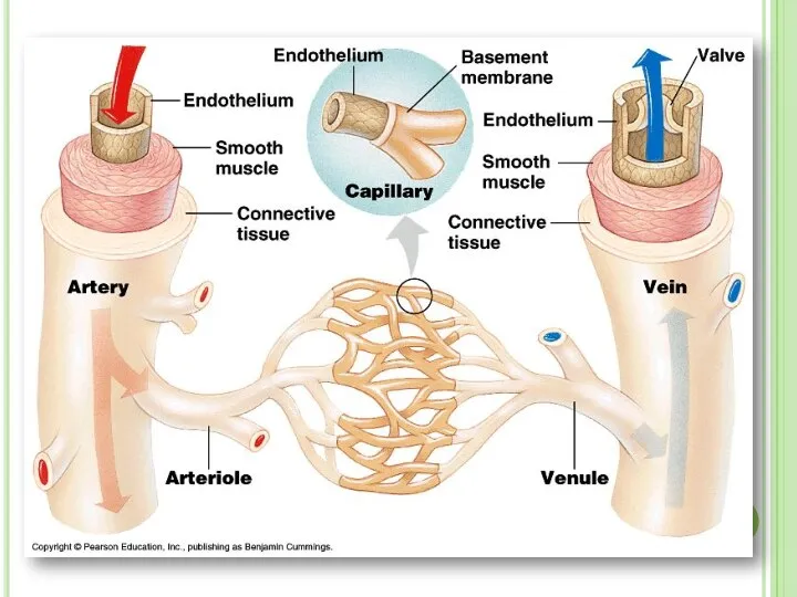
artery
vein
arteriole
venule
capillary
artery
vein
arteriole
venule
capillary
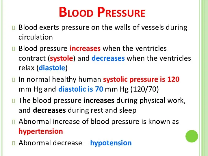
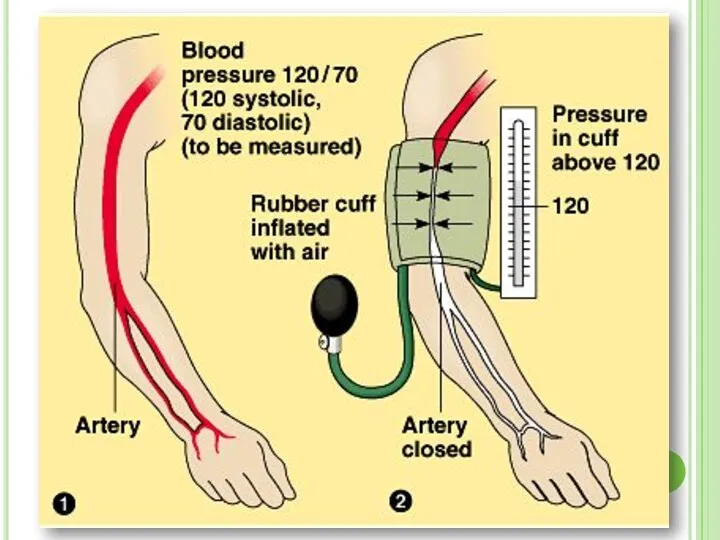
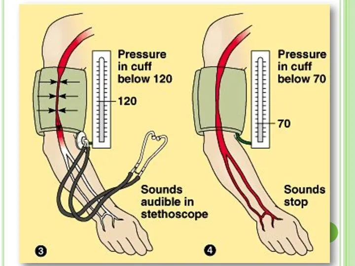
Blood Pressure
Blood exerts pressure on the walls of vessels during circulation
Blood
Blood Pressure
Blood exerts pressure on the walls of vessels during circulation
Blood
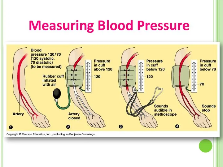
Measuring Blood Pressure
Measuring Blood Pressure
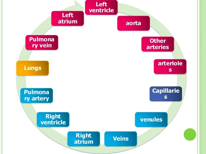
Blood Circulation
There two types of circulation in human body:
1. Pulmonary Circulation:
Blood Circulation
There two types of circulation in human body:
1. Pulmonary Circulation:
Pulmonary
circulation
Systemic
circulation
Pulmonary
circulation
Systemic
circulation
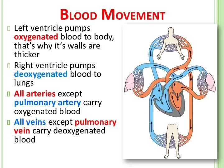
Blood Movement
Left ventricle pumps oxygenated blood to body, that’s why it’s
Blood Movement
Left ventricle pumps oxygenated blood to body, that’s why it’s

Internal Structure
Internal Structure
BLOOD
Blood is a type of tissue that formed by mesoderm layer
BLOOD
Blood is a type of tissue that formed by mesoderm layer
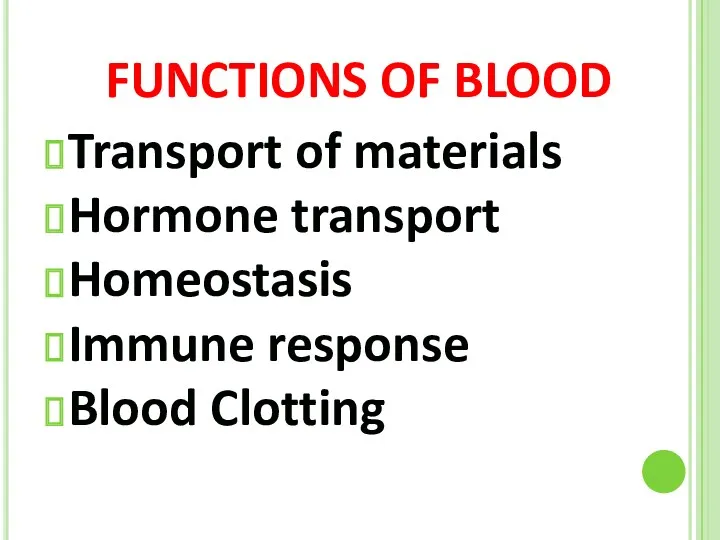
FUNCTIONS OF BLOOD
Transport of materials
Hormone transport
Homeostasis
Immune response
Blood Clotting
FUNCTIONS OF BLOOD
Transport of materials
Hormone transport
Homeostasis
Immune response
Blood Clotting
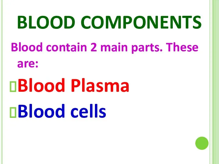
BLOOD COMPONENTS
Blood contain 2 main parts. These are:
Blood Plasma
Blood cells
BLOOD COMPONENTS
Blood contain 2 main parts. These are:
Blood Plasma
Blood cells
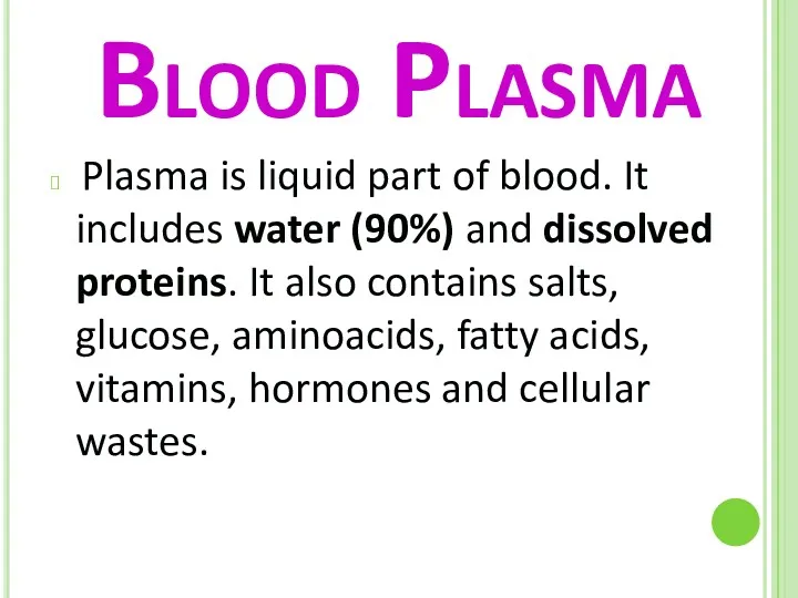
Blood Plasma
Plasma is liquid part of blood. It includes water
Blood Plasma
Plasma is liquid part of blood. It includes water
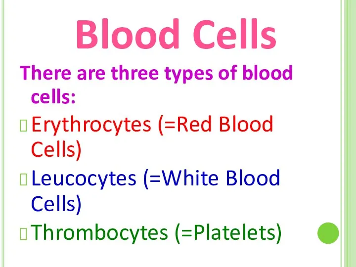
Blood Cells
There are three types of blood cells:
Erythrocytes (=Red Blood Cells)
Leucocytes
Blood Cells
There are three types of blood cells:
Erythrocytes (=Red Blood Cells)
Leucocytes
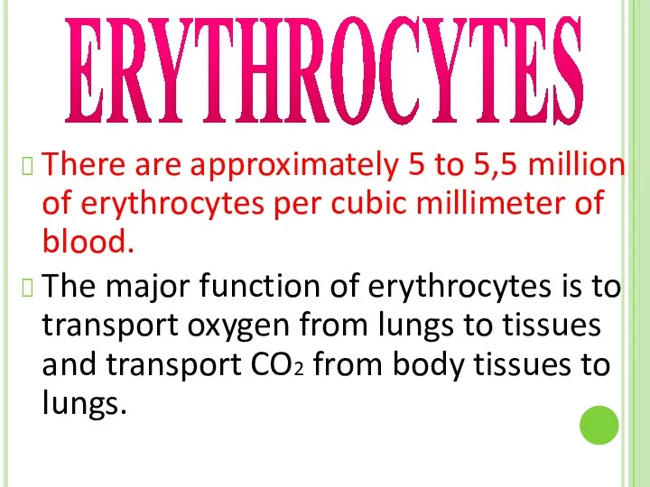
There are approximately 5 to 5,5 million of erythrocytes per cubic
There are approximately 5 to 5,5 million of erythrocytes per cubic
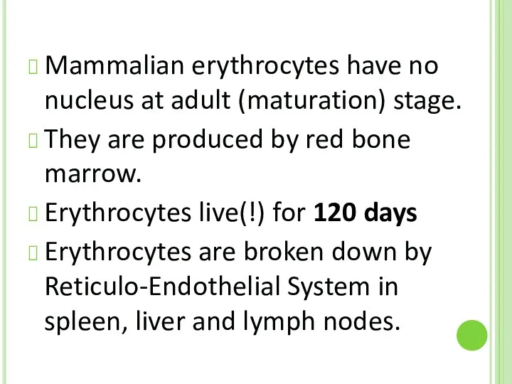
Mammalian erythrocytes have no nucleus at adult (maturation) stage.
They are
Mammalian erythrocytes have no nucleus at adult (maturation) stage.
They are
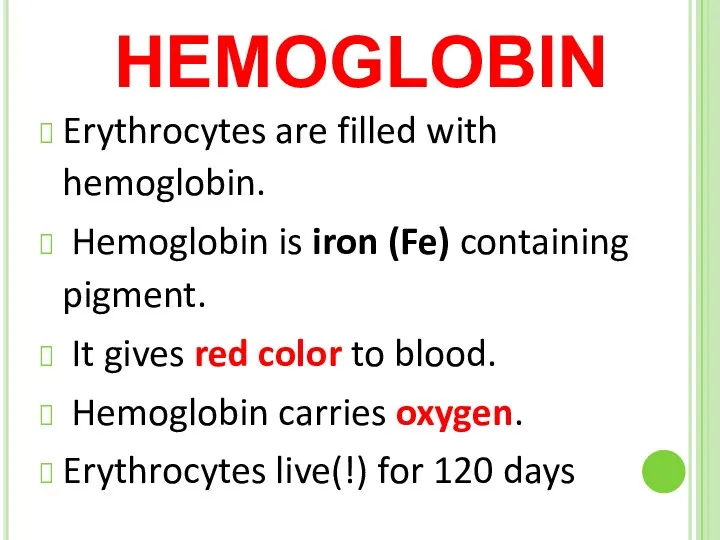
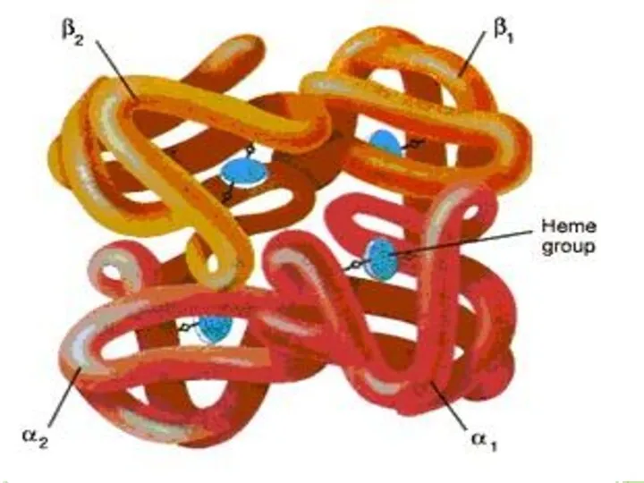
HEMOGLOBIN
Erythrocytes are filled with hemoglobin.
Hemoglobin is iron (Fe) containing pigment.
HEMOGLOBIN
Erythrocytes are filled with hemoglobin.
Hemoglobin is iron (Fe) containing pigment.

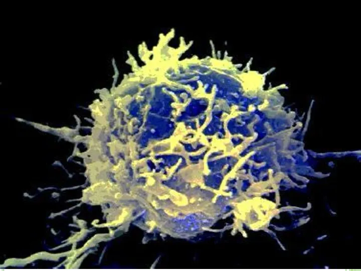
LEUCOCYTES
Leucocytes protect the body from infections.
They are produced by red
LEUCOCYTES
Leucocytes protect the body from infections.
They are produced by red
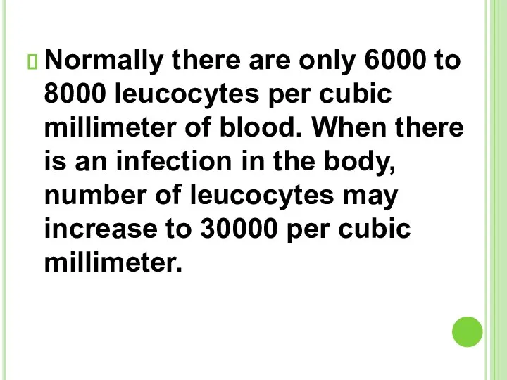
Normally there are only 6000 to 8000 leucocytes per cubic millimeter
Normally there are only 6000 to 8000 leucocytes per cubic millimeter
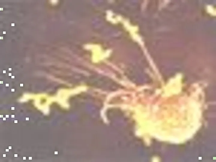
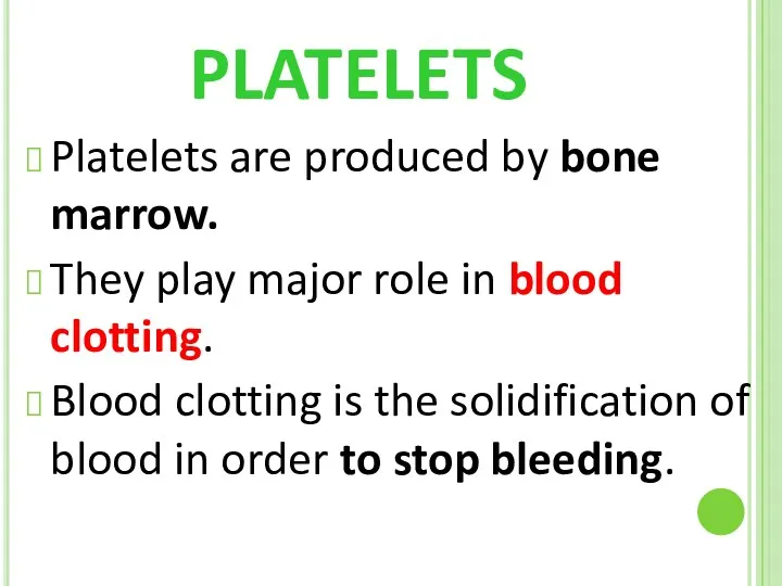
PLATELETS
Platelets are produced by bone marrow.
They play major role in blood
PLATELETS
Platelets are produced by bone marrow.
They play major role in blood
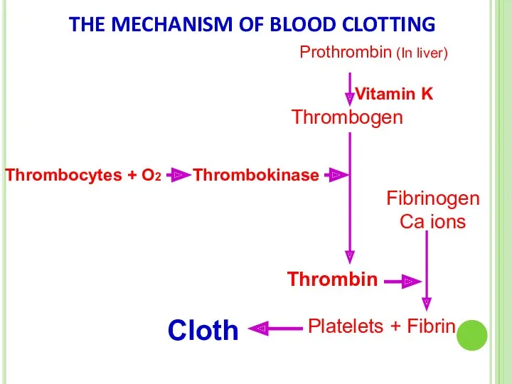
THE MECHANISM OF BLOOD CLOTTING
Prothrombin (In liver)
Vitamin K
Thrombogen
Thrombocytes + O2
Thrombokinase
Thrombin
Fibrinogen Ca
THE MECHANISM OF BLOOD CLOTTING
Prothrombin (In liver)
Vitamin K
Thrombogen
Thrombocytes + O2
Thrombokinase
Thrombin
Fibrinogen Ca
Diseases related to circulatory system
Anemia
Leukemia
Arteriosclerosis
Diseases related to circulatory system
Anemia
Leukemia
Arteriosclerosis
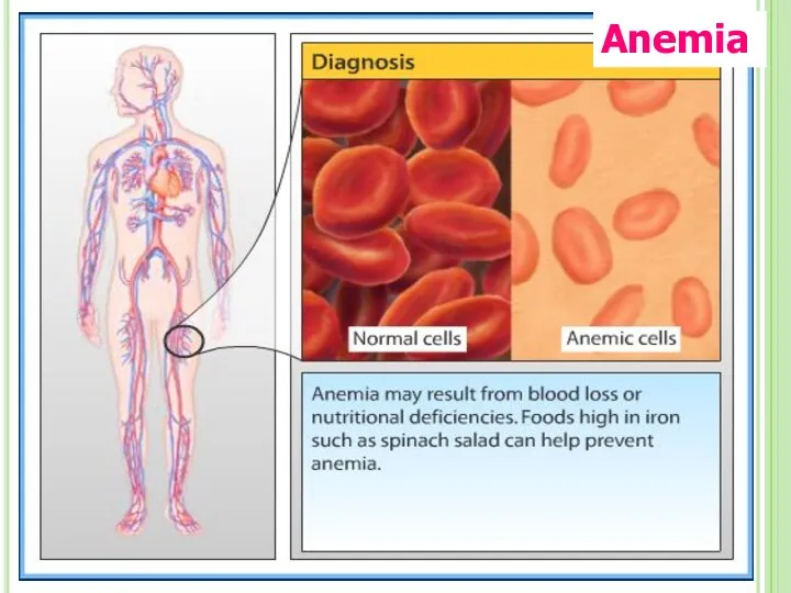
Anemia
Anemia
Arteriosclerosis
When blood vessels become narrow and lose their elasticity
Fats and
Arteriosclerosis
When blood vessels become narrow and lose their elasticity
Fats and
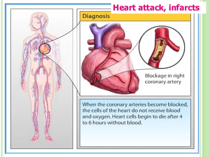
Heart attack, infarcts
Heart attack, infarcts
 Рентгеноанатомия черепа. Обозначьте кости мозгового черепа
 Водорастворимые витамины
 Птицы водоемов
 Опора и движение. Опорно-двигательная система
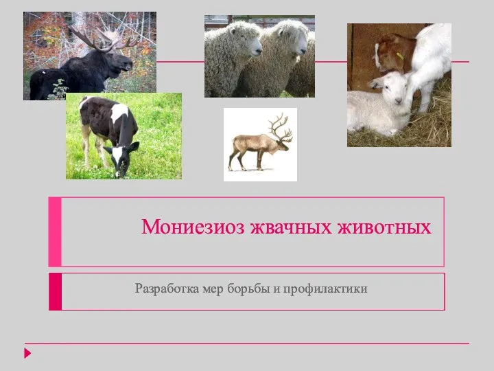 Мониезиоз жвачных животных. Разработка мер борьбы и профилактики
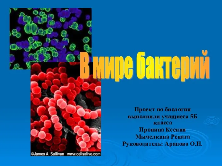 Презентация В мире бактерий
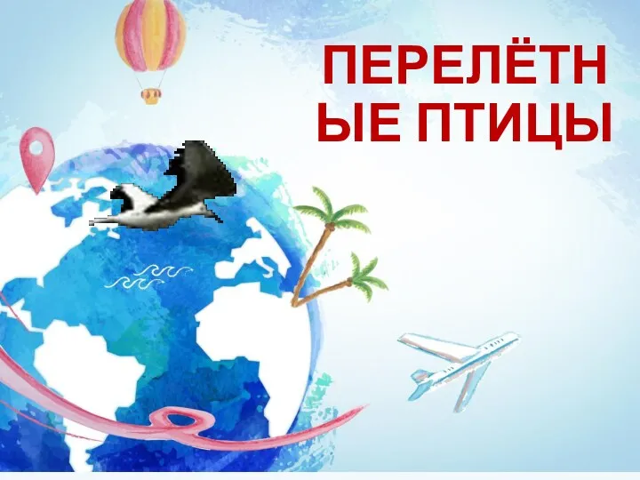 Перелётные птицы
 Пространственная организация белков. Конформации полипептидной цепи. Карты Рамачандрана
 Всасывание. Роль печени. Функции толстого кишечника
 Викторина. Тема Птицы.
 Функциональная асимметрия полушарий
 Черты приспособленности организмов к среде обитания
 Ядовитые грибы и ягоды
 Презентация Плоды
 Урок по биологии в 7 классе по теме Семейство злаковые по программе Сонина Н.И. (содержит презентацию и текстовую разработку урока)
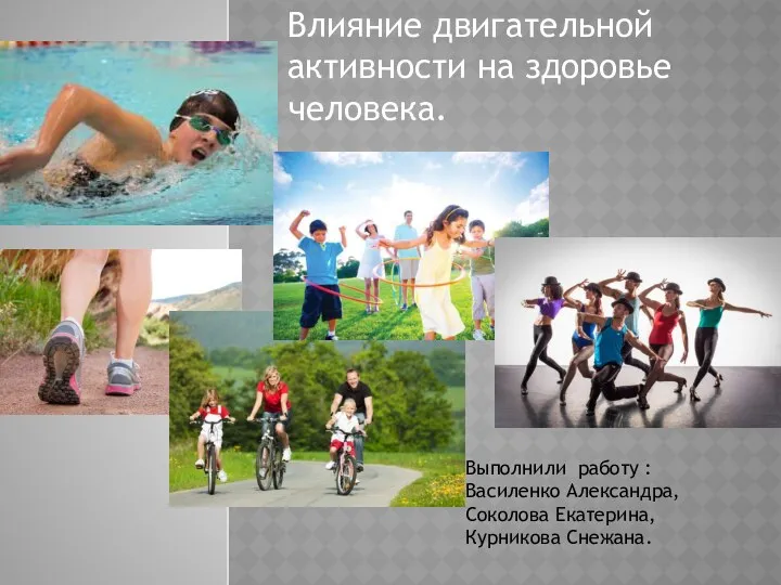 Влияние двигательной активности на здоровье человека
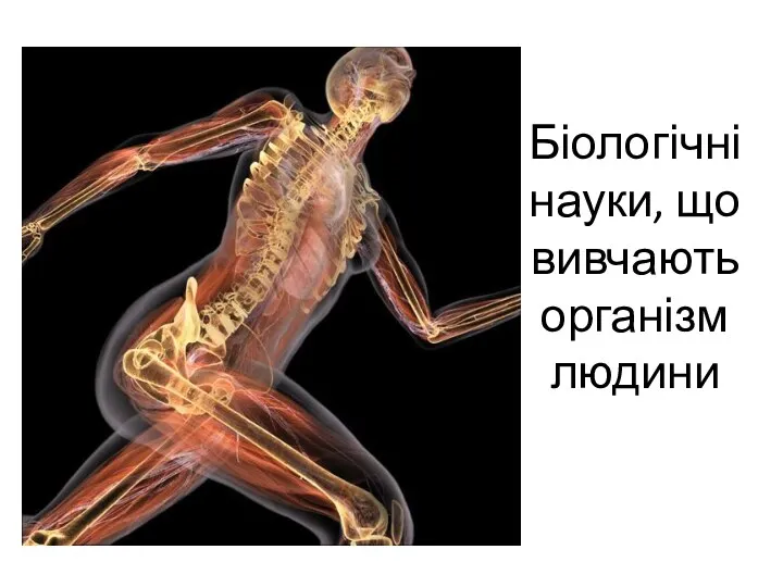 Біологічні науки, що вивчають організм людини
 Доклад на тему: Формирование универсальных умений и универсальных учебных действий учащихся в свете требований ФГОС на уроках биологии .
  Бактерии. История открытия. Строение бактерий
 Проект Разнообразие природы родного края
 Класс насекомые
 Викторина Анализаторы
 История развития знаний о строении и функциях организма человека
 Класс Пресмыкающиеся, или Рептилии. Отряд Чешуйчатые
 Альбом по эмбриологии
 Социально-биологические основы физической культуры
 Методика обучения биологии
 Кольчатые черви