- Immune system

Содержание
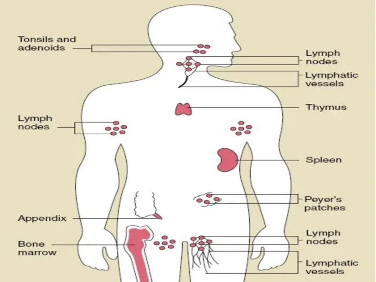
- 4. The major organs of the immune system are: Central: Bone marrow Thymus Peripheral: Spleen Lymph nodes
- 6. In central organs antigen-independent production of uncommitted T lymphocyte (thymus) or B lymphocyte (bone marrow) precursors
- 7. Bone Marrow is a soft tissue occupying the medullary cavity of a long bone There are
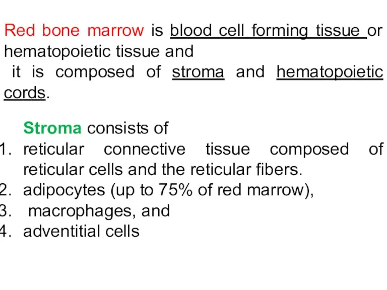
- 8. Red bone marrow is blood cell forming tissue and it is composed of stroma (reticular tissue)
- 9. Red bone marrow is blood cell forming tissue or hematopoietic tissue and it is composed of
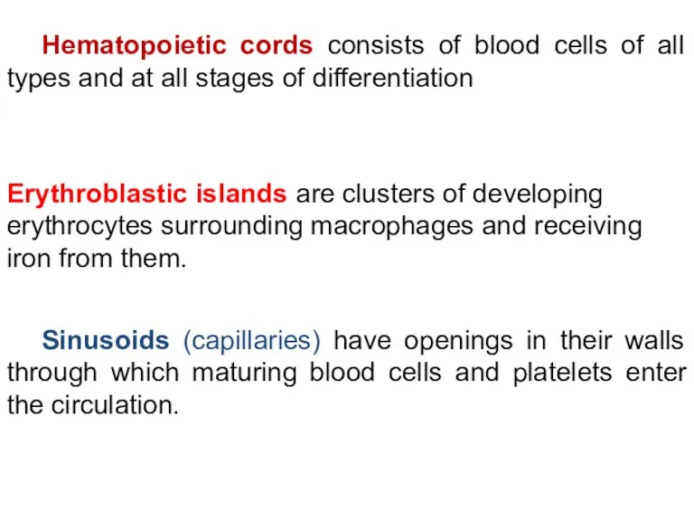
- 10. Erythroblastic islands are clusters of developing erythrocytes surrounding macrophages and receiving iron from them. Sinusoids (capillaries)
- 11. Bone marrow functions 1. Hematopoiesis. 2. Bone marrow helps destroy old red blood cells. 3. Recirculation
- 13. Thymus Functions: 1. Production of T- lymphocyte. 2. Production of hormone - thymosin Consists of epithelial
- 14. Thymus
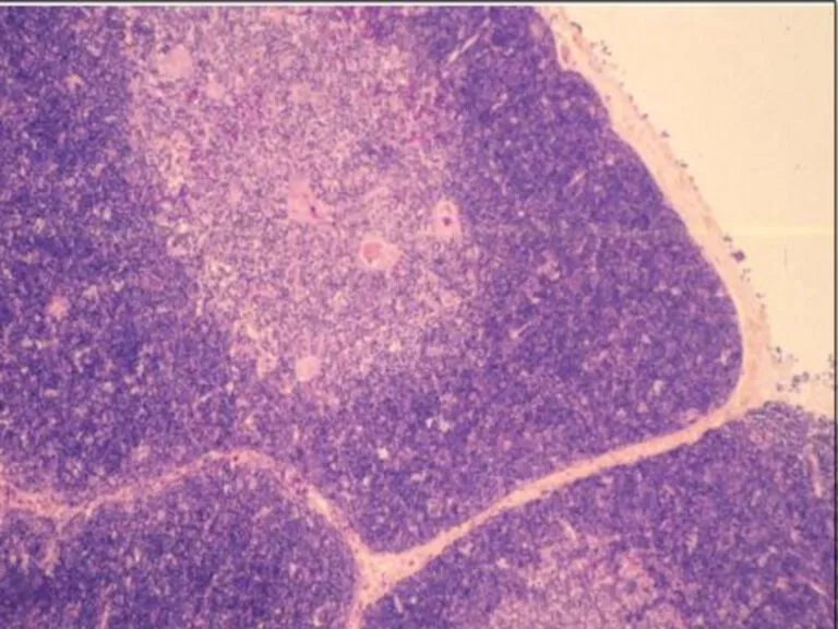
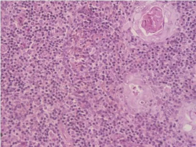
- 15. Thymus Capsule Lobules Cortex Medulla Hassal’s Corpuscles
- 16. Cortex--- dark-staining periphery of each lobule. Small lymphocytes predominate Medulla is the light core of each
- 17. Figure 5-3 part 1 of 2 Differentiation Immature thymocytes are here More mature thymocytes are here
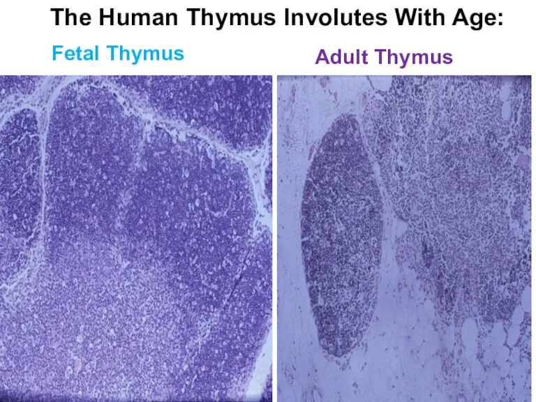
- 20. Adult Thymus Fetal Thymus The Human Thymus Involutes With Age:
- 21. INVOLUTION OF THE THYMUS Two types:1. Age dependent 2. Accidental involution due to some exogenous agent,
- 22. Peripheral part of I. S.
- 23. 1. Lymphoid (= Lymph, Lymphatic) Nodules (Follicles)
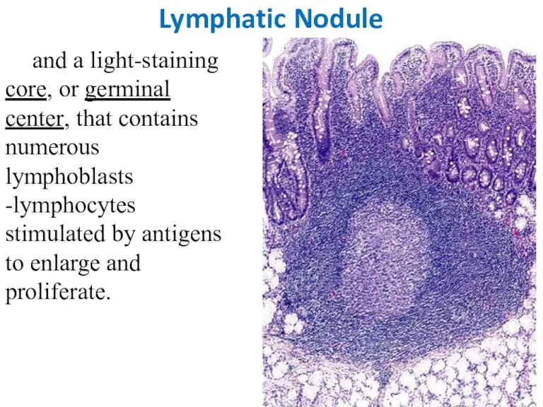
- 24. Lymphatic Nodule - have a dark-staining periphery, or mantle zone, that contains tightly packed small lymphocytes,
- 25. Lymphatic Nodule and a light-staining core, or germinal center, that contains numerous lymphoblasts -lymphocytes stimulated by
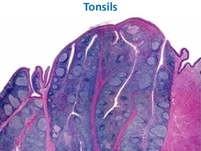
- 26. TONSILS underlie the epithelial lining of the mouth and pharynx. palatine tonsils (2), pharyngeal tonsil (1),
- 27. Tonsils
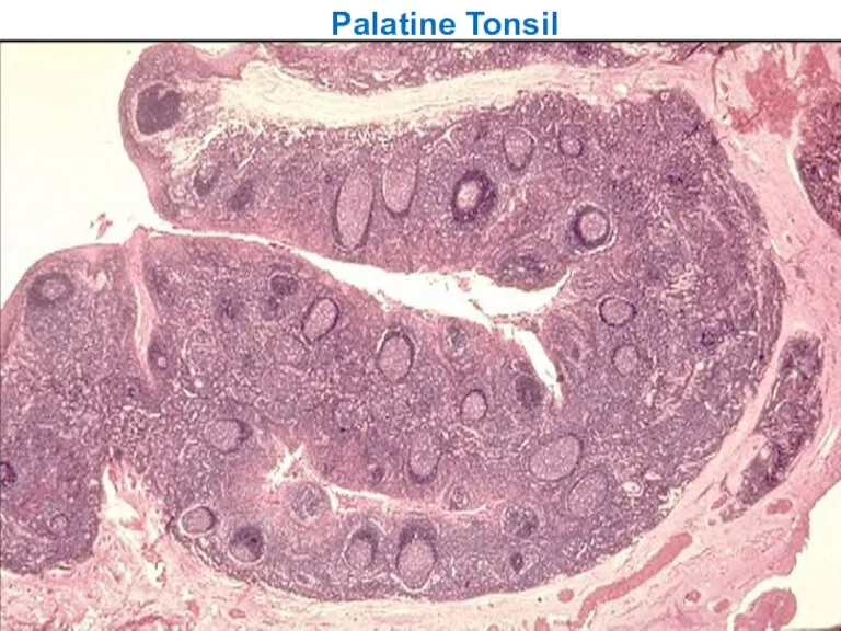
- 28. Palatine Tonsil
- 29. Peyer’s Patches Smaller aggregates present under mucous membrane: “Mucosa Associated Lymphoid Tissue” or MALT (in Digestive
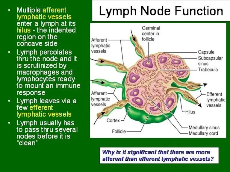
- 31. Capsulated Afferent lymphatics ? “subcapsular sinus” Hilum – blood vessels, efferent lymphatic Cortex and medulla Cortex
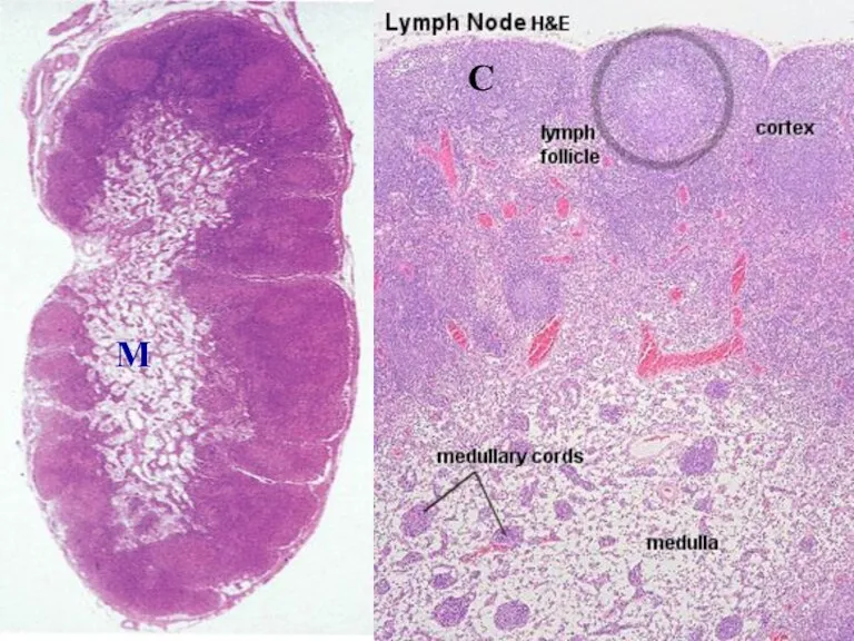
- 32. LYMPH NODES These are the smallest but most numerous encapsulated lymphoid organs. Lie in groups along
- 33. C M
- 34. LYMPH NODES -- Inner space consists of reticular connective tissue and has 3 zones: 1. cortex,
- 35. 2. Paracortical zone. This is the T-dependent region, It contains mainly T-lymphocytes. 3. Medulla. is composed
- 36. Lymphatic vessels inside LN are Sinuses. Types: subcapsular, peritrabecular, medullary
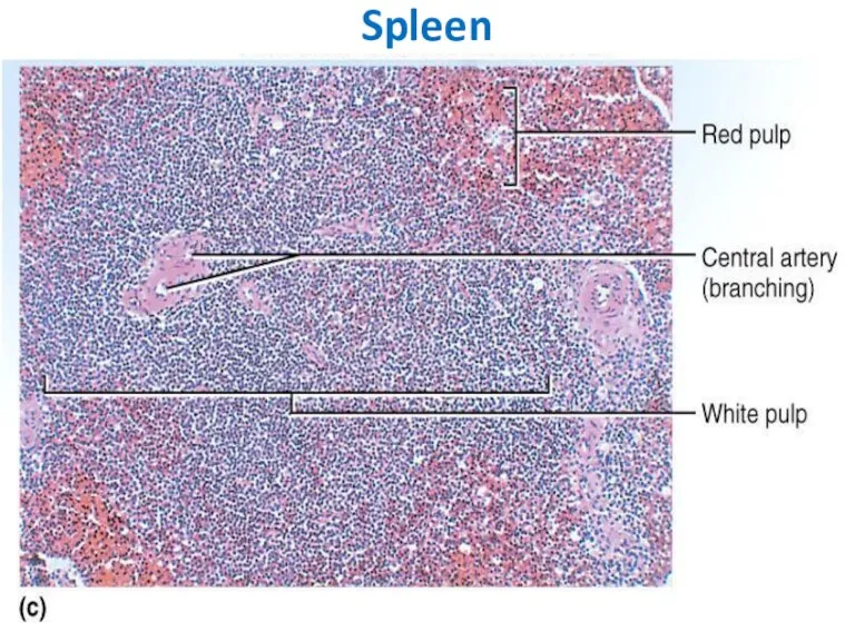
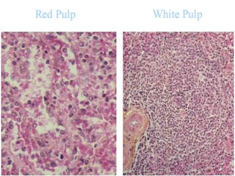
- 39. SPLEEN -- -- Is the largest of the lymphoid organs Functions: 1. Filtration of blood. 2.
- 40. Inner space -- Splenic pulp -- is composed of: reticular tissue consisting of reticular cells and
- 41. White pulp - consists of lymphocytes; -- surround small arteries; --- has 2 major components: Periarterial
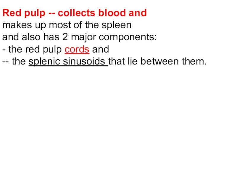
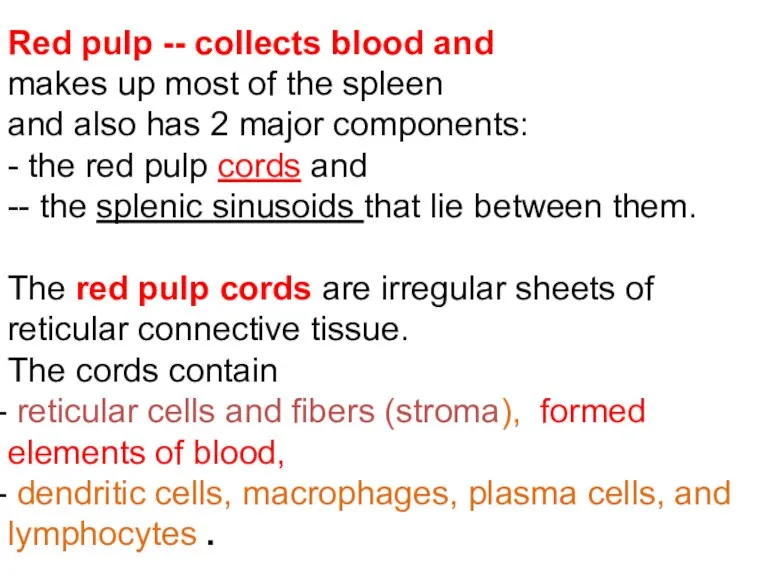
- 42. Red pulp -- collects blood and makes up most of the spleen and also has 2
- 43. Red pulp -- collects blood and makes up most of the spleen and also has 2
- 44. Splenic sinusoids differ from common capillaries: - the lumen is wider and more irregular; - small
- 45. Spleen
- 48. Скачать презентацию


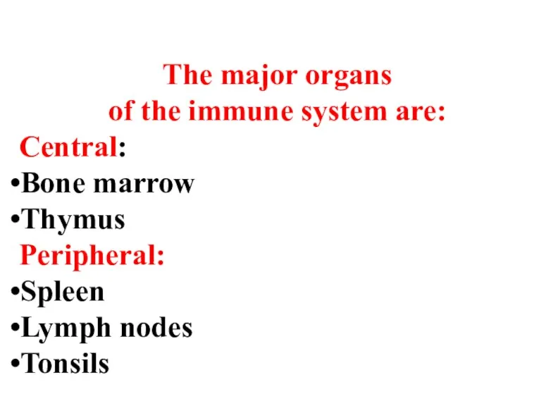
The major organs
of the immune system are:
Central:
Bone marrow
Thymus
Peripheral:
Spleen
Lymph nodes
Tonsils
The major organs
of the immune system are:
Central:
Bone marrow
Thymus
Peripheral:
Spleen
Lymph nodes
Tonsils
In central organs
antigen-independent production of uncommitted T lymphocyte (thymus) or
In central organs
antigen-independent production of uncommitted T lymphocyte (thymus) or

Bone Marrow
is a soft tissue occupying the medullary cavity of a
Bone Marrow
is a soft tissue occupying the medullary cavity of a
Red bone marrow is blood cell forming tissue and
it
it
Red bone marrow is blood cell forming tissue or hematopoietic tissue
Erythroblastic islands are clusters of developing erythrocytes surrounding macrophages and receiving
Erythroblastic islands are clusters of developing erythrocytes surrounding macrophages and receiving
Bone marrow functions
1. Hematopoiesis.
2. Bone marrow helps destroy old
Bone marrow functions
1. Hematopoiesis.
2. Bone marrow helps destroy old
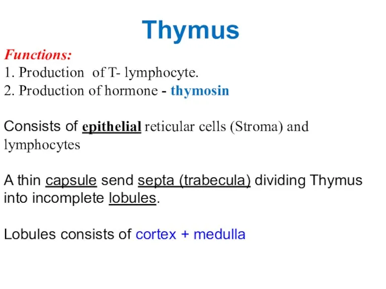
Thymus
Functions:
1. Production of T- lymphocyte.
2. Production of hormone - thymosin
Consists
Thymus
Functions:
1. Production of T- lymphocyte.
2. Production of hormone - thymosin
Consists
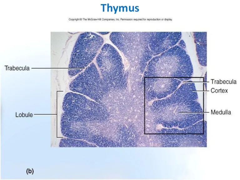
Thymus
Thymus
Thymus
Capsule
Lobules
Cortex
Medulla
Hassal’s Corpuscles
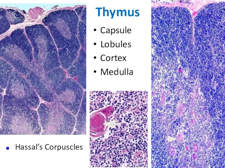
Thymus
Capsule
Lobules
Cortex
Medulla
Hassal’s Corpuscles
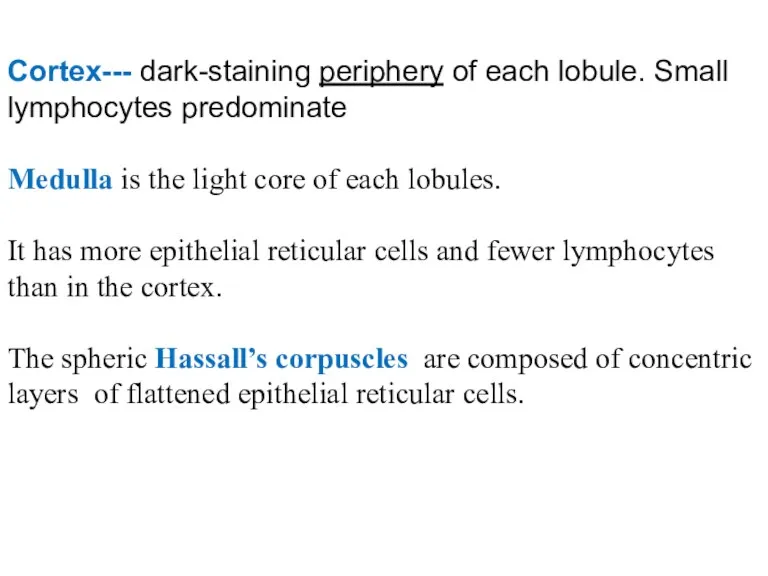
Cortex--- dark-staining periphery of each lobule. Small lymphocytes predominate
Medulla is the
Cortex--- dark-staining periphery of each lobule. Small lymphocytes predominate
Medulla is the
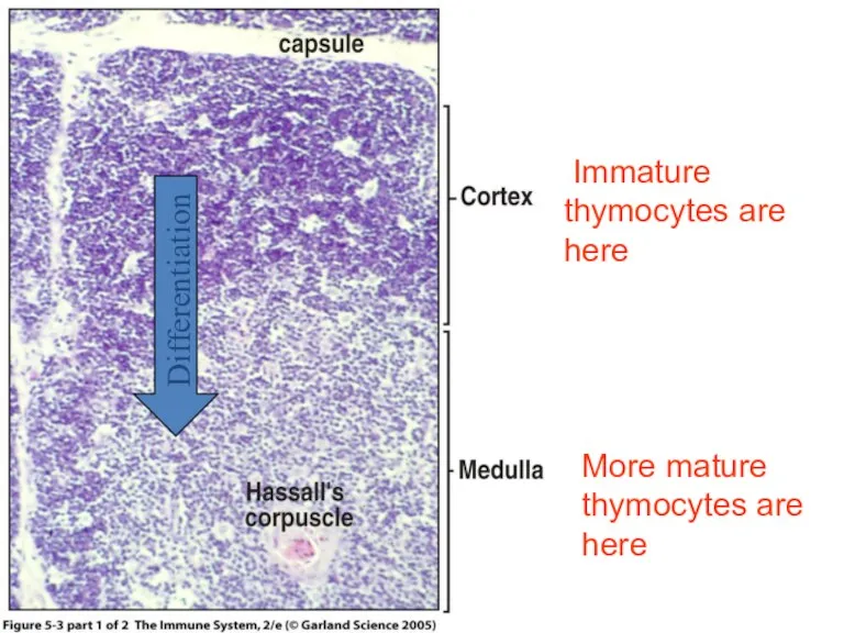
Figure 5-3 part 1 of 2
Differentiation
Immature
thymocytes are here
More mature
Figure 5-3 part 1 of 2
Differentiation
Immature
thymocytes are here
More mature
Adult Thymus
Fetal Thymus
The Human Thymus Involutes With Age:
Adult Thymus
Fetal Thymus
The Human Thymus Involutes With Age:
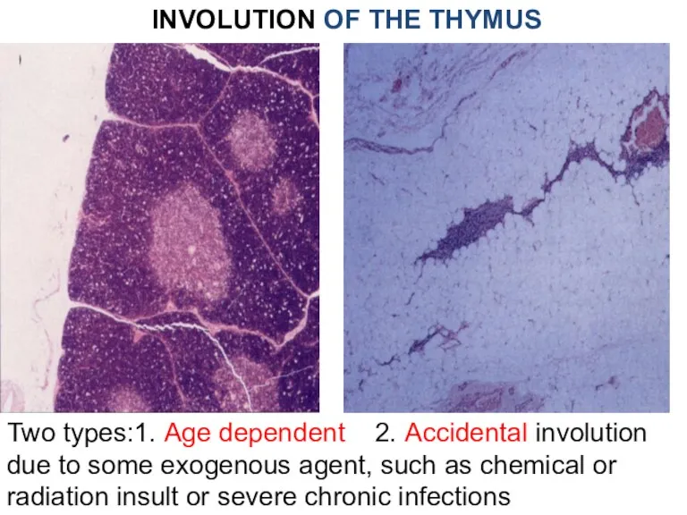
INVOLUTION OF THE THYMUS
Two types:1. Age dependent 2. Accidental involution due
INVOLUTION OF THE THYMUS
Two types:1. Age dependent 2. Accidental involution due
Peripheral part of I. S.
Peripheral part of I. S.
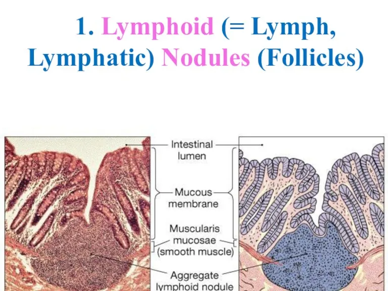
1. Lymphoid (= Lymph, Lymphatic) Nodules (Follicles)
1. Lymphoid (= Lymph, Lymphatic) Nodules (Follicles)
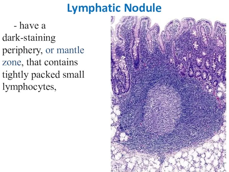
Lymphatic Nodule
- have a dark-staining periphery, or mantle zone, that contains
Lymphatic Nodule
- have a dark-staining periphery, or mantle zone, that contains
Lymphatic Nodule
and a light-staining core, or germinal center, that contains numerous
Lymphatic Nodule
and a light-staining core, or germinal center, that contains numerous
TONSILS
underlie the epithelial lining of the mouth and pharynx.
TONSILS
underlie the epithelial lining of the mouth and pharynx.
Tonsils
Tonsils
Palatine Tonsil
Palatine Tonsil
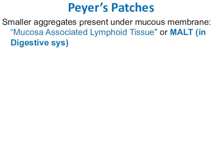
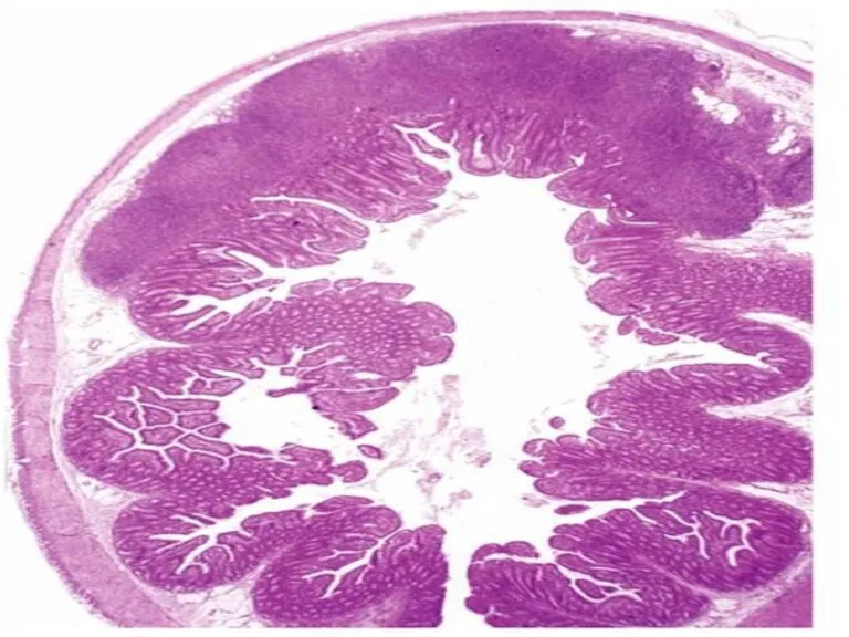
Peyer’s Patches
Smaller aggregates present under mucous membrane: “Mucosa Associated Lymphoid Tissue”
Peyer’s Patches
Smaller aggregates present under mucous membrane: “Mucosa Associated Lymphoid Tissue”
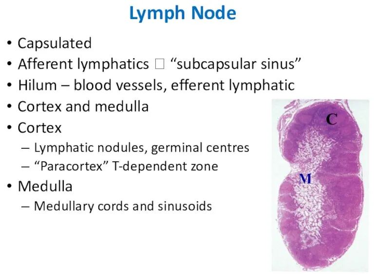
Capsulated
Afferent lymphatics ? “subcapsular sinus”
Hilum – blood vessels, efferent lymphatic
Cortex and
Capsulated
Afferent lymphatics ? “subcapsular sinus”
Hilum – blood vessels, efferent lymphatic
Cortex and
LYMPH NODES
These are
the smallest but most numerous encapsulated lymphoid organs.
LYMPH NODES
These are
the smallest but most numerous encapsulated lymphoid organs.
C
M
C
M
LYMPH NODES
-- Inner space consists of reticular connective tissue and has
LYMPH NODES
-- Inner space consists of reticular connective tissue and has
2. Paracortical zone.
This is the T-dependent region, It contains mainly
2. Paracortical zone.
This is the T-dependent region, It contains mainly
Lymphatic vessels inside LN are Sinuses.
Types: subcapsular,
peritrabecular,
Lymphatic vessels inside LN are Sinuses.
Types: subcapsular,
peritrabecular,

SPLEEN --
-- Is the largest of the lymphoid organs
Functions:
1. Filtration of
-- Is the largest of the lymphoid organs
Functions:
1. Filtration of
Inner space -- Splenic pulp -- is composed of:
reticular tissue
reticular tissue
White pulp
- consists of lymphocytes;
-- surround small arteries;
--- has
White pulp
- consists of lymphocytes;
-- surround small arteries;
--- has
Red pulp -- collects blood and
makes up most of the
Red pulp -- collects blood and
makes up most of the
Red pulp -- collects blood and
makes up most of the
Red pulp -- collects blood and
makes up most of the
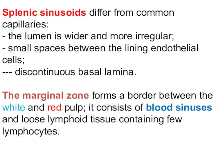
Splenic sinusoids differ from common capillaries:
- the lumen is wider
Splenic sinusoids differ from common capillaries:
- the lumen is wider
Spleen
Spleen
 Строение стебля
 Лекция № 1 Наука экология биосфера
 Размножение мхов
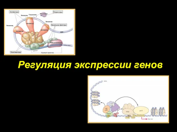 Регуляция экспрессии генов
 Классификация гормонов
 Незаконное истребление животных
 Питание и пищеварение
 Строение нервной системы
 Эмбриональное развитие организмов
 Вендские жители Земли
 Движение крови в организме
 Історія відкриття вітамінів
 Вегетативное размножение покрытосеменных растений
 Chromosomes. (Chapter 6.1)
 Регуляция пищеварения. Заболевания органов пищеварения и их предупреждение. Биология 8 кл (Пасечник)
 Органы выделения у животных
 Голосовой аппарат
 Биология как наука. Экосистемы. Факультатив по биологии 10 -11 классы
 Живые клетки
 Разбор заданий ВПР 5 класс
 Борьба за существование
 Роль покрытосеменных в жизни человека
 Введение в биохимию. Структура и функции белков
 Система двигательных действий и организация управления ими
 Цитогенетические основы размножения
 Работа мышц
 Животные саванн и полупустынь Южной Америки
 Бионика. Биология – техника