- Integumentary system

Содержание
- 2. Objectives: 1- Describe the functions of the integumentary system. 2- Identify the major structures found in
- 3. This system is divided into: 1- skin 2- hair 3- glands 4- nails 5- nerve endings
- 5. Anatomy (structure) Epidermis (thinner outer layer of skin) Dermis (thicker connective tissue layer) Hypodermis (subcutaneous layer
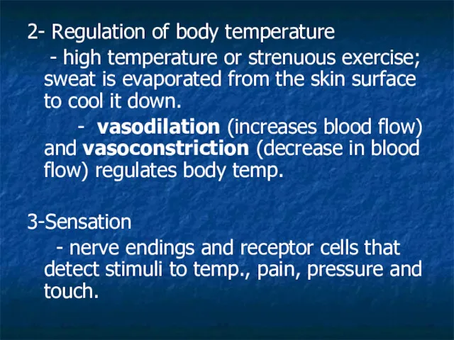
- 6. 2- Regulation of body temperature - high temperature or strenuous exercise; sweat is evaporated from the
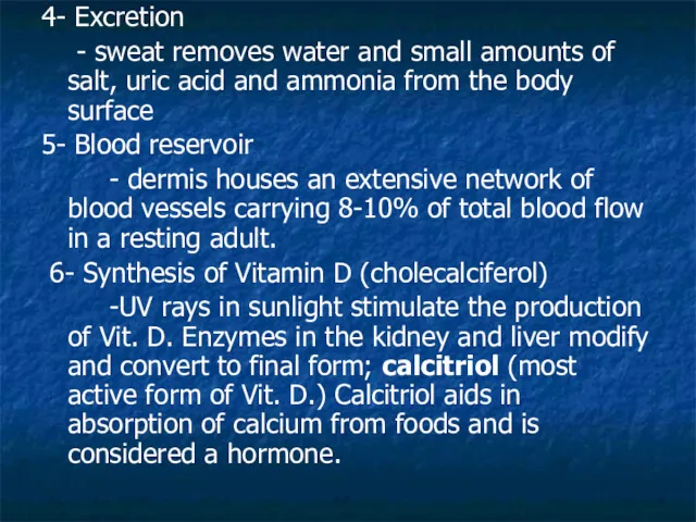
- 7. 4- Excretion - sweat removes water and small amounts of salt, uric acid and ammonia from
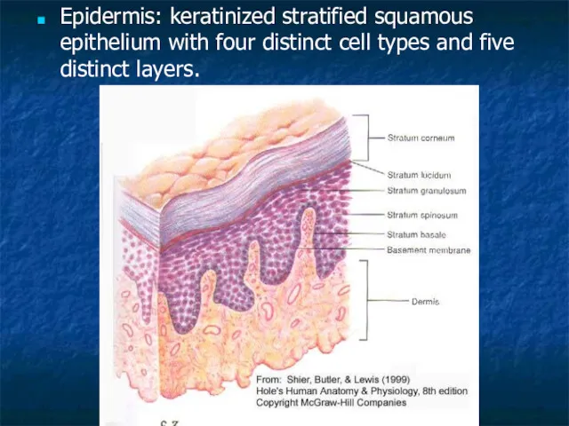
- 8. Epidermis: keratinized stratified squamous epithelium with four distinct cell types and five distinct layers.
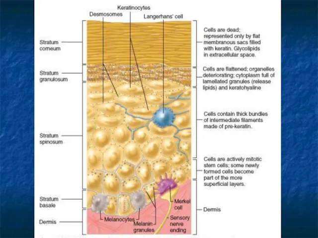
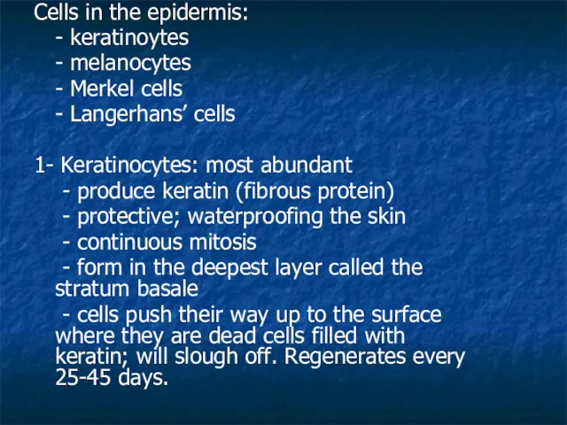
- 10. Cells in the epidermis: - keratinoytes - melanocytes - Merkel cells - Langerhans’ cells 1- Keratinocytes:
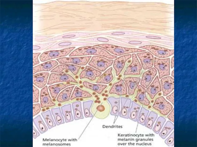
- 12. 2- Melanocytes: - cells produce brownish/black pigment called melanin. (8% of epidermal cells) - stratum basale
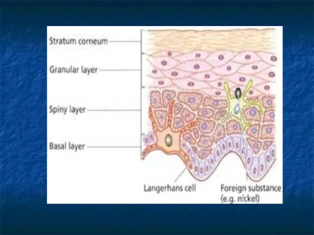
- 14. 3- Merkel cells: - stratum basale - epidermis of hairless skin - attach to keratinocytes by
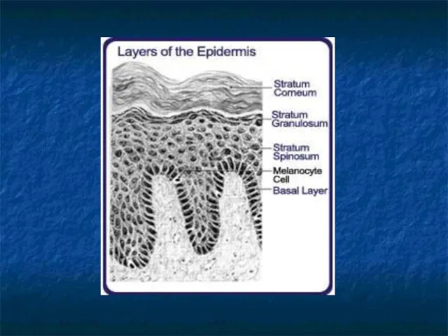
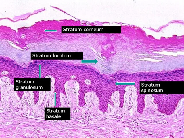
- 17. Stratum corneum Stratum lucidum Stratum spinosum Stratum granulosum Stratum basale
- 18. 5 layers of the epidermis: 1- Stratum corneum (horny layer) - layer has many rows of
- 19. 2- Stratum lucidum - seen in thick skin of the palms and soles of feet. -
- 20. 4- Stratum spinosum: “spiny layer” - 8-10 rows of polyhedral (many sided) cells - appearance of
- 22. 5- Stratum basale: deepest epidermal layer - attached to dermis - single row of cells -
- 25. Dermis: - flexible and strong connective tissue - elastic, reticular and collagen fibers - cells: fibroblasts,
- 26. 1- Papillary layer: - loose connective tissue with nipple like surface projection called dermal papilla. -
- 27. 2- Reticular layer: - dense irregular c.t. - collagen fibers offer strength - holds water -
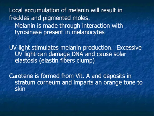
- 28. Local accumulation of melanin will result in freckles and pigmented moles. Melanin is made through interaction
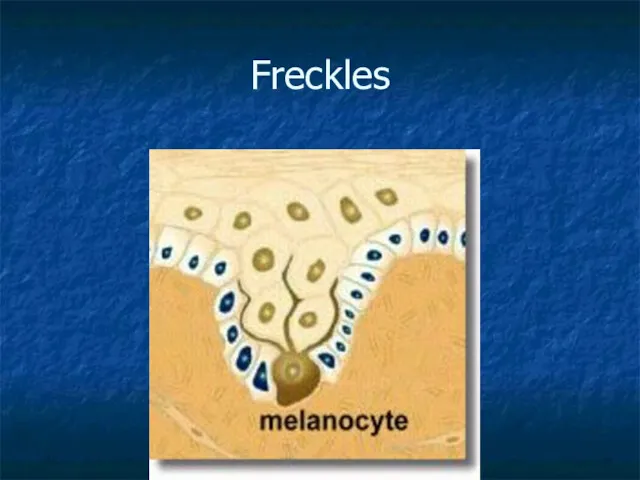
- 29. Freckles
- 30. Hemoglobin (blood) will impart pinkish tones to skin. Blushing 1- Redness (erythema) - reddened skin, embarrassment,
- 31. Hair color: Dark hair: mostly melanin Blond and red hair: melanin with Fe and S. Gray
- 32. Hair (pili) - main function is protection - hair root nerve plexus for touch - normal
- 35. Hair anatomy: - composed of dead columns of keratinized cells. - shaft: is the superficial portion
- 36. Vellus hair: fine hair Terminal hair : coarser hair; axillary and pubic region. Grow in response
- 37. Hair follicle surrounds the root. Bulb is the enlargement at the end of the follicle. -
- 38. Arrector pili (pl. pilorum) is smooth muscle located in the dermis and is attached to the
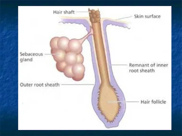
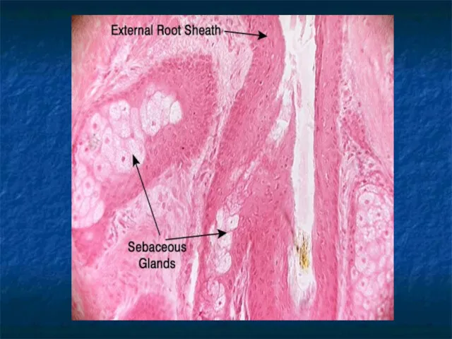
- 40. Glands: Two types of glands exist in the integument. - Sebaceous glands (oil glands) - Sudoriferous
- 43. Whitehead: When the trapped sebum and bacteria stay below the skin surface, a whitehead is formed.
- 44. Blackhead: A blackhead occurs when the trapped sebum and bacteria partially open to the surface and
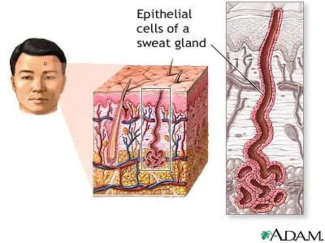
- 45. Sudoriferous glands: exocrine glands - millions located throughout the skin - two types: - eccrine: more
- 47. - apocrine: axillary and pubic region - duct empties onto hair follicle - viscous fluid -
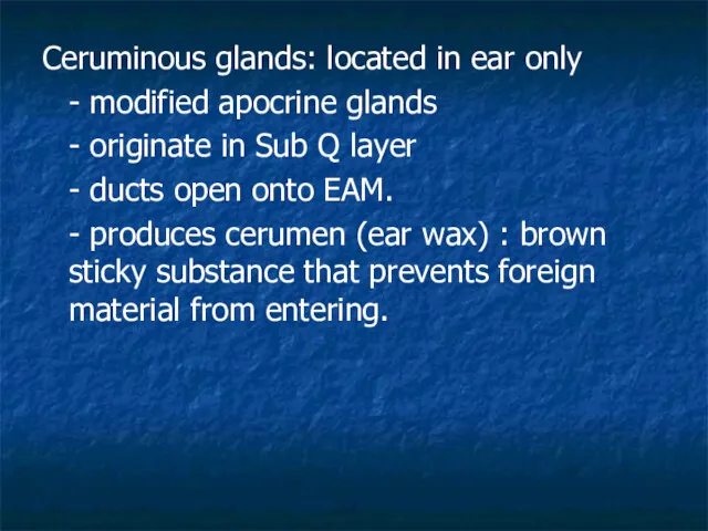
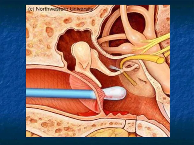
- 48. Ceruminous glands: located in ear only - modified apocrine glands - originate in Sub Q layer
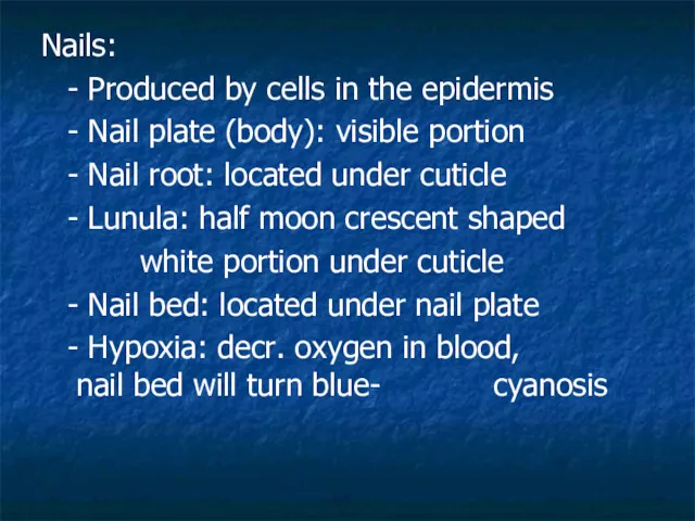
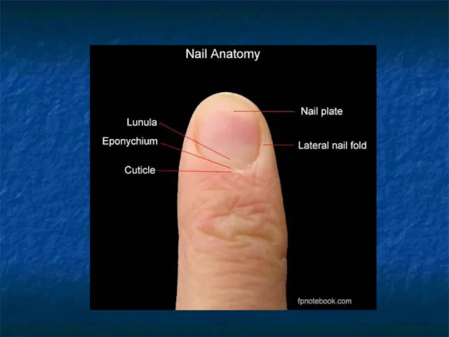
- 50. Nails: - Produced by cells in the epidermis - Nail plate (body): visible portion - Nail
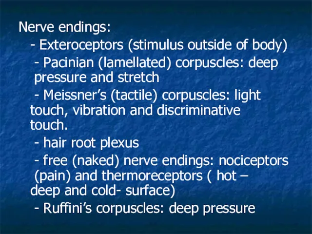
- 52. Nerve endings: - Exteroceptors (stimulus outside of body) - Pacinian (lamellated) corpuscles: deep pressure and stretch
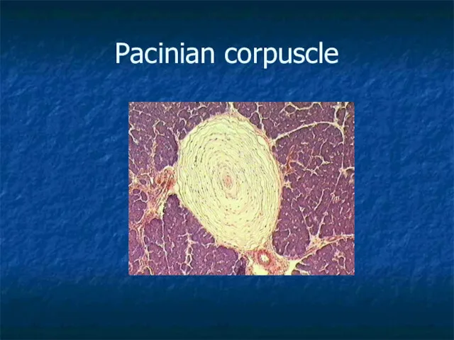
- 53. Pacinian corpuscle
- 54. Hypodermis - called subcutaneous, Sub-Q or superficial fascia - anchors skin to underlying structures - contains
- 55. Dermatopathological terms Macule – flat spot on skin with color (freckle) Wheal – round and temp.
- 56. Sebaceous hyperplasia - enlargement of the sebaceous gland Pruritis - irritating itching sensation of the skin
- 58. Скачать презентацию
Objectives:
1- Describe the functions of the integumentary system.
2- Identify the major
Objectives:
1- Describe the functions of the integumentary system.
2- Identify the major
This system is divided into:
1- skin
2- hair
3- glands
4- nails
5- nerve endings
I)
This system is divided into:
1- skin
2- hair
3- glands
4- nails
5- nerve endings
I)
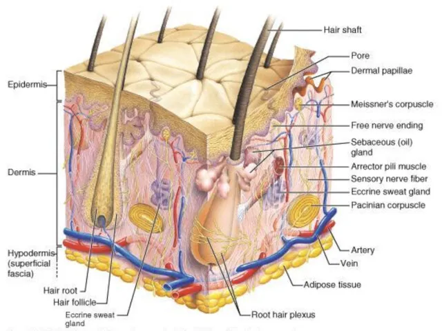
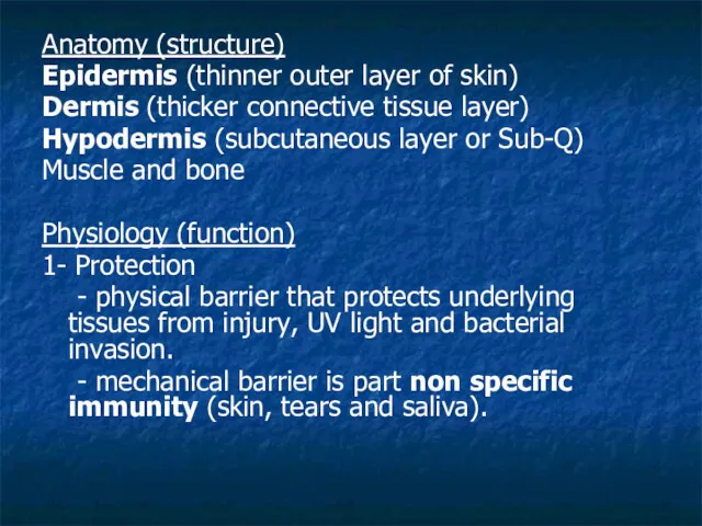
Anatomy (structure)
Epidermis (thinner outer layer of skin)
Dermis (thicker connective tissue layer)
Hypodermis
Anatomy (structure)
Epidermis (thinner outer layer of skin)
Dermis (thicker connective tissue layer)
Hypodermis
2- Regulation of body temperature
- high temperature or strenuous exercise; sweat
2- Regulation of body temperature
- high temperature or strenuous exercise; sweat
4- Excretion
- sweat removes water and small amounts of salt,
4- Excretion
- sweat removes water and small amounts of salt,
Epidermis: keratinized stratified squamous epithelium with four distinct cell types and
Epidermis: keratinized stratified squamous epithelium with four distinct cell types and
Cells in the epidermis:
- keratinoytes
- melanocytes
- Merkel cells
- Langerhans’ cells
1- Keratinocytes:
Cells in the epidermis:
- keratinoytes
- melanocytes
- Merkel cells
- Langerhans’ cells
1- Keratinocytes:
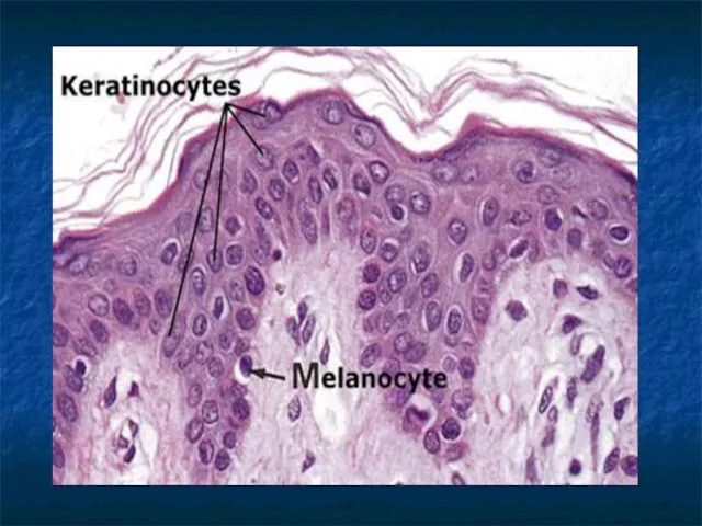
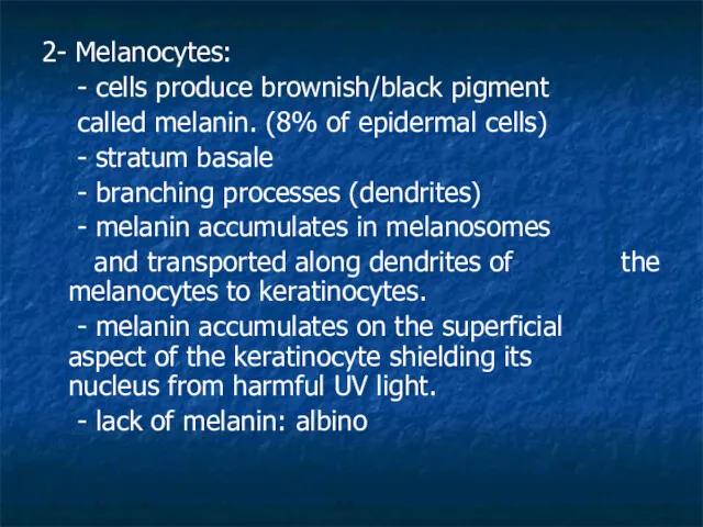
2- Melanocytes:
- cells produce brownish/black pigment
called melanin. (8% of epidermal
2- Melanocytes:
- cells produce brownish/black pigment
called melanin. (8% of epidermal
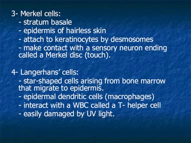
3- Merkel cells:
- stratum basale
- epidermis of hairless skin
- attach to
3- Merkel cells:
- stratum basale
- epidermis of hairless skin
- attach to
Stratum corneum
Stratum lucidum
Stratum spinosum
Stratum granulosum
Stratum basale
Stratum corneum
Stratum lucidum
Stratum spinosum
Stratum granulosum
Stratum basale
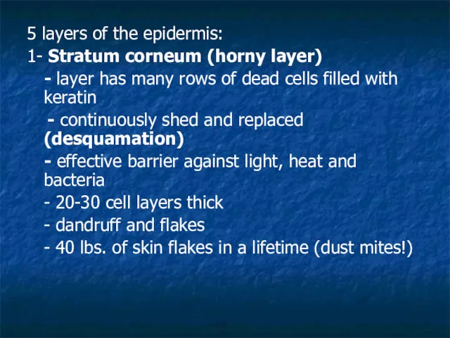
5 layers of the epidermis:
1- Stratum corneum (horny layer)
- layer has
5 layers of the epidermis:
1- Stratum corneum (horny layer)
- layer has
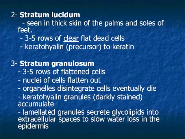
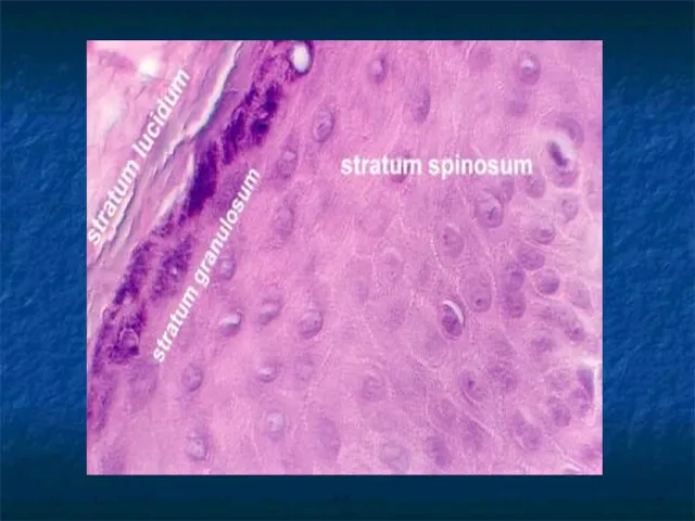
2- Stratum lucidum
- seen in thick skin of the palms
2- Stratum lucidum
- seen in thick skin of the palms
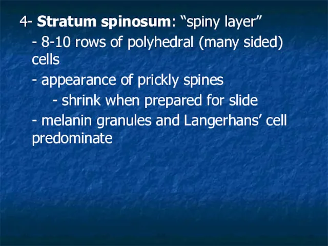
4- Stratum spinosum: “spiny layer”
- 8-10 rows of polyhedral (many sided)
4- Stratum spinosum: “spiny layer”
- 8-10 rows of polyhedral (many sided)
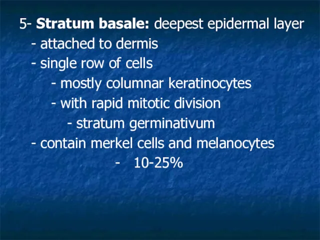
5- Stratum basale: deepest epidermal layer
- attached to dermis
- single row
5- Stratum basale: deepest epidermal layer
- attached to dermis
- single row


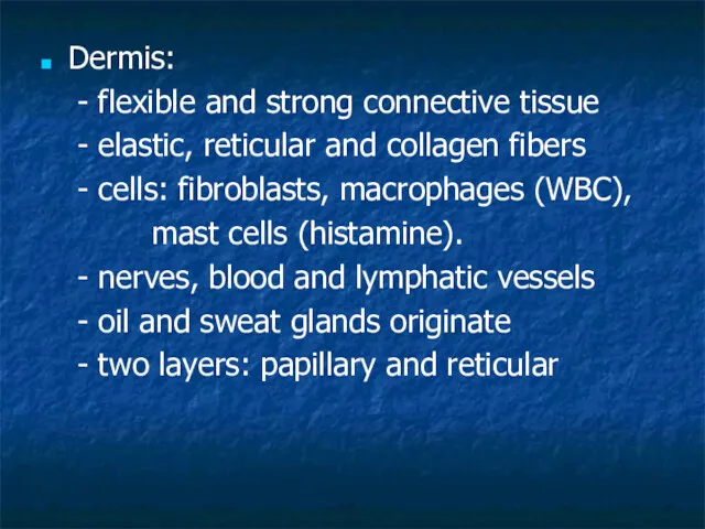
Dermis:
- flexible and strong connective tissue
- elastic, reticular and collagen fibers
-
Dermis:
- flexible and strong connective tissue
- elastic, reticular and collagen fibers
-
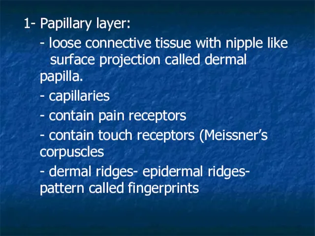
1- Papillary layer:
- loose connective tissue with nipple like
1- Papillary layer:
- loose connective tissue with nipple like
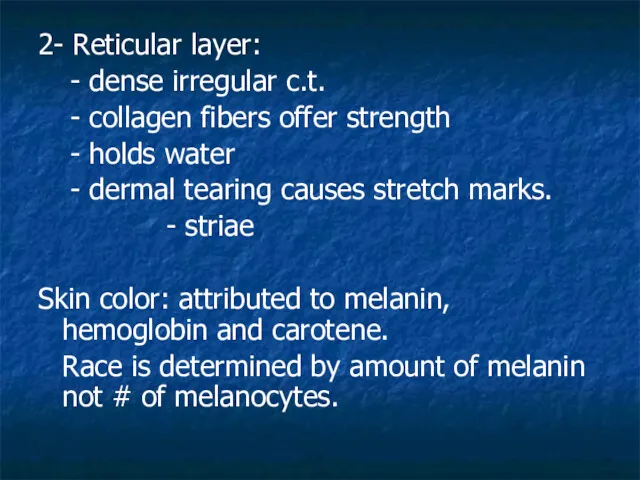
2- Reticular layer:
- dense irregular c.t.
- collagen fibers offer strength
- holds
2- Reticular layer:
- dense irregular c.t.
- collagen fibers offer strength
- holds
Local accumulation of melanin will result in
freckles and pigmented moles.
Melanin is
Local accumulation of melanin will result in
freckles and pigmented moles.
Melanin is
Freckles
Freckles
Hemoglobin (blood) will impart pinkish tones to skin. Blushing
1- Redness (erythema)
Hemoglobin (blood) will impart pinkish tones to skin. Blushing
1- Redness (erythema)
Hair color:
Dark hair: mostly melanin
Blond and red hair: melanin with Fe
Hair color:
Dark hair: mostly melanin
Blond and red hair: melanin with Fe
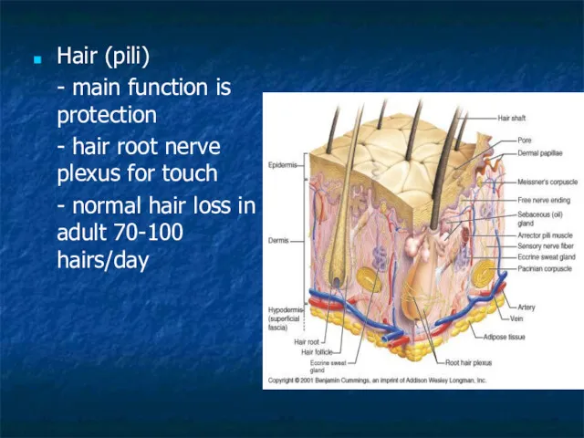
Hair (pili)
- main function is protection
- hair root nerve plexus for
Hair (pili)
- main function is protection
- hair root nerve plexus for
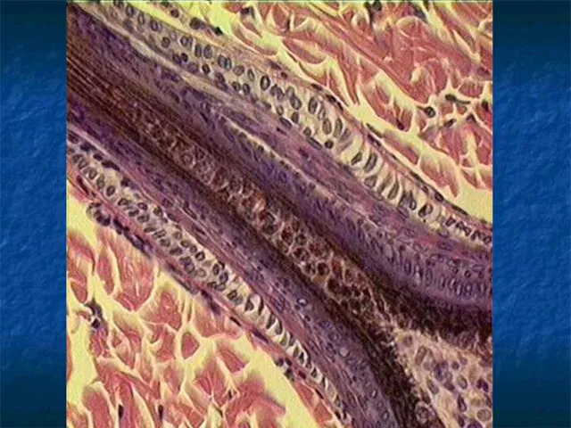
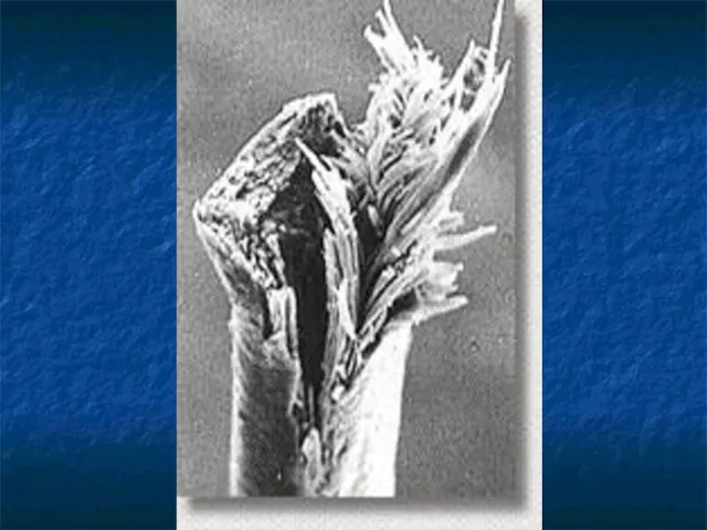
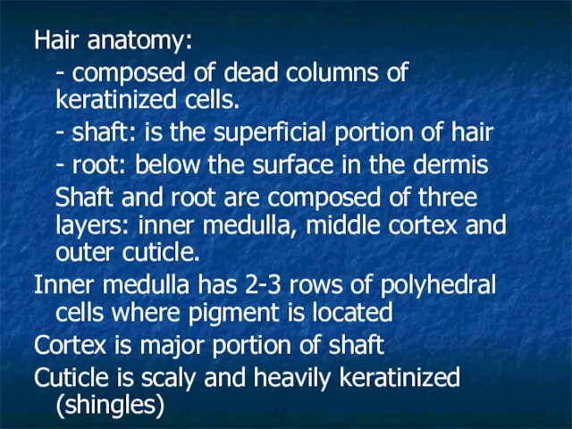
Hair anatomy:
- composed of dead columns of keratinized cells.
- shaft: is
Hair anatomy:
- composed of dead columns of keratinized cells.
- shaft: is
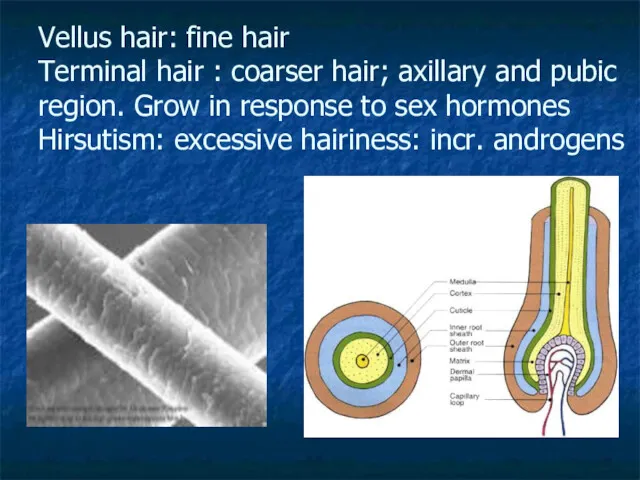
Vellus hair: fine hair
Terminal hair : coarser hair; axillary and
Vellus hair: fine hair Terminal hair : coarser hair; axillary and
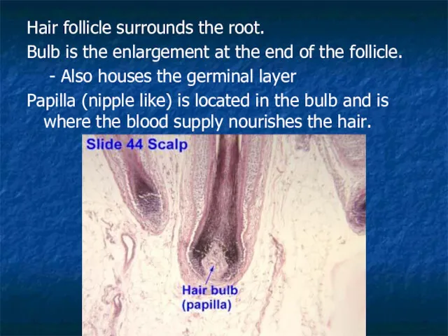
Hair follicle surrounds the root.
Bulb is the enlargement at the end
Hair follicle surrounds the root.
Bulb is the enlargement at the end
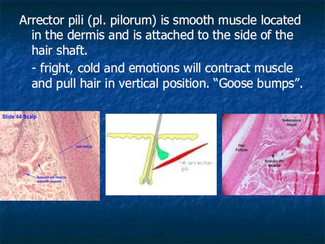
Arrector pili (pl. pilorum) is smooth muscle located in the dermis
Arrector pili (pl. pilorum) is smooth muscle located in the dermis
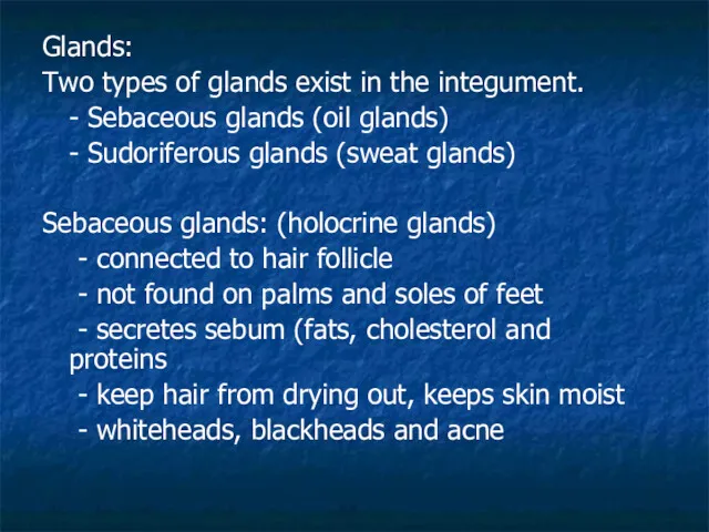
Glands:
Two types of glands exist in the integument.
- Sebaceous glands (oil
Glands:
Two types of glands exist in the integument.
- Sebaceous glands (oil
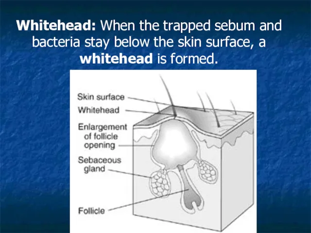
Whitehead: When the trapped sebum and bacteria stay below the skin
Whitehead: When the trapped sebum and bacteria stay below the skin
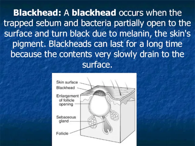
Blackhead: A blackhead occurs when the trapped sebum and bacteria partially
Blackhead: A blackhead occurs when the trapped sebum and bacteria partially
Sudoriferous glands: exocrine glands
- millions located throughout the skin
- two types:
-
Sudoriferous glands: exocrine glands
- millions located throughout the skin
- two types:
-
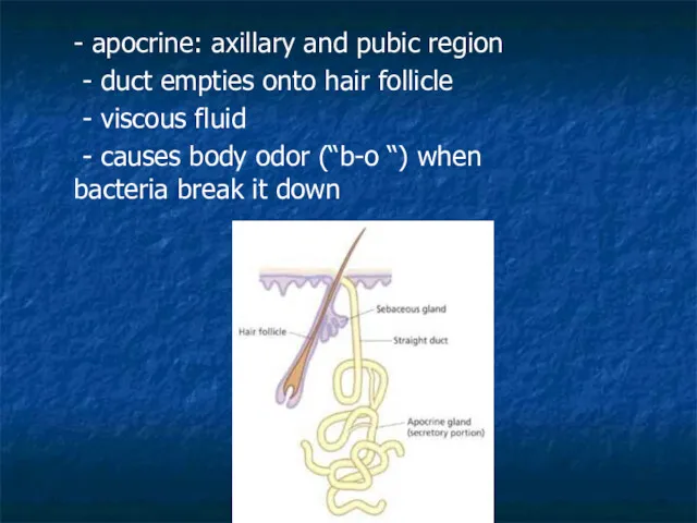
- apocrine: axillary and pubic region
- duct empties onto hair follicle
-
- apocrine: axillary and pubic region
- duct empties onto hair follicle
-
Ceruminous glands: located in ear only
- modified apocrine glands
- originate in
Ceruminous glands: located in ear only
- modified apocrine glands
- originate in
Nails:
- Produced by cells in the epidermis
- Nail plate (body): visible
Nails:
- Produced by cells in the epidermis
- Nail plate (body): visible
Nerve endings:
- Exteroceptors (stimulus outside of body)
- Pacinian (lamellated) corpuscles:
Nerve endings:
- Exteroceptors (stimulus outside of body)
- Pacinian (lamellated) corpuscles:
Pacinian corpuscle
Pacinian corpuscle
Hypodermis
- called subcutaneous, Sub-Q or superficial fascia
- anchors skin to underlying
Hypodermis
- called subcutaneous, Sub-Q or superficial fascia
- anchors skin to underlying
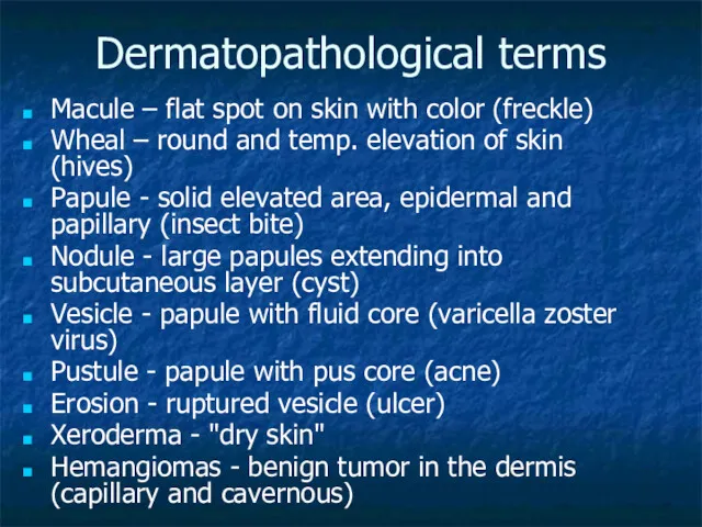
Dermatopathological terms
Macule – flat spot on skin with color (freckle)
Wheal
Dermatopathological terms
Macule – flat spot on skin with color (freckle)
Wheal
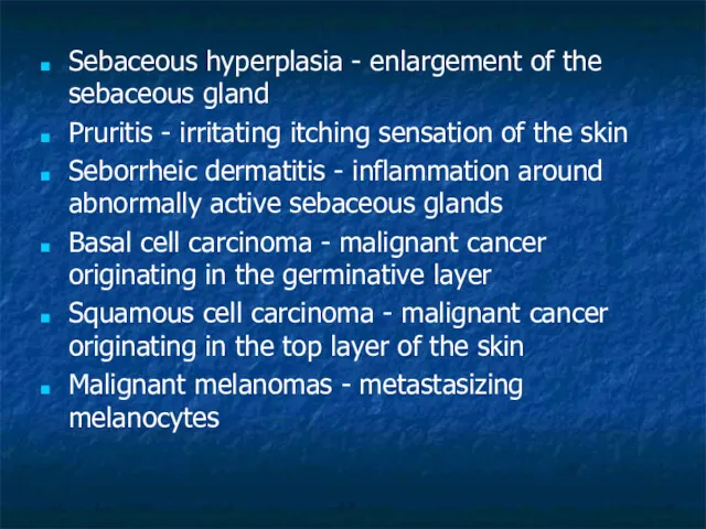
Sebaceous hyperplasia - enlargement of the sebaceous gland
Pruritis - irritating
Sebaceous hyperplasia - enlargement of the sebaceous gland
Pruritis - irritating
 Рекомбинантные антитела для диагностики и терапии
 Презентация к уроку биологии Взаимоотношения у животных 2 часть Диск
 Механизмы передачи сигнала: фермент-связывающие и фермент-содержащие рецепторы
 Органы цветковых растений
 Презентация к уроку Факторы эволюции
 Тип Иглокожие
 Севооборот
 Разнообразие рептилий России
 Урок-презентация. Подцарство одноклеточные
 Экологический проект Письма животным
 Собственно- соединительные ткани
 Грибы. Обобщающий познавательно-развлекательный урок
 Скелет и мышцы
 Внешнее строение листа.
 Тема Тип Членистоногие
 Пауки
Пауки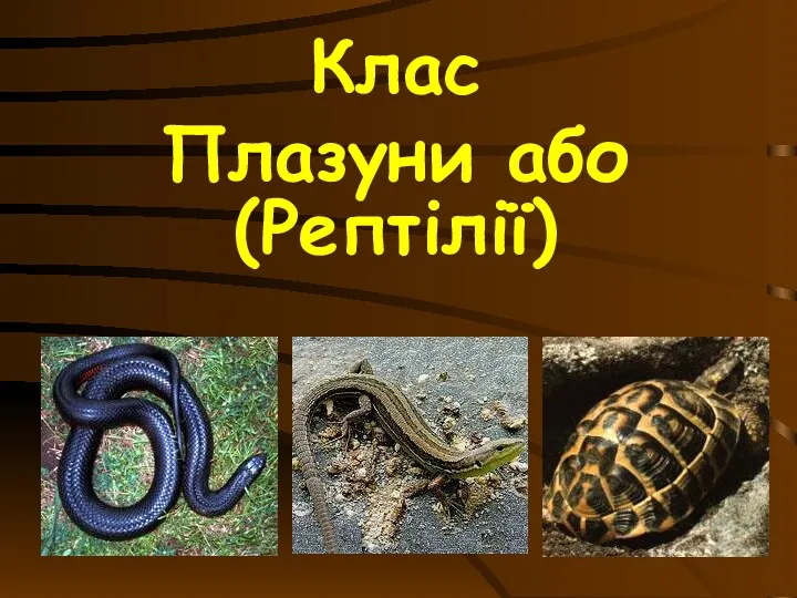 Клас плазуни. (Рептілії)
 Наследственность и изменчивость как основа способности к развитию и эволюции
 Анализ типичных ошибок при выполнении заданий ЕГЭ по биологии 2019 года
 Многообразие и значение грибов
 Электронный портфолио
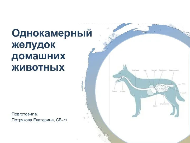 Однокамерный желудок домашних животных
 Метод классификации организмов, применение двойных названий организмов
 Насекомое. Фото
 Только ли лист кормит растение? 5 класс
 Микробиологическая лаборатория, оснащение, правила работы
 Черепно-мозговые нервы
 Белки, жиры и углеводы