- Introduction. Essential Cytology

Содержание
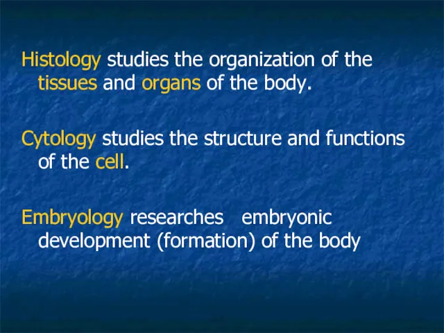
- 2. Histology studies the organization of the tissues and organs of the body. Cytology studies the structure
- 3. Cytology
- 4. Note: 1. The cell is the smallest structural and functional unit of the body 2. Cells
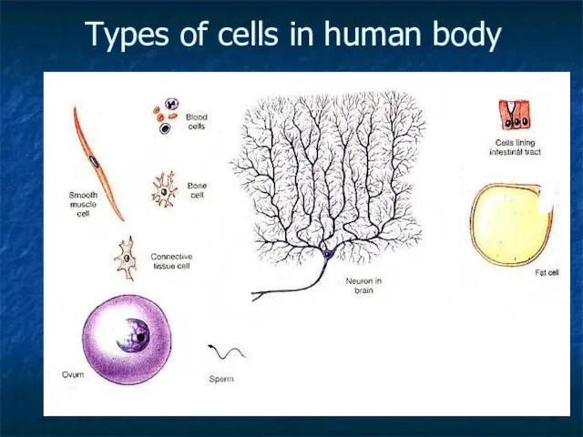
- 5. Types of cells in human body
- 6. Cells produce matrix
- 7. Methods of investigation
- 8. Microscopy – basic method Light microscope: Histological slide:
- 9. Electron microscopy
- 10. Electron microscopy researches Ultrastructure of cells (organelles) and organisation of intercellular matrix
- 11. Light and electron microscopy - are 2 mane methods in histology
- 12. Levels of biological systems Biomolecules Membranes Organelles CELL
- 13. Phospholipids structure : Phosphate group (hydrophilic heads) Glycerol Fatty acids (hydrophobic tails)
- 14. Membrane contents: A. Phospholipids: (1 – hydrophilic head, 2 – hydrophobic tails) B. (3 ) –
- 15. Lipids may be: Phospholipids – triglycerides (polar) Cholesterol (non-polar)
- 16. Proteins may constitute close to 50% of membrane content
- 17. Proteins function: 1- channels, 2- pumps, 3- receptors, 4- enzymes, 5- integrative, 6- structural
- 18. Carbohydrates Present in the outer cell membrane Form Receptors
- 19. Outer cell membrane – cytolemma or plasmalemma
- 20. Membranes form: Outer cell membrane Organelles Vesicles Nuclear envelop
- 21. Cell consists of: - Outer cell membrane, - Cytoplasm and - Nucleus
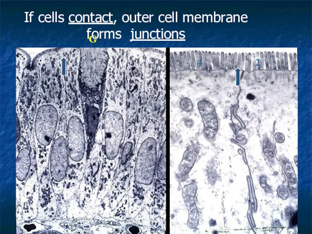
- 22. 1 2 G If cells contact, outer cell membrane forms junctions
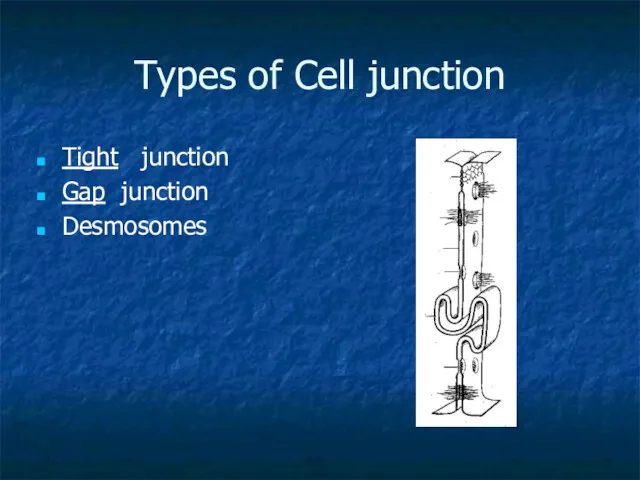
- 23. Types of Cell junction Tight junction Gap junction Desmosomes
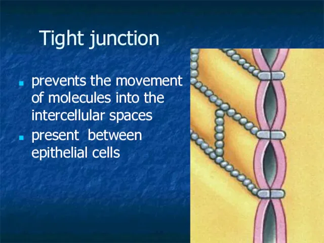
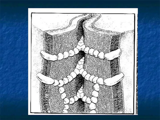
- 24. Tight junction prevents the movement of molecules into the intercellular spaces present between epithelial cells
- 26. Gap junction channels between cells
- 27. Desmosomes - Provide cell attachment
- 28. Inside the cell … Cytoplasm consists of: Matrix (hialoplasm, cytozol) Organelles Inclusions
- 29. Inclusions - granules with secretions, pigment granules, lipid and glycogen droplets
- 30. Organelles: (classification by structure) Membranous Non-membranous
- 31. Organelles: (classification by function) General (present in every cell, perform general function) Ex.: Mitochondrion Special (in
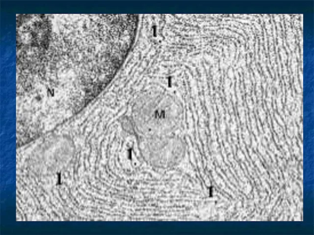
- 32. Rough endoplasmic reticulum Membranes form a network of sac-like structures called cisternae . Ribosomes lie on
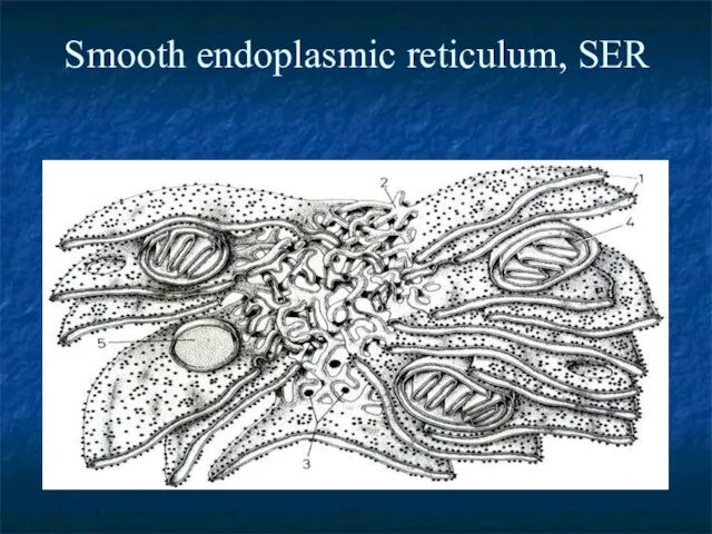
- 34. Smooth endoplasmic reticulum, SER
- 35. SER structure: membranes form tubules without ribosomes. Function: 1. synthesizis of lipids. 2. metabolism of carbohydrates
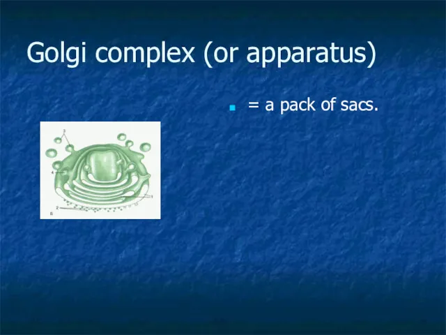
- 36. Golgi complex (or apparatus) = a pack of sacs.
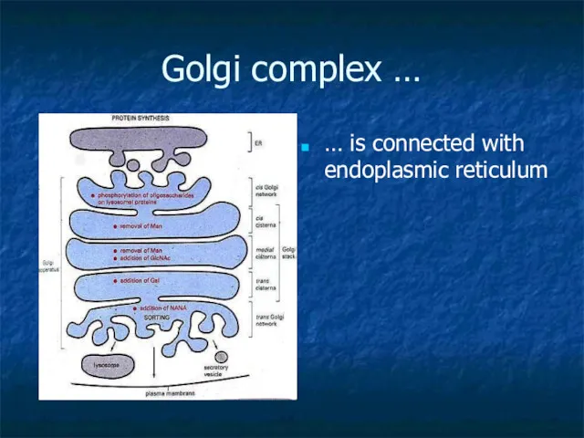
- 37. Golgi complex … … is connected with endoplasmic reticulum
- 38. Golgi apparatus functions: 1. formation of compound molecules – glycoproteins, lipoproteins. 2. production of lysosomes and
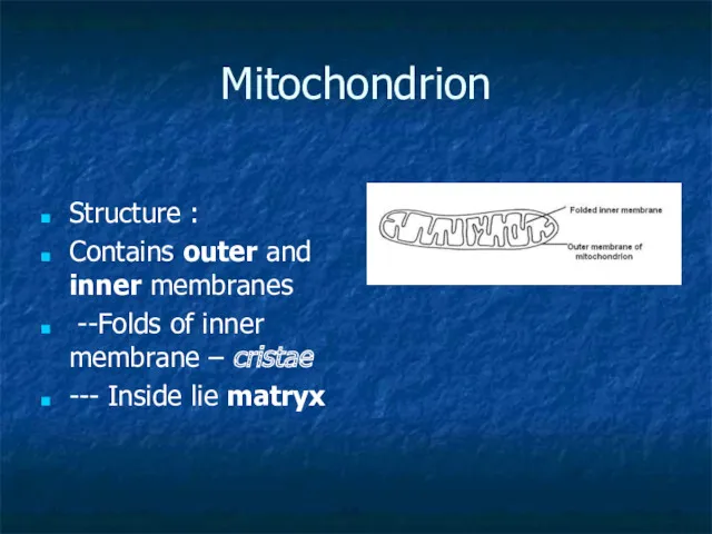
- 39. Mitochondrion Structure : Contains outer and inner membranes --Folds of inner membrane – cristae --- Inside
- 40. Mitochondrion Produce ATP molecules (energy) by Krebs cycle
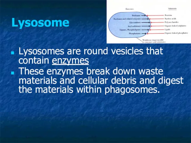
- 41. Lysosome Lysosomes are round vesicles that contain enzymes These enzymes break down waste materials and cellular
- 42. Non-membranous organelles: Microfilaments Microtubules Centrioles (Cell Center) Ribosomes
- 43. Note: Microfilaments, Microtubules form “Skeleton” of the cell
- 44. Cell center Consists of 2 centrioles Centriole = 9 x 3 = 27 microtubules; Function -
- 45. Nucleus consists of: Nucleolemma = nuclear envelope Nucleoplasm Nucleolus Chromatin
- 46. Nuclear envelope - Consists of two membranes: outer and inner
- 47. In the nuclear envelope there are gaps, called nuclear pores, provide transport from nucleus into cytoplasm
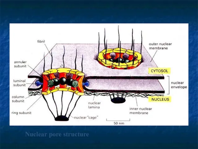
- 48. Nuclear pore structure
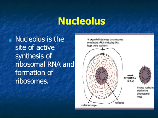
- 49. Nucleolus Nucleolus is the site of active synthesis of ribosomal RNA and formation of ribosomes.
- 50. Chromatin is the combination of DNA and proteins that make up the contents of the nucleus
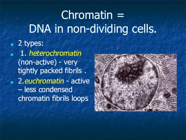
- 51. Chromatin = DNA in non-dividing cells. 2 types: 1. heterochromatin (non-active) - very tightly packed fibrils
- 52. Euchromatin predominates in metabolically active nuclei, Heterochromatin predominates in metabolically inactive nuclei
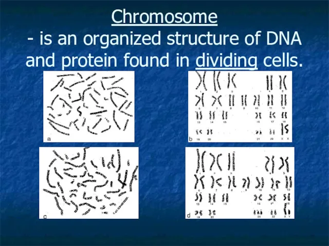
- 53. Chromosome - is an organized structure of DNA and protein found in dividing cells.
- 54. Cell cycle
- 55. The life of a somatic cell is a cyclic process It is called cell cycle It
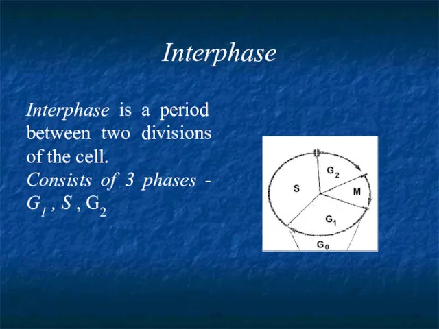
- 56. Interphase Interphase is a period between two divisions of the cell. Consists of 3 phases -
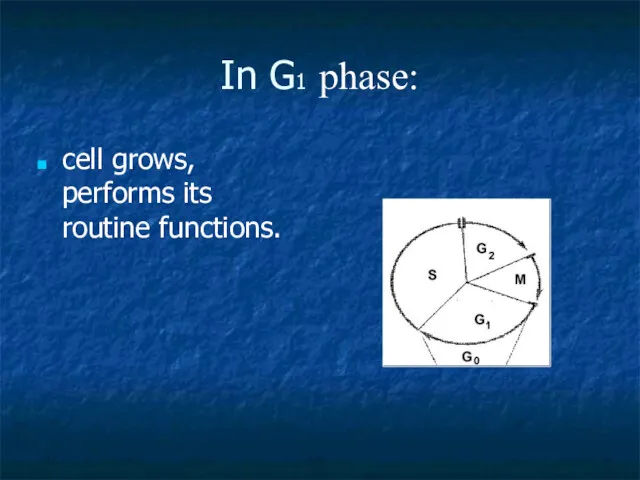
- 57. In G1 phase: cell grows, performs its routine functions.
- 58. S- phase (S- synthesis) DNA molecules are duplicated NOTE: At the beginning of this phase the
- 59. G2 phase In this phase synthesis of proteins, which are required for cell division, takes place.
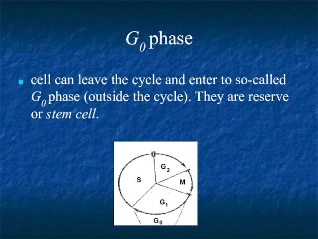
- 60. G0 phase cell can leave the cycle and enter to so-called G0 phase (outside the cycle).
- 61. Mitosis is the process of somatic cells division. Mitosis consists of four phase: prophase, metaphase, anaphase,
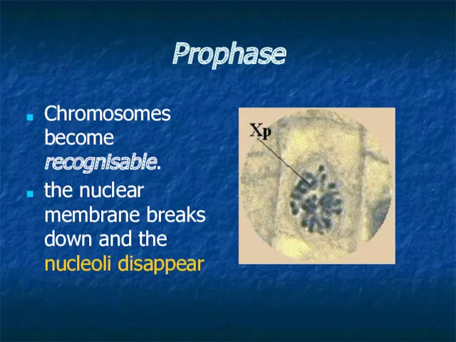
- 62. Prophase Chromosomes become recognisable. the nuclear membrane breaks down and the nucleoli disappear
- 63. Two centrioles separate and move to opposite poles of the cell. microtubules pass from one centriole
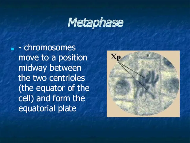
- 64. Metaphase - chromosomes move to a position midway between the two centrioles (the equator of the
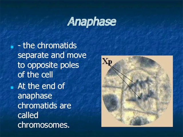
- 65. Anaphase - the chromatids separate and move to opposite poles of the cell At the end
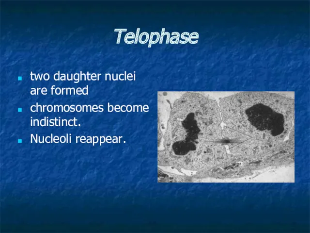
- 66. Telophase two daughter nuclei are formed chromosomes become indistinct. Nucleoli reappear.
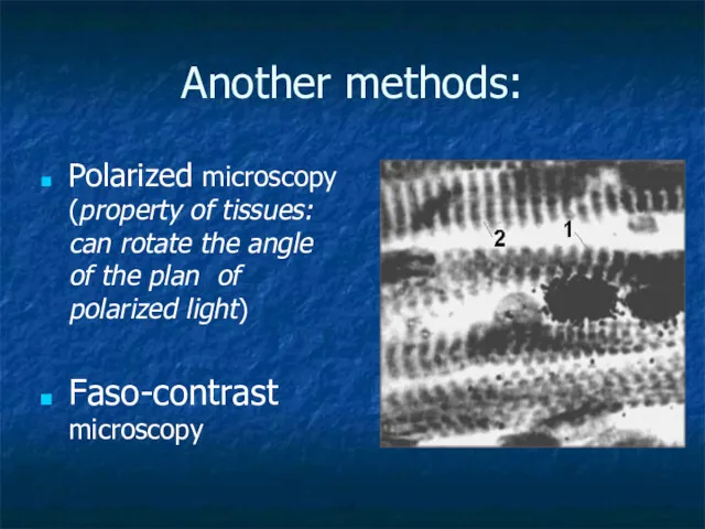
- 67. Another methods: Polarized microscopy (property of tissues: can rotate the angle of the plan of polarized
- 69. Скачать презентацию
Histology studies the organization of the tissues and organs of the
Cytology
Cytology
Note:
1. The cell is the smallest structural and functional unit of
Note:
1. The cell is the smallest structural and functional unit of
Types of cells in human body
Types of cells in human body
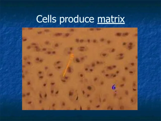
Cells produce matrix
Cells produce matrix
Methods of investigation
Methods of investigation
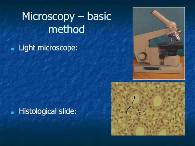
Microscopy – basic method
Light microscope:
Histological slide:
Microscopy – basic method
Light microscope:
Histological slide:
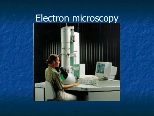
Electron microscopy
Electron microscopy
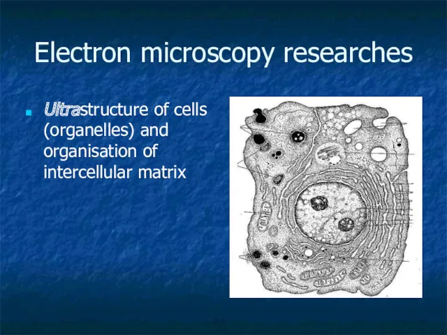
Electron microscopy researches
Ultrastructure of cells (organelles) and organisation of intercellular
Electron microscopy researches
Ultrastructure of cells (organelles) and organisation of intercellular
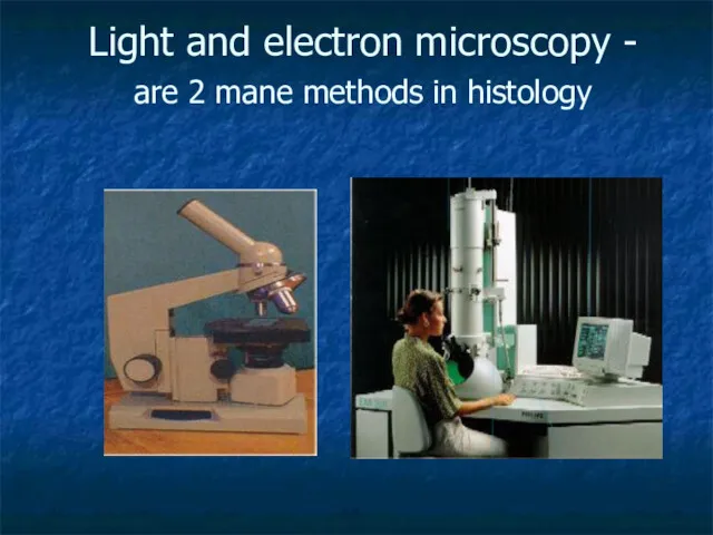
Light and electron microscopy -
are 2 mane methods in histology
Light and electron microscopy -
are 2 mane methods in histology
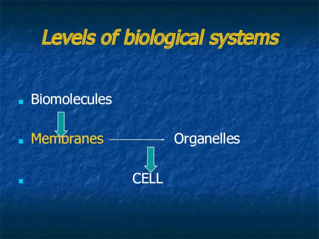
Levels of biological systems
Biomolecules
Membranes Organelles
CELL
Levels of biological systems
Biomolecules
Membranes Organelles
CELL
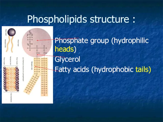
Phospholipids structure :
Phosphate group (hydrophilic heads)
Glycerol
Fatty acids (hydrophobic tails)
Phospholipids structure :
Phosphate group (hydrophilic heads)
Glycerol
Fatty acids (hydrophobic tails)
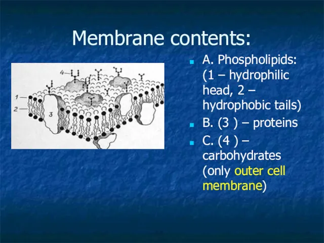
Membrane contents:
A. Phospholipids: (1 – hydrophilic head, 2 – hydrophobic tails)
B.
Membrane contents:
A. Phospholipids: (1 – hydrophilic head, 2 – hydrophobic tails)
B.
Lipids
may be:
Phospholipids – triglycerides (polar)
Cholesterol (non-polar)
Lipids
may be:
Phospholipids – triglycerides (polar)
Cholesterol (non-polar)
Proteins
may constitute close to 50% of membrane content
Proteins
may constitute close to 50% of membrane content
Proteins
function:
1- channels,
2- pumps,
3- receptors,
4- enzymes,
5- integrative,
Proteins
function:
1- channels,
2- pumps,
3- receptors,
4- enzymes,
5- integrative,
Carbohydrates
Present in the outer cell membrane
Form Receptors
Carbohydrates
Present in the outer cell membrane
Form Receptors
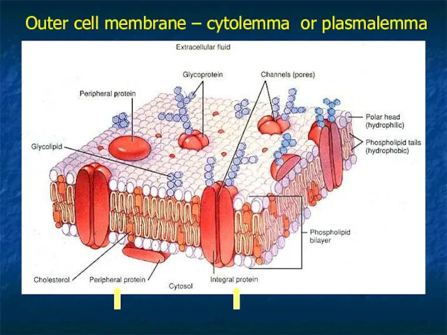
Outer cell membrane – cytolemma or plasmalemma
Outer cell membrane – cytolemma or plasmalemma
Membranes form:
Outer cell membrane
Organelles
Vesicles
Nuclear envelop
Membranes form:
Outer cell membrane
Organelles
Vesicles
Nuclear envelop
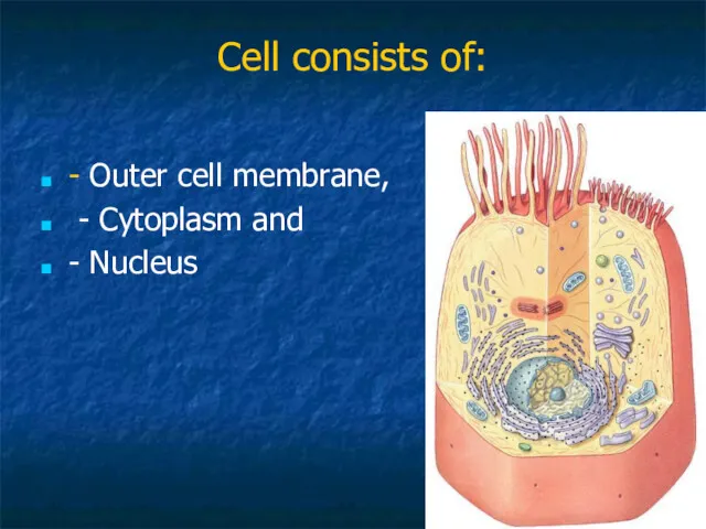
Cell consists of:
- Outer cell membrane,
- Cytoplasm and
-
Cell consists of:
- Outer cell membrane,
- Cytoplasm and
-
1
2
G
If cells contact, outer cell membrane forms junctions
1
2
G
If cells contact, outer cell membrane forms junctions
Types of Cell junction
Tight junction
Gap junction
Desmosomes
Types of Cell junction
Tight junction
Gap junction
Desmosomes
Tight junction
prevents the movement of molecules into the intercellular spaces
Tight junction
prevents the movement of molecules into the intercellular spaces
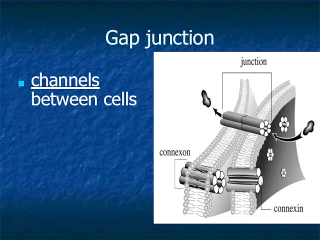
Gap junction
channels between cells
Gap junction
channels between cells
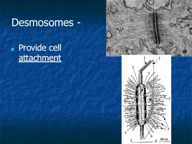
Desmosomes -
Provide cell attachment
Desmosomes -
Provide cell attachment
Inside the cell …
Cytoplasm consists of:
Matrix (hialoplasm, cytozol)
Organelles
Inclusions
Inside the cell …
Cytoplasm consists of:
Matrix (hialoplasm, cytozol)
Organelles
Inclusions
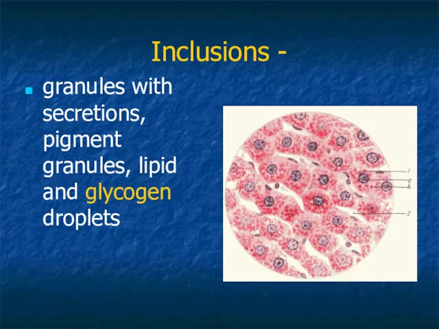
Inclusions -
granules with secretions, pigment granules, lipid and glycogen droplets
Inclusions -
granules with secretions, pigment granules, lipid and glycogen droplets
Organelles:
(classification by structure)
Membranous
Non-membranous
Organelles:
(classification by structure)
Membranous
Non-membranous
Organelles:
(classification by function)
General
(present in every cell, perform general function)
Ex.:
Organelles:
(classification by function)
General
(present in every cell, perform general function)
Ex.:
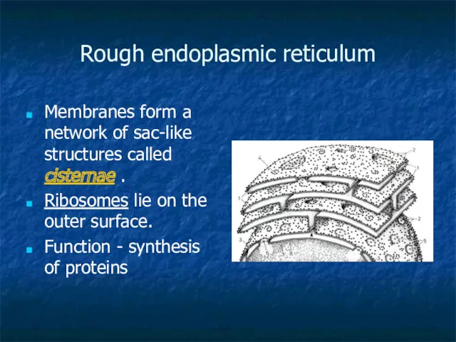
Rough endoplasmic reticulum
Membranes form a network of sac-like structures called cisternae
Rough endoplasmic reticulum
Membranes form a network of sac-like structures called cisternae
Smooth endoplasmic reticulum, SER
Smooth endoplasmic reticulum, SER
SER structure: membranes form tubules without ribosomes.
Function:
1. synthesizis of
Function:
1. synthesizis of
Golgi complex (or apparatus)
= a pack of sacs.
Golgi complex (or apparatus)
= a pack of sacs.
Golgi complex …
… is connected with endoplasmic reticulum
Golgi complex …
… is connected with endoplasmic reticulum
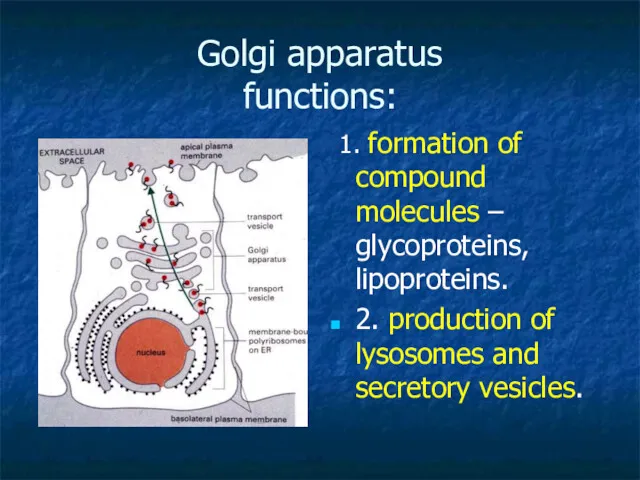
Golgi apparatus
functions:
1. formation of compound molecules – glycoproteins, lipoproteins.
Golgi apparatus
functions:
1. formation of compound molecules – glycoproteins, lipoproteins.
Mitochondrion
Structure :
Contains outer and inner membranes
--Folds of
Mitochondrion
Structure :
Contains outer and inner membranes
--Folds of
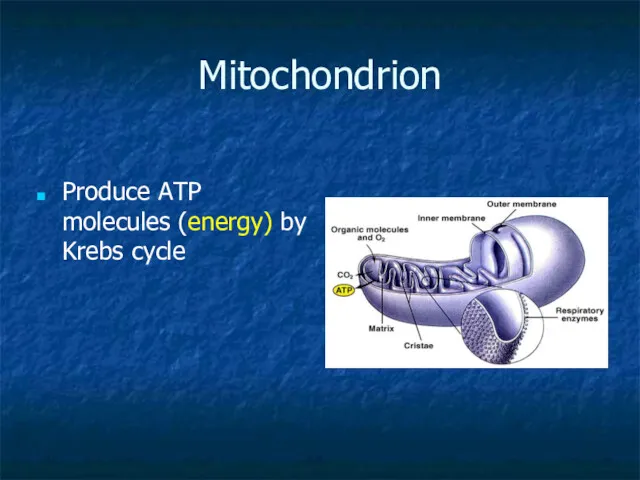
Mitochondrion
Produce ATP molecules (energy) by Krebs cycle
Mitochondrion
Produce ATP molecules (energy) by Krebs cycle
Lysosome
Lysosomes are round vesicles that contain enzymes
These enzymes break down waste
Lysosome
Lysosomes are round vesicles that contain enzymes
These enzymes break down waste
Non-membranous organelles:
Microfilaments
Microtubules
Centrioles (Cell Center)
Ribosomes
Non-membranous organelles:
Microfilaments
Microtubules
Centrioles (Cell Center)
Ribosomes
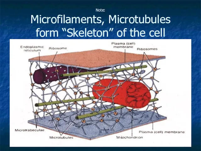
Note:
Microfilaments, Microtubules
form “Skeleton” of the cell
Note:
Microfilaments, Microtubules
form “Skeleton” of the cell
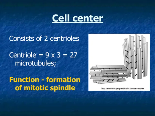
Cell center
Consists of 2 centrioles
Centriole = 9 x 3 = 27
Cell center
Consists of 2 centrioles
Centriole = 9 x 3 = 27

Nucleus
consists of:
Nucleolemma = nuclear envelope
Nucleoplasm
Nucleolus
Chromatin
Nucleus
consists of:
Nucleolemma = nuclear envelope
Nucleoplasm
Nucleolus
Chromatin
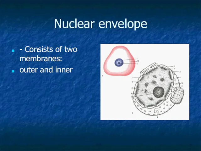
Nuclear envelope
- Consists of two membranes:
outer and inner
Nuclear envelope
- Consists of two membranes:
outer and inner
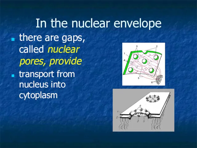
In the nuclear envelope
there are gaps, called nuclear pores, provide
transport
In the nuclear envelope
there are gaps, called nuclear pores, provide
transport
Nuclear pore structure
Nuclear pore structure
Nucleolus
Nucleolus is the site of active synthesis of ribosomal RNA and
Nucleolus
Nucleolus is the site of active synthesis of ribosomal RNA and
Chromatin
is the combination of DNA and proteins that make up
Chromatin
is the combination of DNA and proteins that make up
Chromatin =
DNA in non-dividing cells.
2 types:
1. heterochromatin (non-active)
Chromatin =
DNA in non-dividing cells.
2 types:
1. heterochromatin (non-active)
Euchromatin predominates in metabolically active nuclei,
Heterochromatin predominates in metabolically inactive
Euchromatin predominates in metabolically active nuclei,
Heterochromatin predominates in metabolically inactive
Chromosome
- is an organized structure of DNA and protein found
Chromosome - is an organized structure of DNA and protein found
Cell cycle
Cell cycle
The life of a somatic cell is a cyclic process
It
The life of a somatic cell is a cyclic process
It
Interphase
Interphase is a period between two divisions of the cell.
Interphase
Interphase is a period between two divisions of the cell.
In G1 phase:
cell grows, performs its routine functions.
In G1 phase:
cell grows, performs its routine functions.
S- phase (S- synthesis)
DNA molecules are duplicated
NOTE: At the
S- phase (S- synthesis)
DNA molecules are duplicated
NOTE: At the
G2 phase
In this phase synthesis of proteins, which are required for
G2 phase
In this phase synthesis of proteins, which are required for
G0 phase
cell can leave the cycle and enter to so-called
G0 phase
cell can leave the cycle and enter to so-called
Mitosis
is the process of somatic cells division.
Mitosis consists of four
Mitosis
is the process of somatic cells division.
Mitosis consists of four
Prophase
Chromosomes become recognisable.
the nuclear membrane breaks down and the nucleoli
Prophase
Chromosomes become recognisable.
the nuclear membrane breaks down and the nucleoli
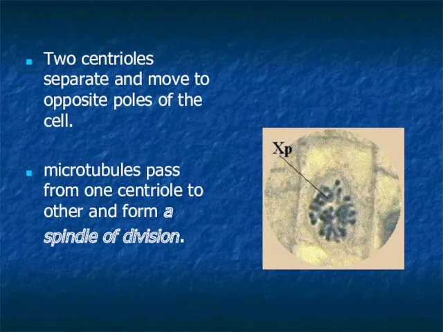
Two centrioles separate and move to opposite poles of the cell.
Two centrioles separate and move to opposite poles of the cell.
Metaphase
- chromosomes move to a position midway between the two centrioles
Metaphase
- chromosomes move to a position midway between the two centrioles
Anaphase
- the chromatids separate and move to opposite poles of
Anaphase
- the chromatids separate and move to opposite poles of
Telophase
two daughter nuclei are formed
chromosomes become indistinct.
Nucleoli reappear.
Telophase
two daughter nuclei are formed
chromosomes become indistinct.
Nucleoli reappear.
Another methods:
Polarized microscopy (property of tissues: can rotate the angle of
Another methods:
Polarized microscopy (property of tissues: can rotate the angle of
 Удивительный мир живой природы
 Эволюционный процесс
 Энергетический метаболизм микробов. Роль генома в метаболической активности микроорганизмов
 Урок на тему :Совместная жизнь видов в биогеоценозе.
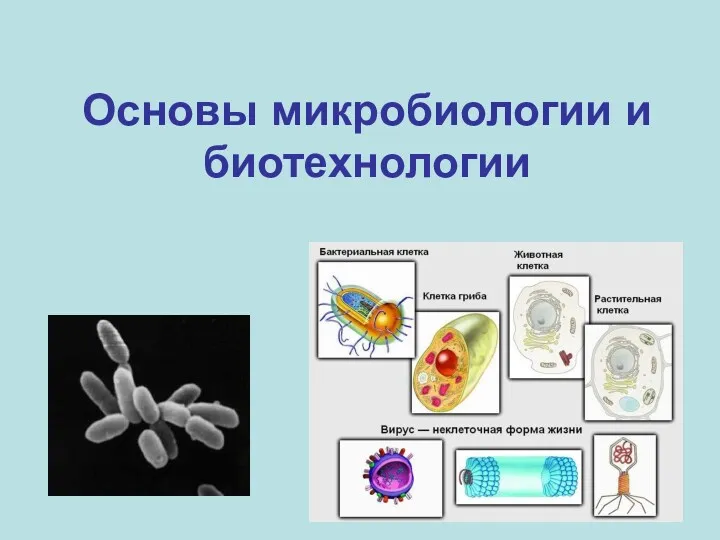 Основы микробиологии и биотехнологии
 Красноухая черепаха
 Нәсілдердің пайда болуы
 Жизнь и творчество Виктора Драгунского
 A Tour of the Cell
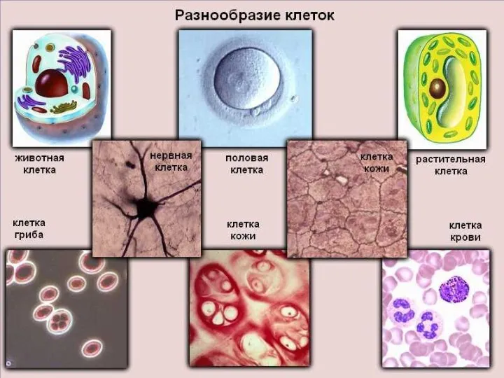 Разнообразие клеток
 Невидимые нити. Окружающий мир 2 класс
 Гербарии растений, содержащих флавоноиды I
 Собака – верный друг
 Приспособленность организмов к определенной среде обитания
 Удивительные животные
 Лишайники. Внешнее и внутреннее строение
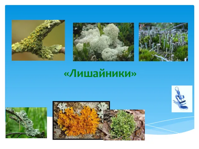 Лишайники. Симбиоз
 изменчивость
 В краю родном. Викторина о природе Ростовской области
 Строение спинного мозга человека
 Рентгеноанатомия черепа. Обозначьте кости мозгового черепа
 Первобытный человек
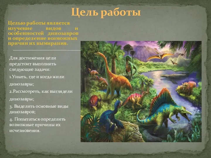 Где и когда жили динозавры?
 Вирусы. (11 класс)
 Ткани, органы и системы органов
 Значення мікробіології в практичній діяльності лікаря-стоматолога
 Физиология возбудимых клеток. Медиаторы (лекция № 5)
 Сердце. Анатомия сердца