- Introduction to Biology. Forms of life. Biology of the cell

Содержание
- 2. Department of Medical Biology
- 3. Biology teacher Svetlana
- 6. Biology teacher Tatyana
- 7. Biology teacher Anna
- 9. https://www.facebook.com/Medical-Biology-299203590209244/
- 10. https://vk.com/club80450232
- 11. Characteristics of Life Biology examines the structure, function, growth, origin, evolution, and distribution of living things.
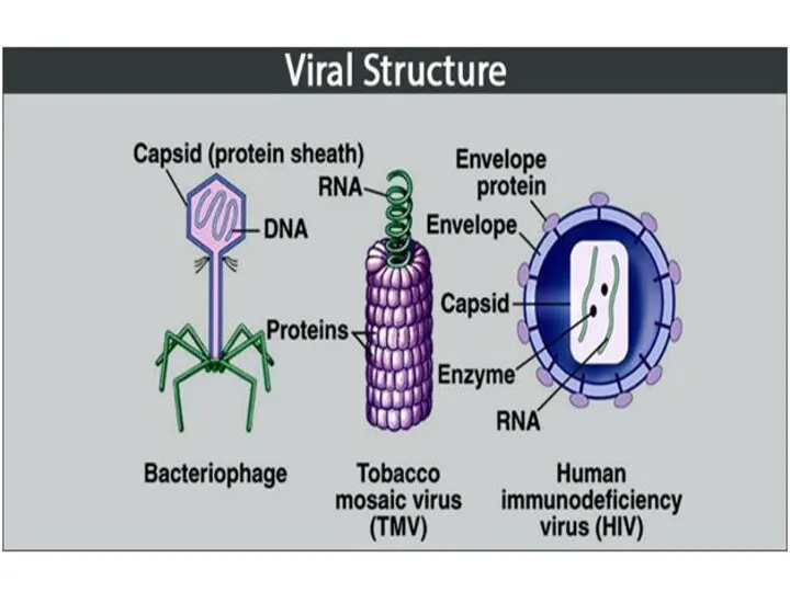
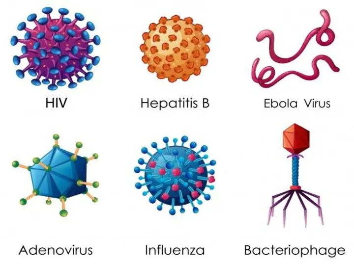
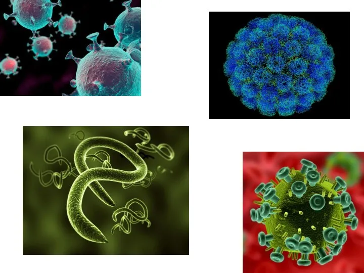
- 13. Forms of life - non-cellular and cellular organisms.
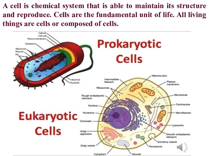
- 17. A cell is chemical system that is able to maintain its structure and reproduce. Cells are
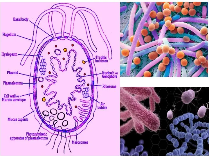
- 18. The first life on Earth came in the form of a prokaryotic cell. For two billion
- 20. Eukaryotic cells are cells that contain a nucleus and organelles, and are enclosed by a plasma
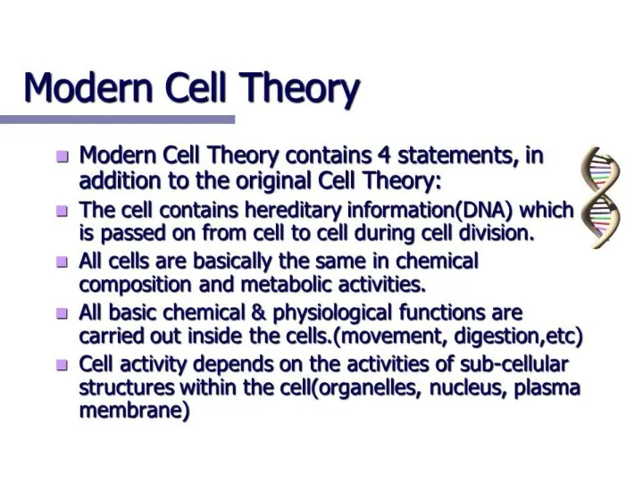
- 21. The cell theory developed in 1839 by microbiologists Schleiden and Schwann describes the properties of cells.
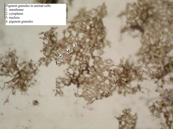
- 25. Representative Animal Cell
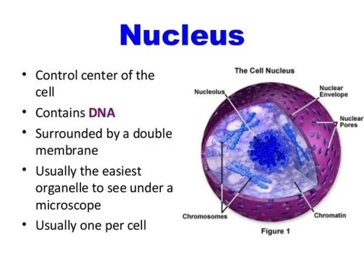
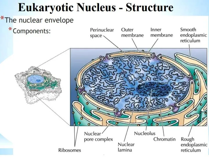
- 27. The nucleus is a highly specialized organelle that serves as the information processing and administrative center
- 30. Nucleolus Within the nucleus is a small subspace known as the nucleolus. It is not bound
- 32. Endoplasmic Reticulum Endoplasmic means inside (endo) the cytoplasm (plasm). Reticulum comes from the Latin word for
- 33. Rough Endoplasmic Reticulum The rough endoplasmic reticulum is so-called because its surface is studded with ribosomes,
- 34. Smooth Endoplasmic Reticulum The smooth endoplasmic reticulum makes lipids and steroids, instead of being involved in
- 35. Ribosome: Situated in two areas of the cytoplasm. They are seen scattered in the cytoplasm and
- 40. Golgi apparatus (aka Golgi body aka Golgi) We mentioned the Golgi apparatus earlier when we discussed
- 41. Different molecules actually have different fates upon entering the Golgi. This determination is done by tagging
- 43. Lysosome The lysosome is the cell’s recycling center. These organelles are spheres full of enzymes ready
- 45. Peroxisome Like the lysosome, the peroxisome is a spherical organelle responsible for destroying its contents. Unlike
- 46. Key Concepts Mitochondria are semi‐autonomous organelles that are descendants of endosymbiotic bacteria. Mitochondria play a pivotal
- 47. In the cell, mitochondria form a continuous and highly dynamic network. In addition, they intimately interact
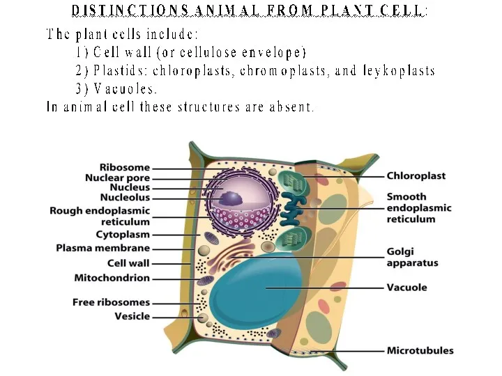
- 50. Cell Walls Found in plants, fungi, & many protists Surrounds plasma membrane Cell Wall Differences Plants
- 51. Cytoskeleton Filaments & fibers Made of 3 fiber types Microfilaments Microtubules Intermediate filaments 3 functions: mechanical
- 52. Cilia & Flagella Provide motility Cilia Short Used to move substances outside human cells Flagella Whip-like
- 53. Centrioles Pairs of microtubular structures Play a role in cell division
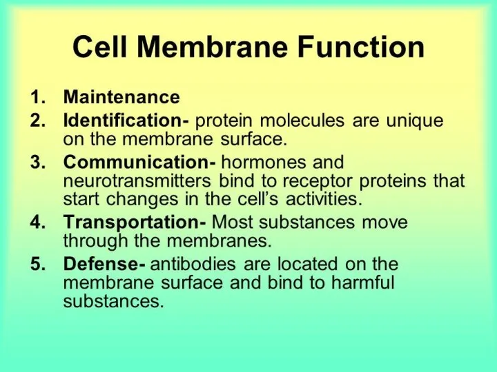
- 55. Plasma membrane can be defined as a biological membrane or an outer membrane of a cell,
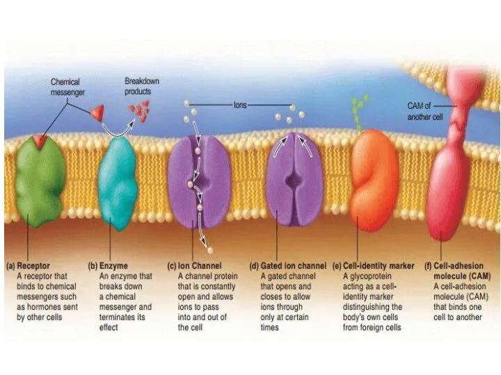
- 59. Membrane Proteins 1. Channels or transporters Move molecules in one direction 2. Receptors Recognize certain chemicals
- 60. Membrane Proteins 3. Glycoproteins Identify cell type 4. Enzymes Catalyze production of substances
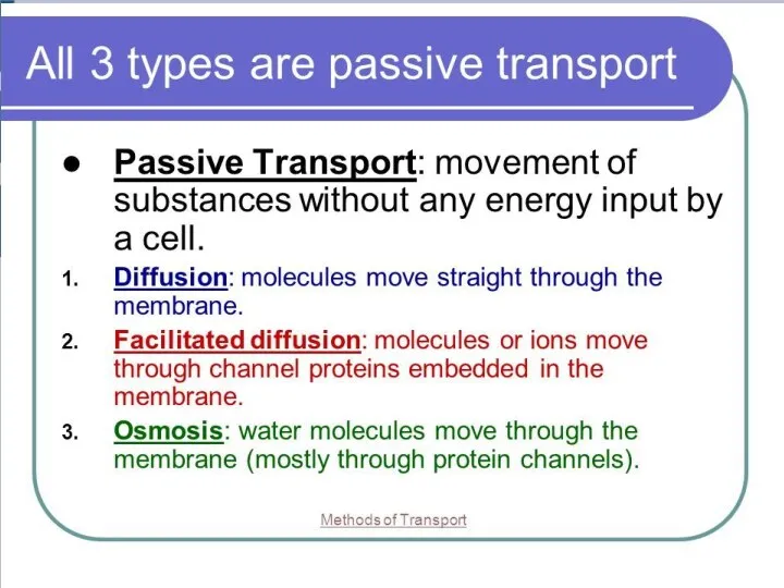
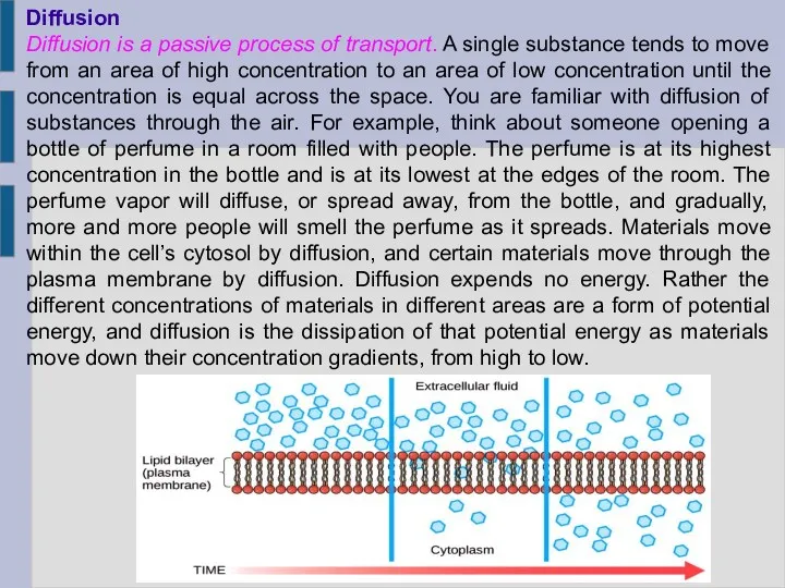
- 77. Diffusion Diffusion is a passive process of transport. A single substance tends to move from an
- 79. Several factors affect the rate of diffusion. Extent of the concentration gradient: The greater the difference
- 80. Facilitated transport In facilitated transport, also called facilitated diffusion, material moves across the plasma membrane with
- 83. Osmosis Osmosis is the diffusion of water through a semipermeable membrane according to the concentration gradient
- 84. Solution Differences & Cells solvent + solute = solution Hypotonic Solutes in cell more than outside
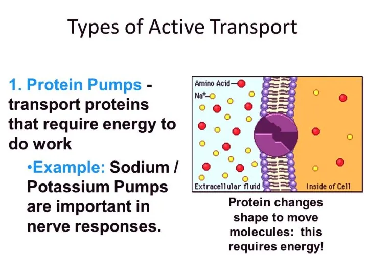
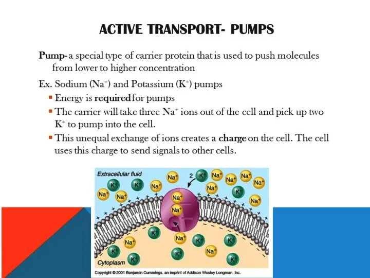
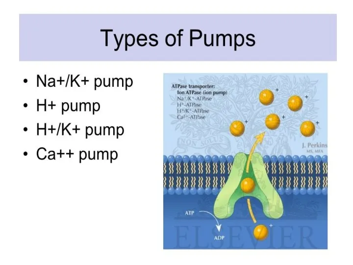
- 89. Active Transport Molecular movement Requires energy (against gradient) Example is sodium-potassium pump
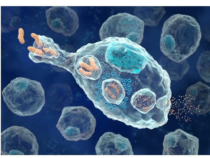
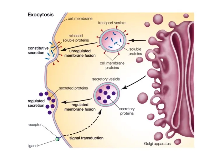
- 94. Endocytosis Movement of large material Particles Organisms Large molecules Movement is into cells Types of endocytosis
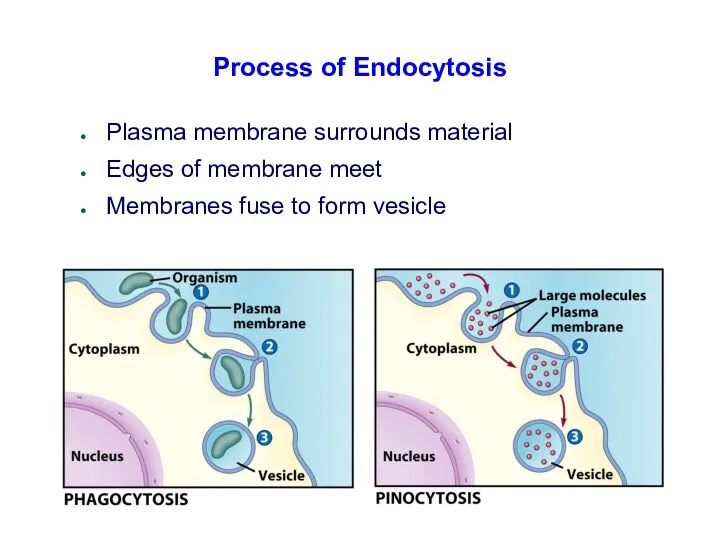
- 95. Process of Endocytosis Plasma membrane surrounds material Edges of membrane meet Membranes fuse to form vesicle
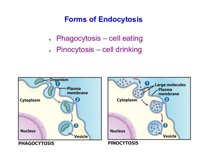
- 96. Forms of Endocytosis Phagocytosis – cell eating Pinocytosis – cell drinking
- 101. Скачать презентацию
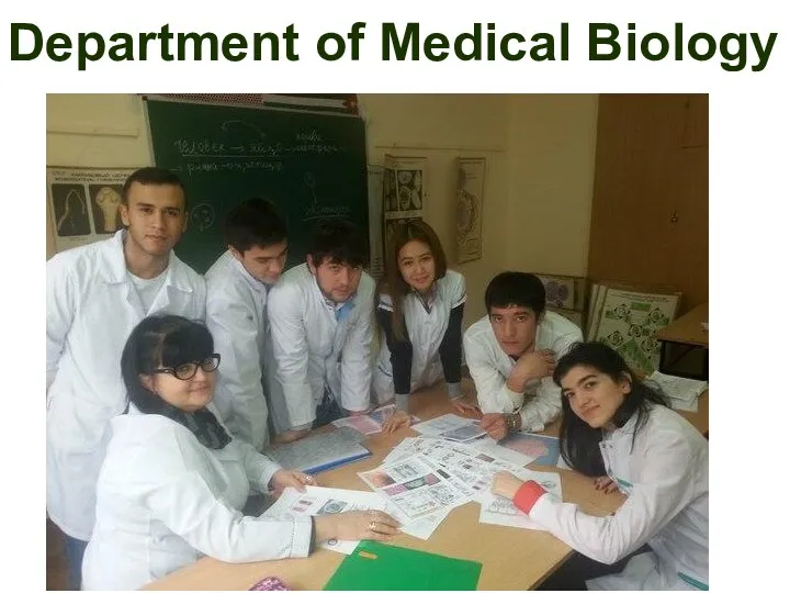
Department of Medical Biology
Department of Medical Biology
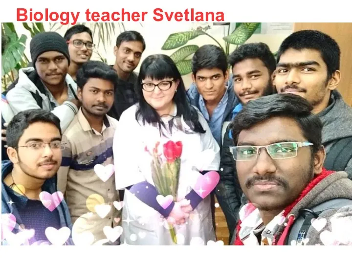
Biology teacher Svetlana
Biology teacher Svetlana
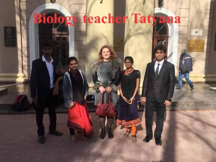
Biology teacher Tatyana
Biology teacher Tatyana
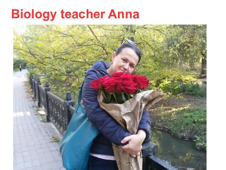
Biology teacher Anna
Biology teacher Anna
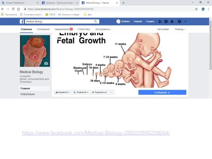
https://www.facebook.com/Medical-Biology-299203590209244/
https://www.facebook.com/Medical-Biology-299203590209244/
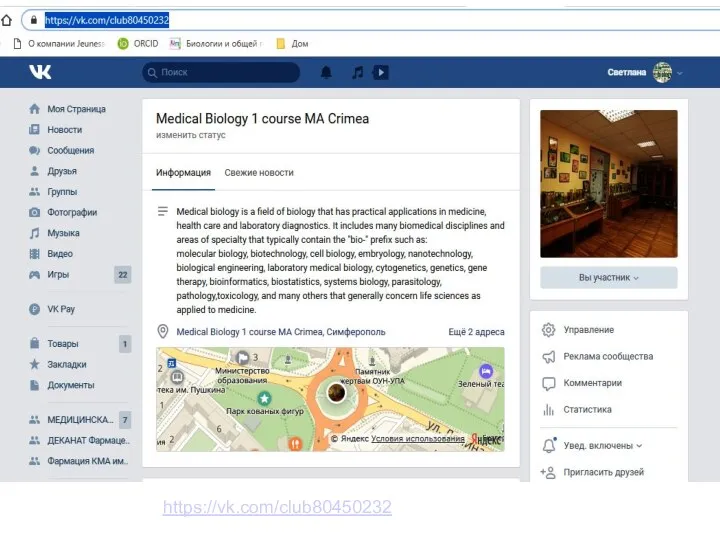
https://vk.com/club80450232
https://vk.com/club80450232
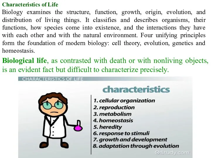
Characteristics of Life
Biology examines the structure, function, growth, origin, evolution, and
Characteristics of Life
Biology examines the structure, function, growth, origin, evolution, and
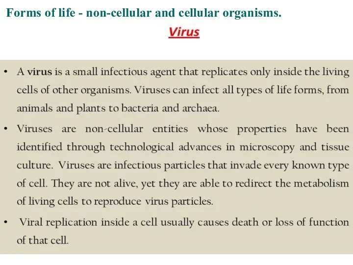
Forms of life - non-cellular and cellular organisms.
Forms of life - non-cellular and cellular organisms.
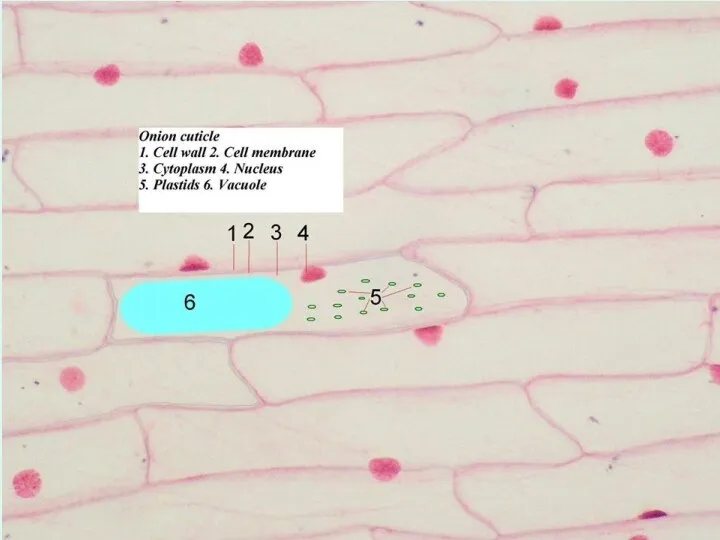
A cell is chemical system that is able to maintain its
A cell is chemical system that is able to maintain its
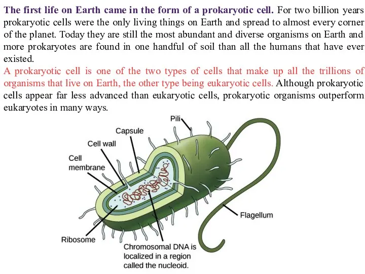
The first life on Earth came in the form of a
The first life on Earth came in the form of a
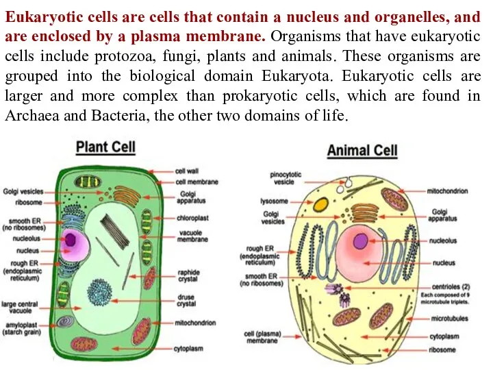
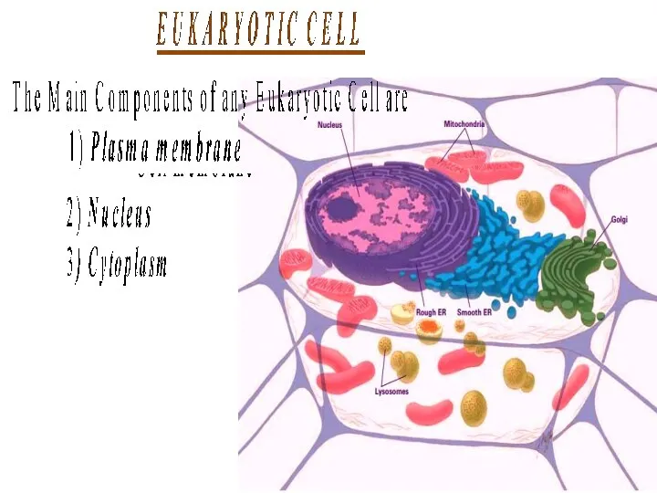
Eukaryotic cells are cells that contain a nucleus and organelles, and
Eukaryotic cells are cells that contain a nucleus and organelles, and
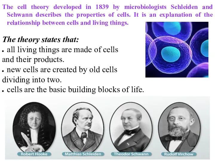
The cell theory developed in 1839 by microbiologists Schleiden and Schwann
The cell theory developed in 1839 by microbiologists Schleiden and Schwann
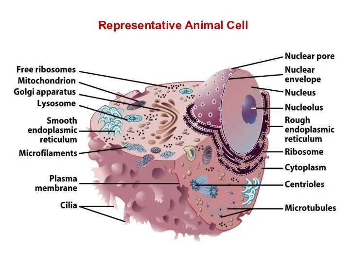
Representative Animal Cell
Representative Animal Cell
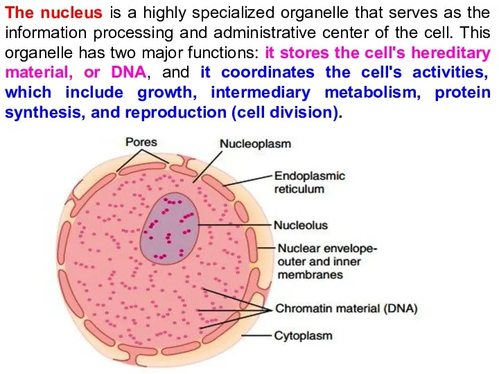
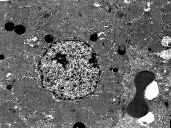
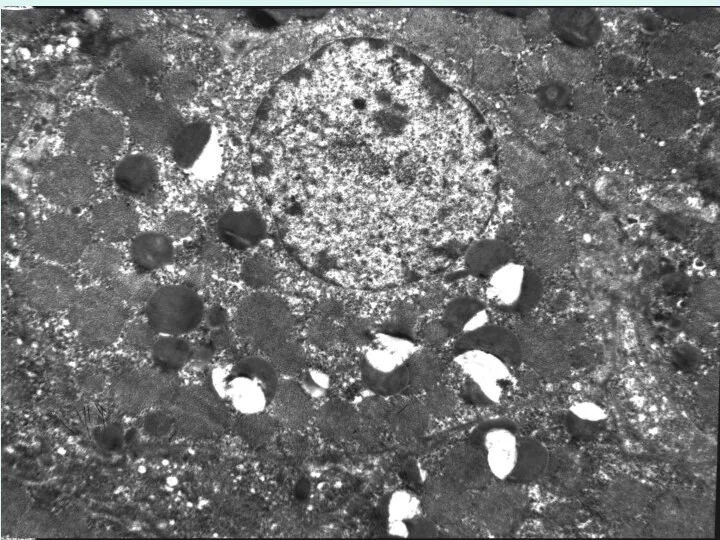
The nucleus is a highly specialized organelle that serves as the
The nucleus is a highly specialized organelle that serves as the
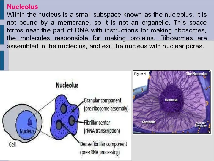
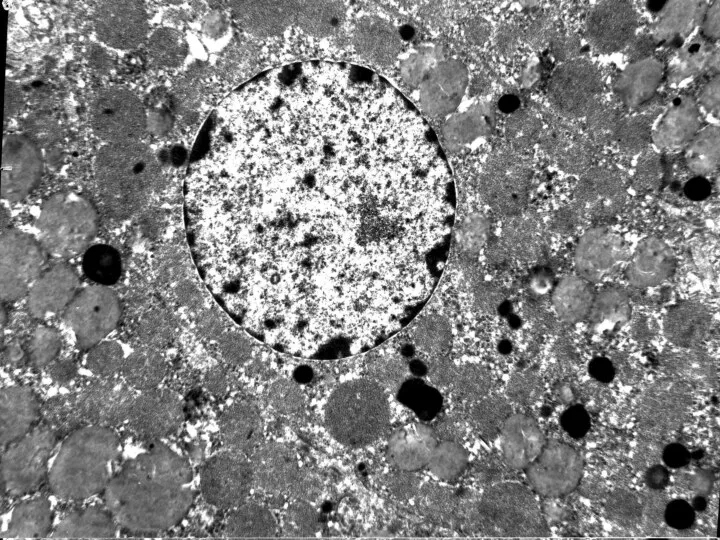
Nucleolus
Within the nucleus is a small subspace known as the nucleolus.
Nucleolus
Within the nucleus is a small subspace known as the nucleolus.
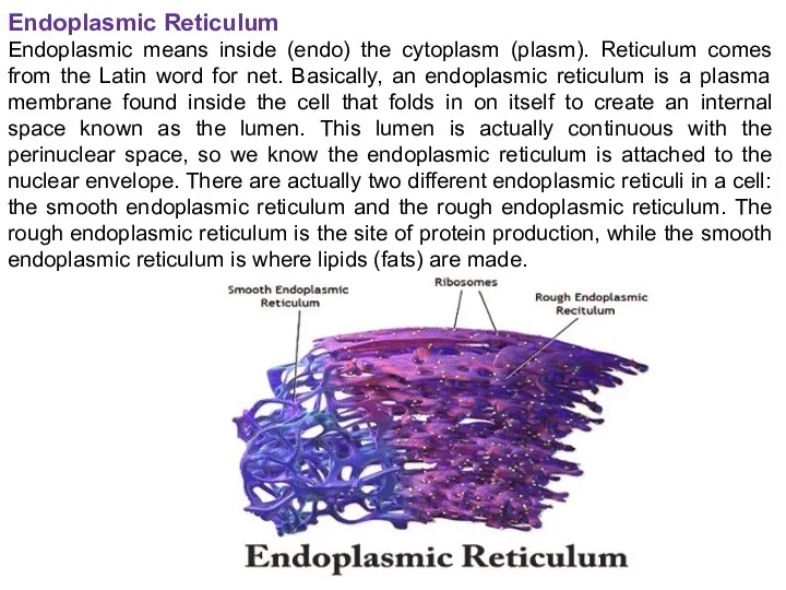
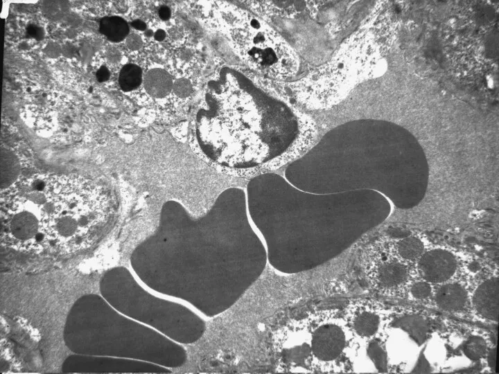
Endoplasmic Reticulum
Endoplasmic means inside (endo) the cytoplasm (plasm). Reticulum comes from
Endoplasmic Reticulum
Endoplasmic means inside (endo) the cytoplasm (plasm). Reticulum comes from
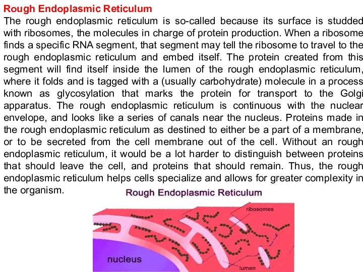
Rough Endoplasmic Reticulum
The rough endoplasmic reticulum is so-called because its surface
Rough Endoplasmic Reticulum
The rough endoplasmic reticulum is so-called because its surface
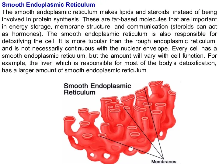
Smooth Endoplasmic Reticulum
The smooth endoplasmic reticulum makes lipids and steroids, instead
Smooth Endoplasmic Reticulum
The smooth endoplasmic reticulum makes lipids and steroids, instead
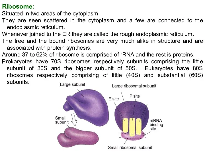
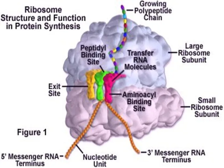
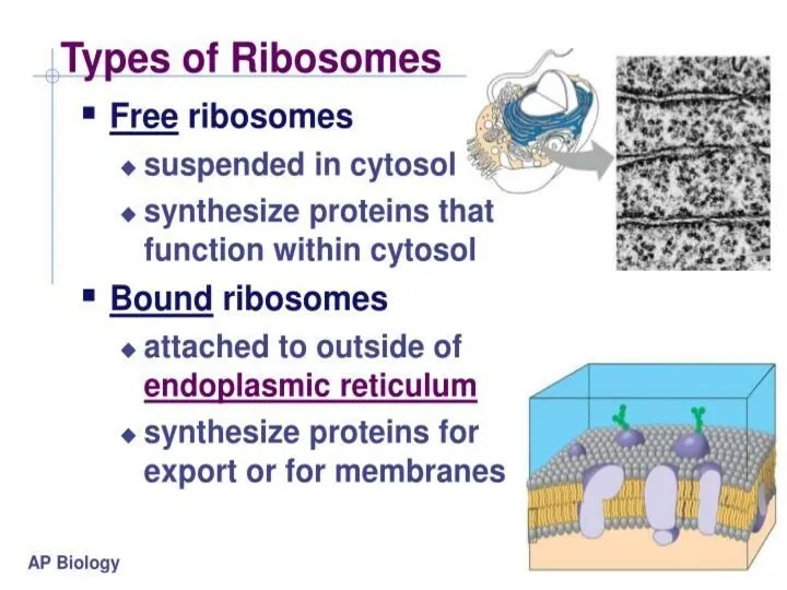
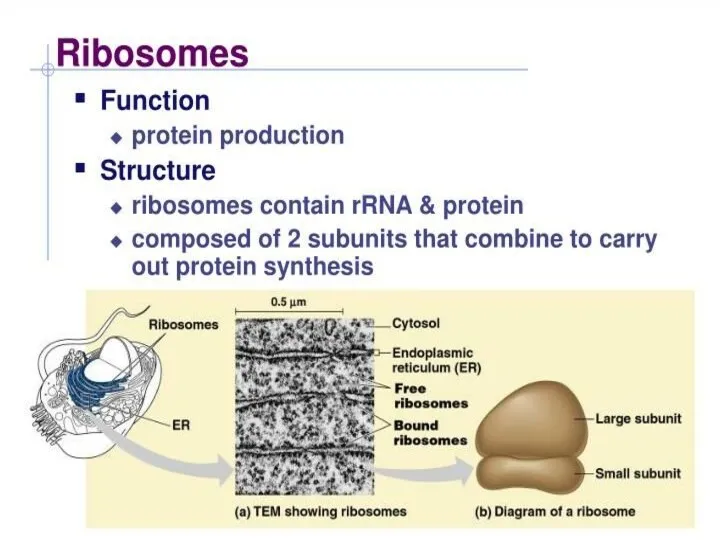
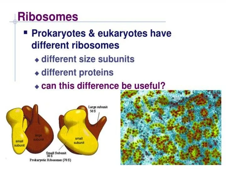
Ribosome:
Situated in two areas of the cytoplasm.
They are seen scattered in
Ribosome:
Situated in two areas of the cytoplasm.
They are seen scattered in
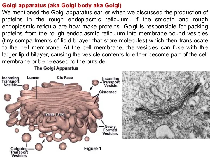
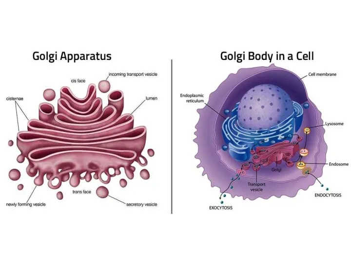
Golgi apparatus (aka Golgi body aka Golgi)
We mentioned the Golgi apparatus
Golgi apparatus (aka Golgi body aka Golgi)
We mentioned the Golgi apparatus
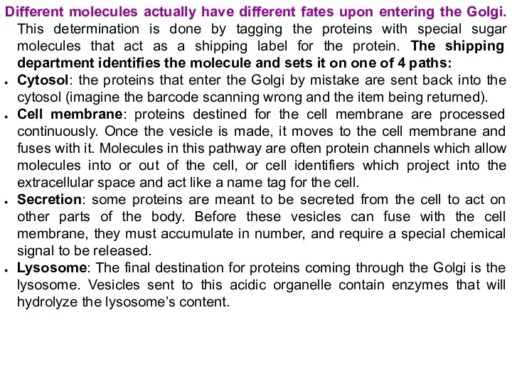
Different molecules actually have different fates upon entering the Golgi. This
Different molecules actually have different fates upon entering the Golgi. This
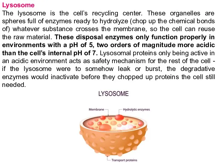
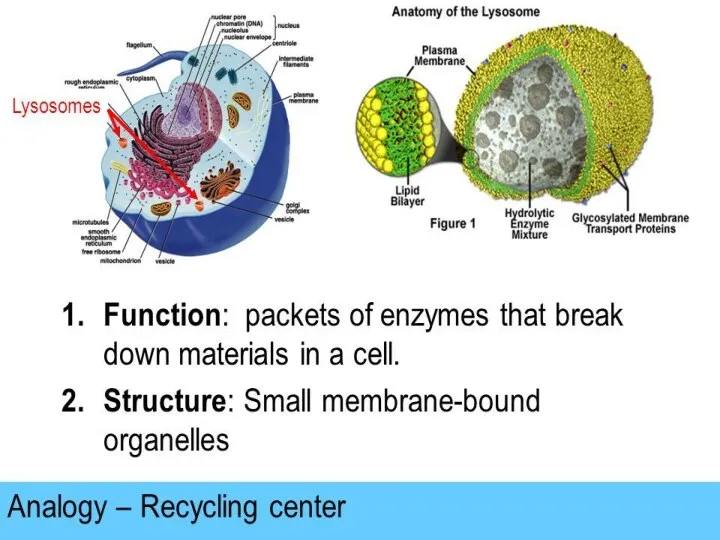
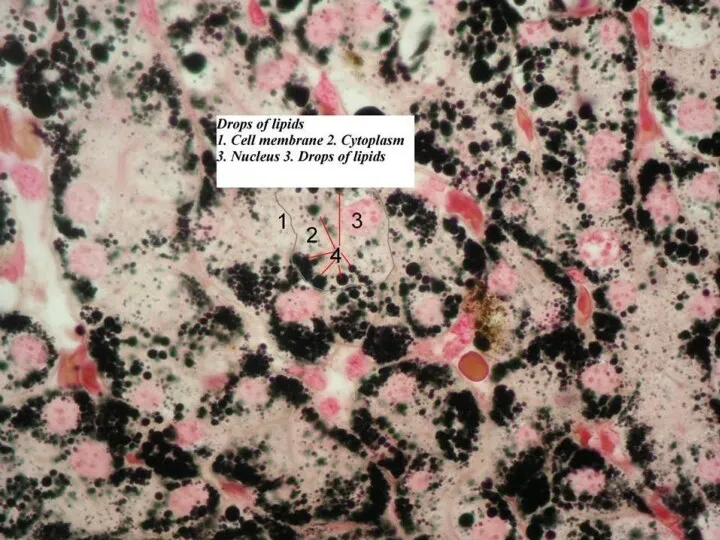
Lysosome
The lysosome is the cell’s recycling center. These organelles are spheres
Lysosome
The lysosome is the cell’s recycling center. These organelles are spheres
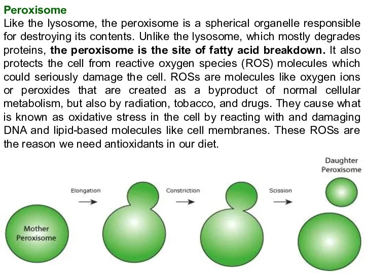
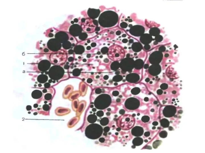
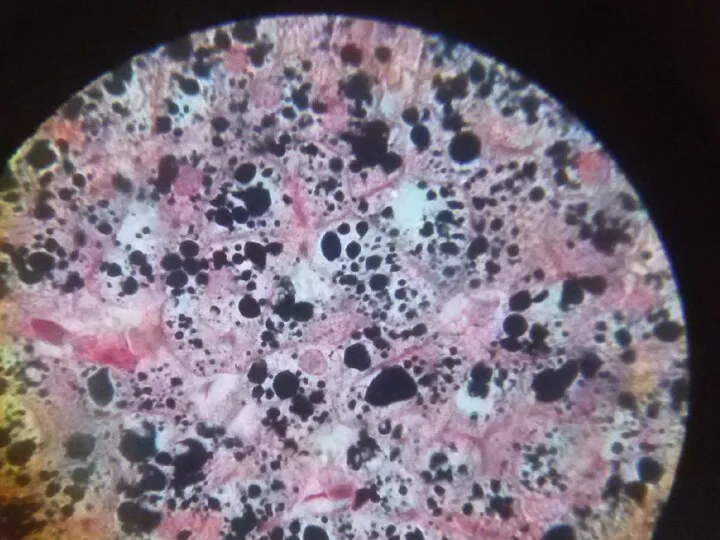
Peroxisome
Like the lysosome, the peroxisome is a spherical organelle responsible for
Peroxisome
Like the lysosome, the peroxisome is a spherical organelle responsible for
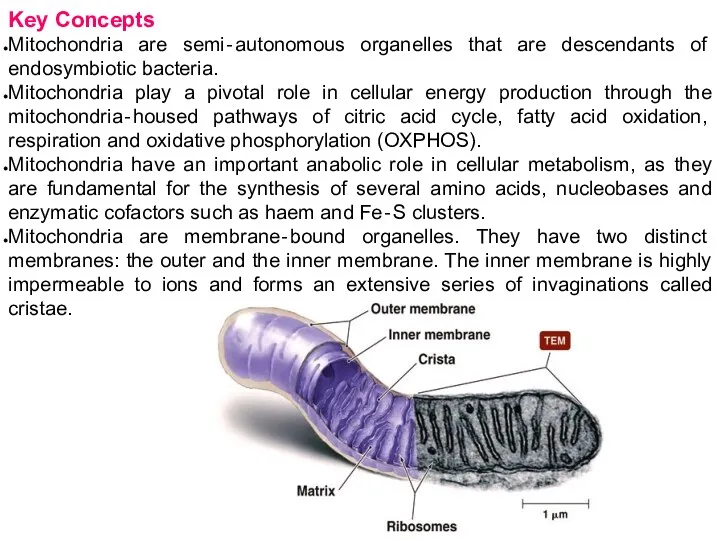
Key Concepts
Mitochondria are semi‐autonomous organelles that are descendants of endosymbiotic bacteria.
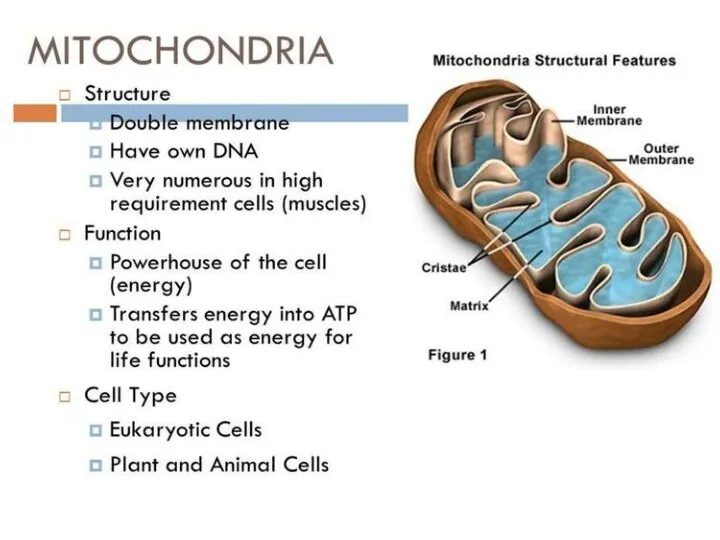
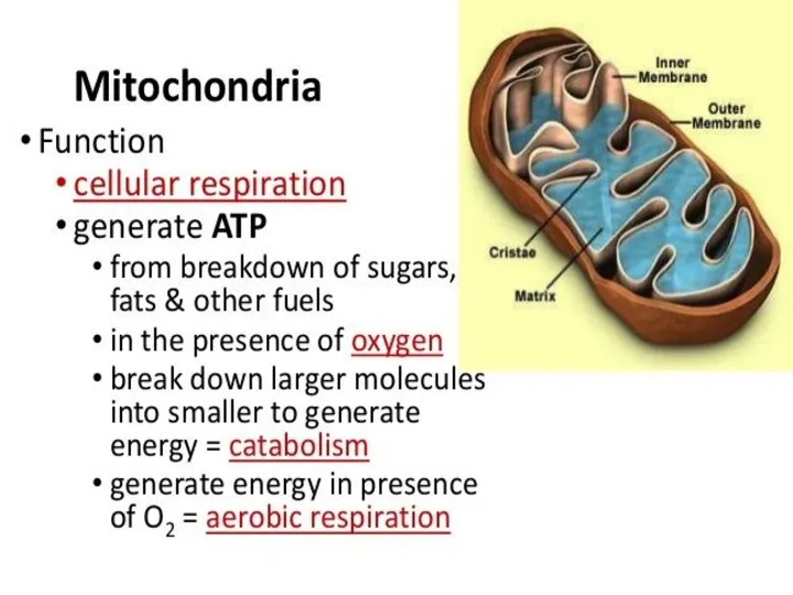
Mitochondria
Key Concepts
Mitochondria are semi‐autonomous organelles that are descendants of endosymbiotic bacteria.
Mitochondria
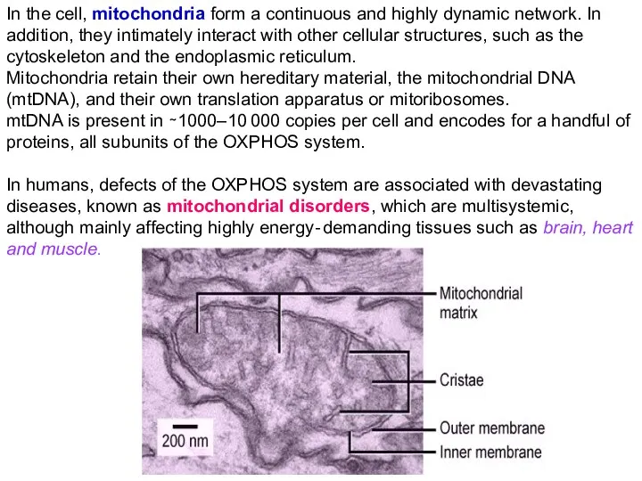
In the cell, mitochondria form a continuous and highly dynamic network.
In the cell, mitochondria form a continuous and highly dynamic network.
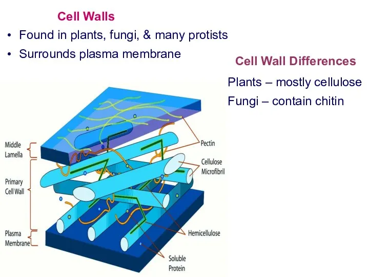
Cell Walls
Found in plants, fungi, & many protists
Surrounds plasma membrane
Cell Wall
Cell Walls
Found in plants, fungi, & many protists
Surrounds plasma membrane
Cell Wall
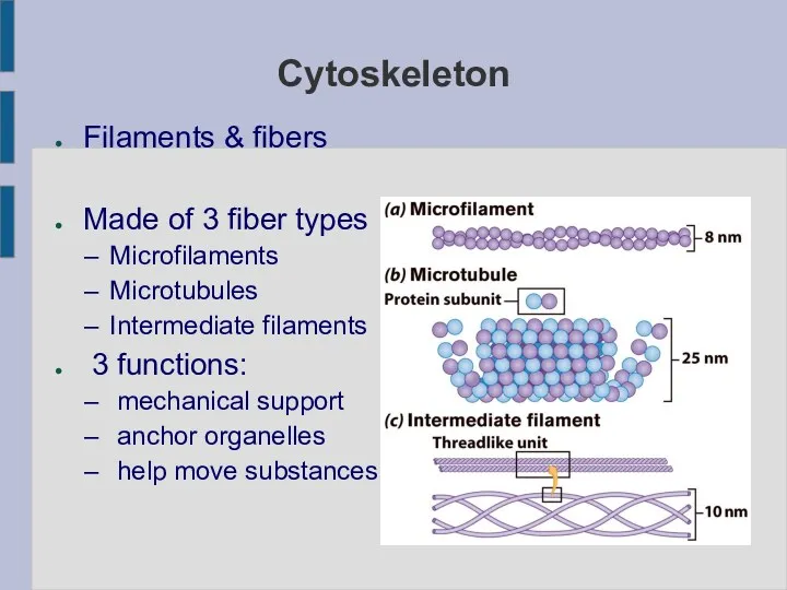
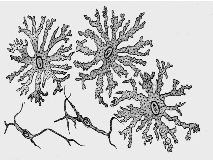
Cytoskeleton
Filaments & fibers
Made of 3 fiber types
Microfilaments
Microtubules
Intermediate filaments
3 functions:
mechanical
Cytoskeleton
Filaments & fibers
Made of 3 fiber types
Microfilaments
Microtubules
Intermediate filaments
3 functions:
mechanical
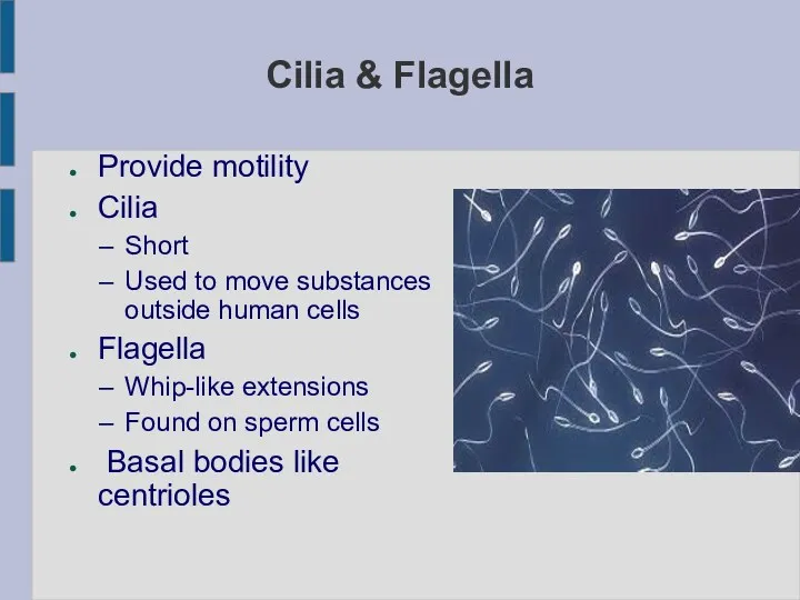
Cilia & Flagella
Provide motility
Cilia
Short
Used to move substances outside human cells
Flagella
Cilia & Flagella
Provide motility
Cilia
Short
Used to move substances outside human cells
Flagella
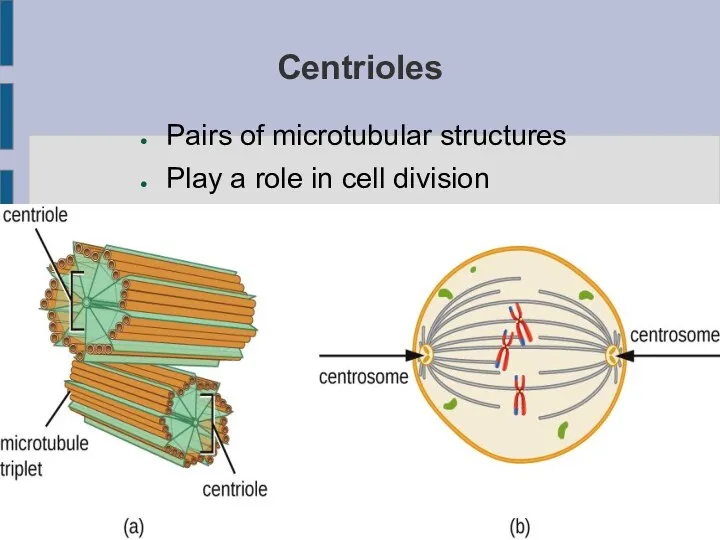
Centrioles
Pairs of microtubular structures
Play a role in cell division
Centrioles
Pairs of microtubular structures
Play a role in cell division
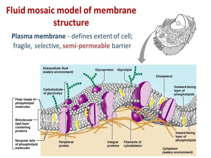
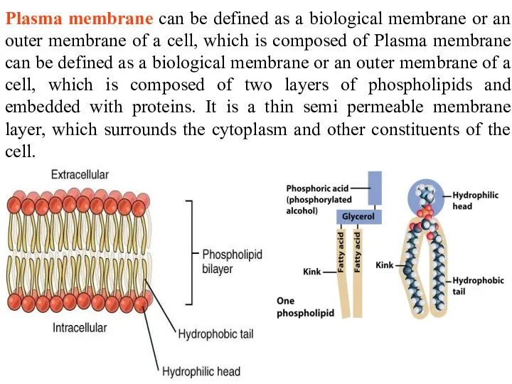
Plasma membrane can be defined as a biological membrane or an
Plasma membrane can be defined as a biological membrane or an
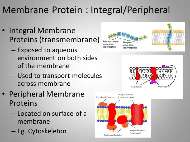
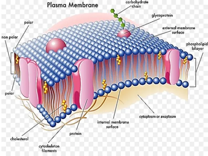
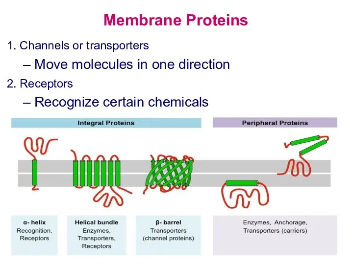
Membrane Proteins
1. Channels or transporters
Move molecules in one direction
2. Receptors
Recognize
Membrane Proteins
1. Channels or transporters
Move molecules in one direction
2. Receptors
Recognize
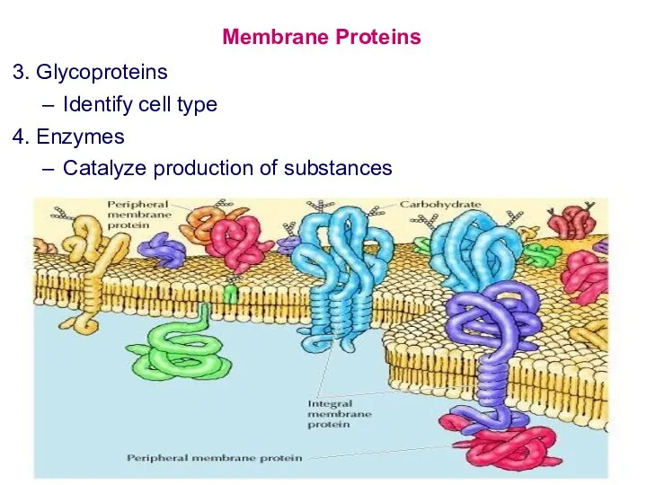
Membrane Proteins
3. Glycoproteins
Identify cell type
4. Enzymes
Catalyze production of substances
Membrane Proteins
3. Glycoproteins
Identify cell type
4. Enzymes
Catalyze production of substances
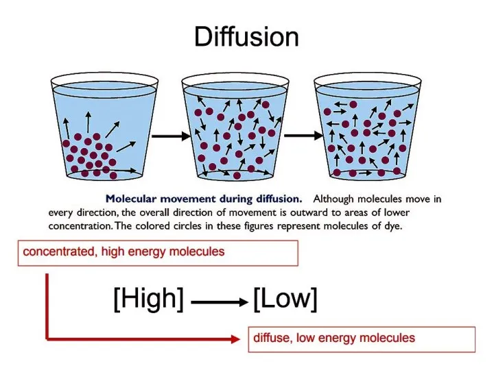
Diffusion
Diffusion is a passive process of transport. A single substance tends
Diffusion
Diffusion is a passive process of transport. A single substance tends
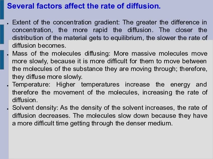
Several factors affect the rate of diffusion.
Extent of the concentration gradient:
Several factors affect the rate of diffusion.
Extent of the concentration gradient:
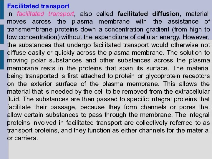
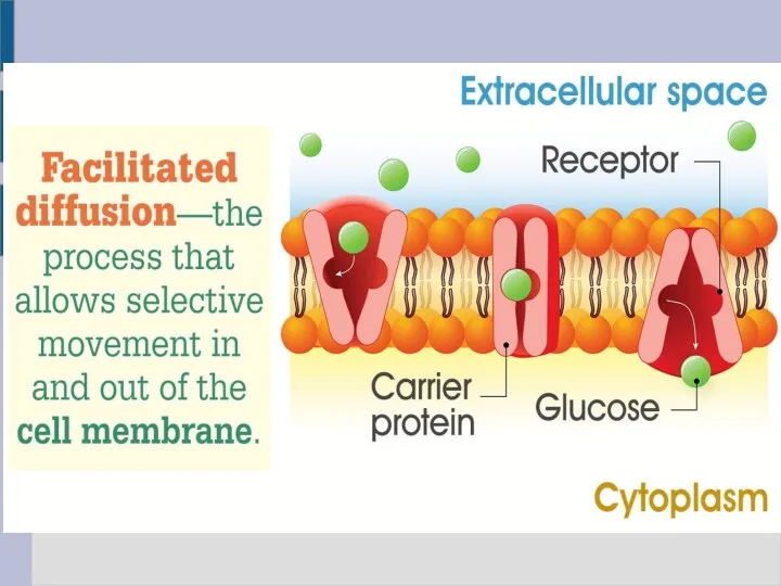
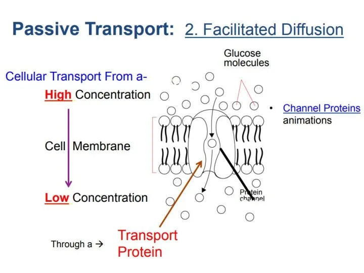
Facilitated transport
In facilitated transport, also called facilitated diffusion, material moves across
Facilitated transport
In facilitated transport, also called facilitated diffusion, material moves across
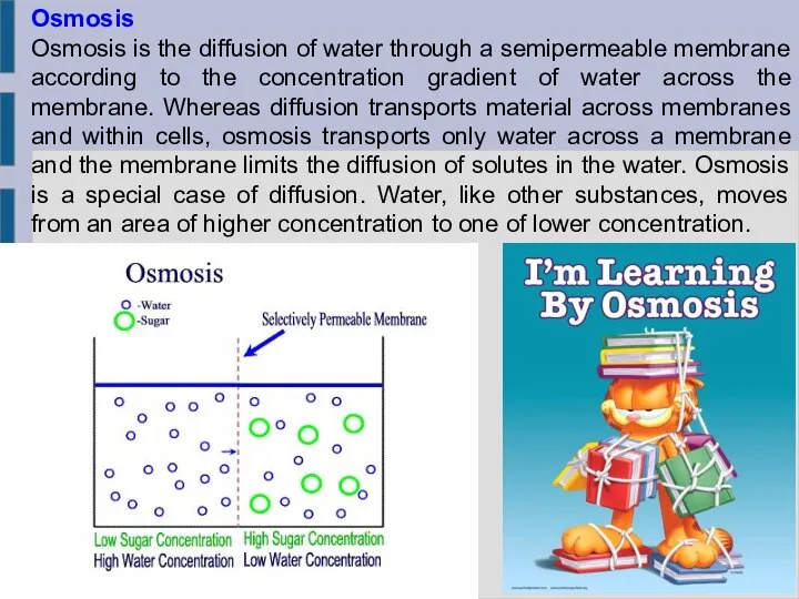
Osmosis
Osmosis is the diffusion of water through a semipermeable membrane according
Osmosis
Osmosis is the diffusion of water through a semipermeable membrane according
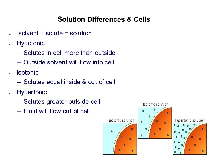
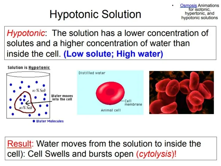
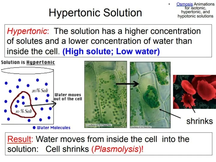
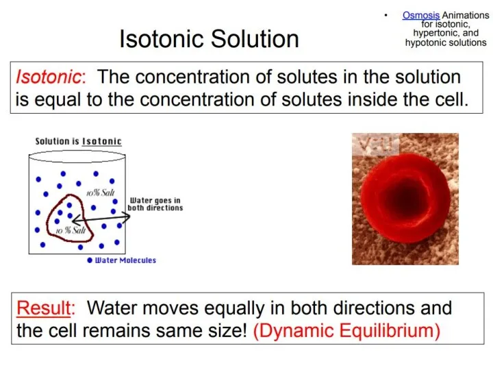
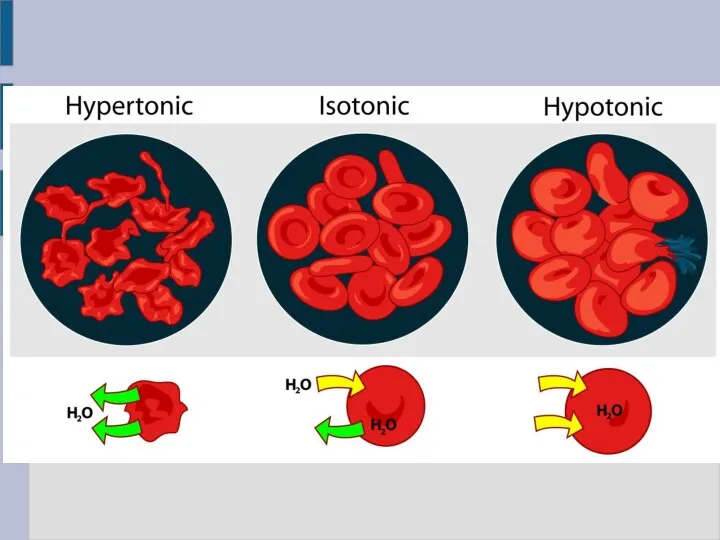
Solution Differences & Cells
solvent + solute = solution
Hypotonic
Solutes in cell
Solution Differences & Cells
solvent + solute = solution
Hypotonic
Solutes in cell
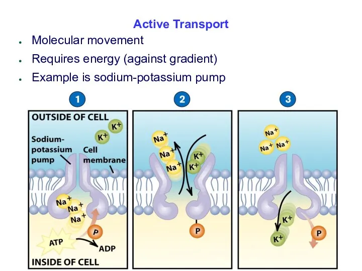
Active Transport
Molecular movement
Requires energy (against gradient)
Example is sodium-potassium pump
Active Transport
Molecular movement
Requires energy (against gradient)
Example is sodium-potassium pump
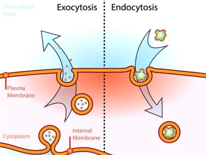
Endocytosis
Movement of large material
Particles
Organisms
Large molecules
Movement is into cells
Types of
Endocytosis
Movement of large material
Particles
Organisms
Large molecules
Movement is into cells
Types of
Process of Endocytosis
Plasma membrane surrounds material
Edges of membrane meet
Membranes fuse to
Process of Endocytosis
Plasma membrane surrounds material
Edges of membrane meet
Membranes fuse to
Forms of Endocytosis
Phagocytosis – cell eating
Pinocytosis – cell drinking
Forms of Endocytosis
Phagocytosis – cell eating
Pinocytosis – cell drinking
 An introduction to metabolism
 Тип Моллюски. Строение, разнообразие и значение
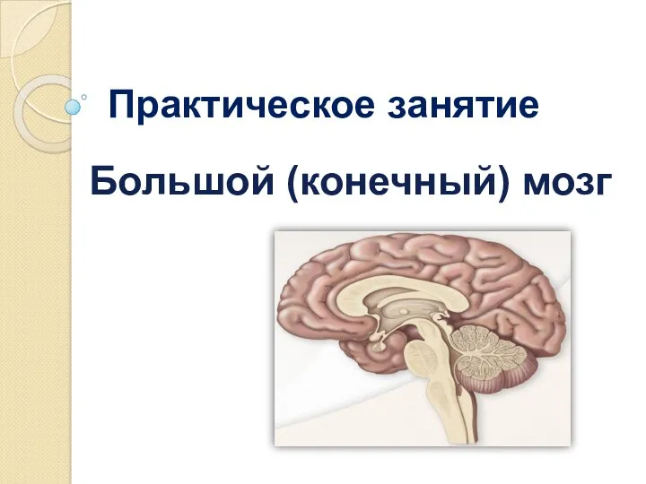 Большой (конечный) мозг
 Классификация микроорганизмов
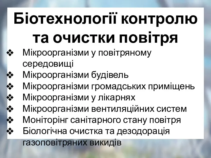 Біотехнології контролю та очистки повітря
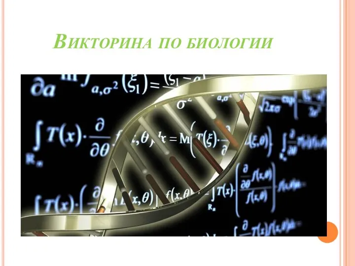 Викторина по биологии
 Биологические ритмы. Сон, его значение. Гигиена сна
 Сердечно – сосудистая система
 Генетика микроорганизмов. Основы биотехнологии
 Вторично чувствующие органы чувств: орган слуха, орган равновесия, орган вкуса
 Особенности строения клетки. 10 класс
 Птицы от А до Я
 Систематика водорослей
 Желто-зеленые водоросли
 Неклассическая наука, конец XIX - первая половина XX века
 Презентация Тайна листа
 Методы исследования в биологии
 Получение ранней продукции кабачков при выращивании посевом семян в открытый грунт и рассадным способом
 Презентация Внешнее строение млекопитающих
 20231227_5_klass_stroenie_kletki
 Класс Земноводные
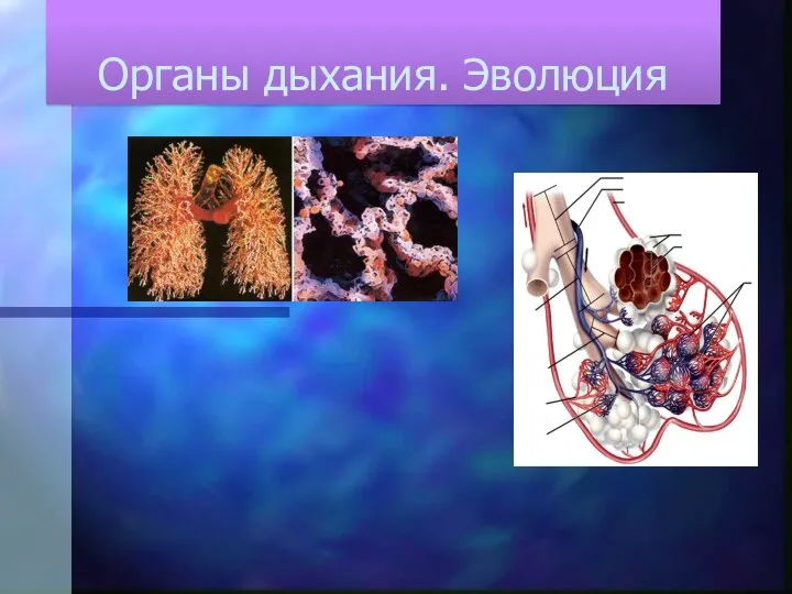 Органы дыхания. Эволюция
 Белый медведь
 Насекомое. Фото
 Ионные каналы
 Открытый урок по темеЛист. Внешнее и внутреннее строение
 Які таємниці приховують хребетні тварини
 Человек как объект генетики. Методы изучения генетики человека