Microbiology. Microbiological laboratory systematics of microorganisms morphology of microorganims презентация
- Microbiology. Microbiological laboratory systematics of microorganisms morphology of microorganims

Содержание
- 2. SAFETY RULES Always wear lab coats and caps Don’t put your bags and personal things on
- 3. IT IS STRICKTLY PROHIBITED to pump fluid into the pipette by mouth to move a burning
- 4. PURPOSES to get acquainted with principles of organization, equipment of microbiology laboratory and rules of work
- 5. MICROBIOLOGY Microbiology (from Greek μῑκρος, mīkros, "small"; βίος, bios, "life"; and -λογία, -logia) is the study
- 8. MICROBIOLOGICAL LABORATORY
- 9. Laboratory rooms and laminar flow cabinets are designed for specific activities in aseptic conditions
- 10. Room for preparation of nutrient media
- 11. Table automatic boiler for the preparation of small volumes of nutrient media
- 12. Specially equipped rooms for sterilization of nutrient media, laboratory glassware, disinfection of infectious material
- 13. Vivarium for laboratory animals
- 14. LABORATORY EQUIPMENT Biological immersion microscope Instruments: inoculation loops, spatulas, tweezers, spirit lamps, etc Laboratory glassware: tubes,
- 15. BIOLOGICAL IMMERSION MICROSCOPE TASK 1 (P. 13) NAME PARTS OF LIGHT MICROSCOPE
- 16. IMMERSION MICROSCOPY
- 17. IMMERSION MICROSCOPY TASK 2 (P. 13) DRAW WAY OF LIGHT IN IMMERSION SYSTEM
- 18. INSTRUMENTS inoculation loops spatula tweezers spirit lamp
- 19. LABORATORY GLASSWARE test tubes Petri dish flasks pipettes
- 20. DEVICES FOR STERILIZATION autoclave Pasteur oven
- 21. NUTRIENT MEDIA Blood agar Endo media
- 22. REAGENTS
- 23. pH Meters
- 24. DISTILLERS
- 25. CENTRIFUGES
- 26. BALANCES technical analytical
- 27. FILTRATION EQUIPMENT
- 29. STUDENT’S LABORATORY EQUIPMENT Microscope Immersion oil Inoculating loop Burner or spirit lamp Staining kits Water for
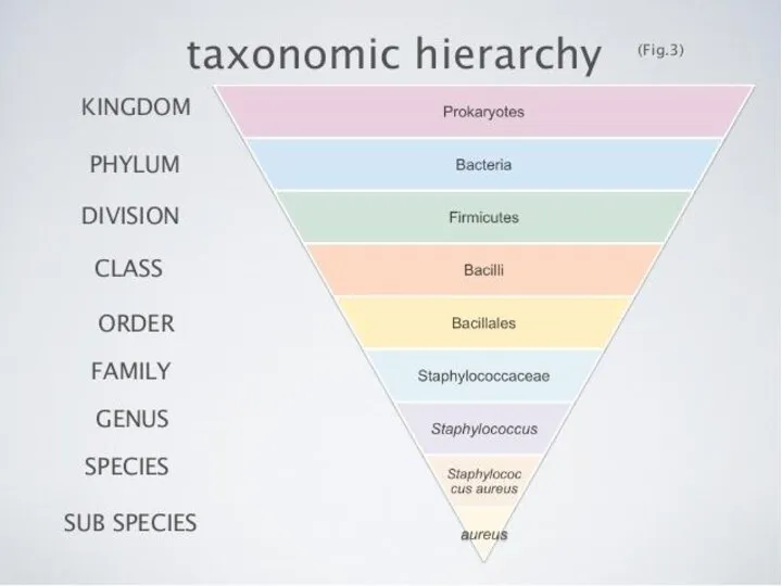
- 30. MORPHOLOGY OF MICROORGANISMS Size of microbial cells Shape of microbial cells Arrangement of microbial cells Bacteria
- 31. SIZE OF MICROORGANISMS Bacteria are of about 0,5—5 µm in size The range of sizes shown
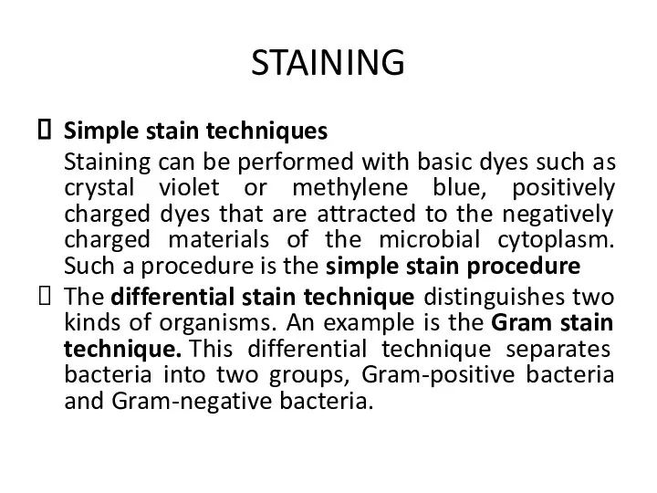
- 35. STAINING Because microbial cytoplasm is usually transparent, it is necessary to stain microorganisms before they can
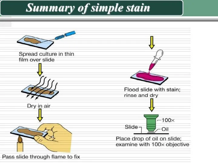
- 37. STAINING Simple stain techniques Staining can be performed with basic dyes such as crystal violet or
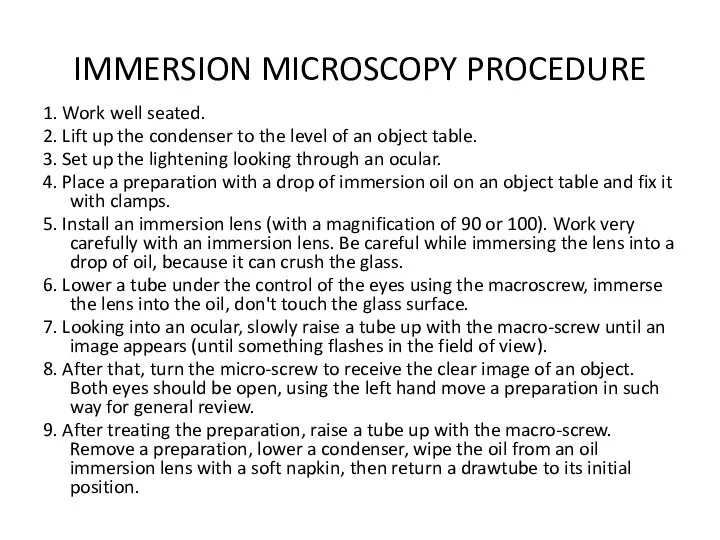
- 39. IMMERSION MICROSCOPY PROCEDURE 1. Work well seated. 2. Lift up the condenser to the level of
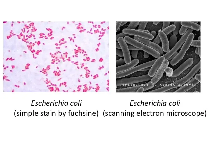
- 40. Escherichia coli (simple stain by fuchsine) Escherichia coli (scanning electron microscope)
- 41. Staphylococcus aureus (simple stain by fuchsine) Staphylococcus aureus (scanning electron microscope)
- 42. Bacillus anthracoides (simple stain by fuchsine)
- 43. Vibrio cholerae (simple stain by fuchsine) Vibrio cholerae (scanning electron microscopy)
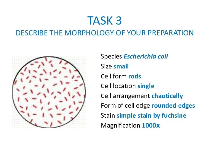
- 44. TASK 3 DESCRIBE THE MORPHOLOGY OF YOUR PREPARATION Species Escherichia coli Size small Cell form rods
- 45. TASK 3 DESCRIBE THE MORPHOLOGY OF YOUR PREPARATION Species Staphylococcus aureus Size large Cell form coccus
- 46. TASK 3 DESCRIBE THE MORPHOLOGY OF YOUR PREPARATION Species Bacillus anthracoides Size large Cell form bacillus
- 47. TASK 3 DESCRIBE THE MORPHOLOGY OF YOUR PREPARATION Species Vibrio cholerae Size small Cell form vibrio
- 49. Скачать презентацию
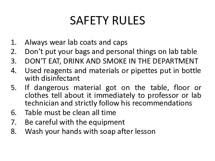
SAFETY RULES
Always wear lab coats and caps
Don’t put your bags and
SAFETY RULES
Always wear lab coats and caps
Don’t put your bags and
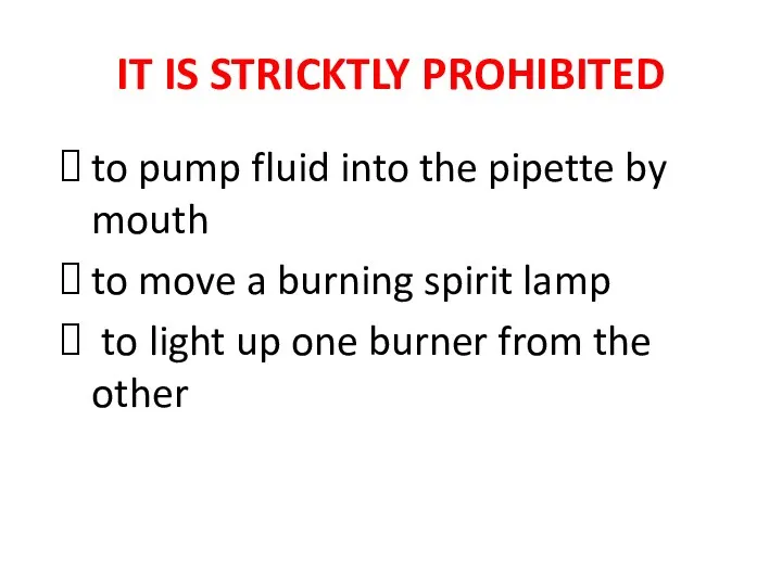
IT IS STRICKTLY PROHIBITED
to pump fluid into the pipette by mouth
to
IT IS STRICKTLY PROHIBITED
to pump fluid into the pipette by mouth
to
PURPOSES
to get acquainted with principles of organization, equipment of microbiology laboratory
PURPOSES
to get acquainted with principles of organization, equipment of microbiology laboratory
MICROBIOLOGY
Microbiology (from Greek μῑκρος, mīkros, "small"; βίος, bios, "life"; and -λογία, -logia) is the study of microorganisms, those being unicellular(single cell), multicellular (cell
MICROBIOLOGY
Microbiology (from Greek μῑκρος, mīkros, "small"; βίος, bios, "life"; and -λογία, -logia) is the study of microorganisms, those being unicellular(single cell), multicellular (cell

MICROBIOLOGICAL LABORATORY
MICROBIOLOGICAL LABORATORY

Laboratory rooms and laminar flow cabinets are designed for specific activities
Laboratory rooms and laminar flow cabinets are designed for specific activities
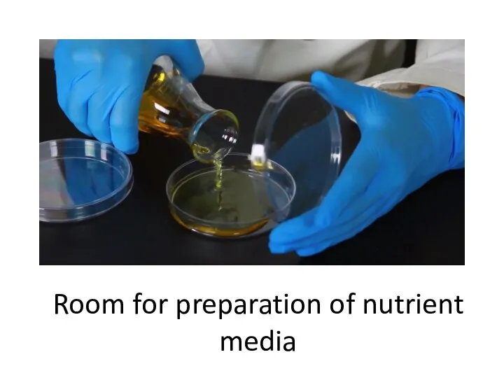
Room for preparation of nutrient media
Room for preparation of nutrient media
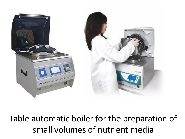
Table automatic boiler for the preparation of small volumes of nutrient
Table automatic boiler for the preparation of small volumes of nutrient
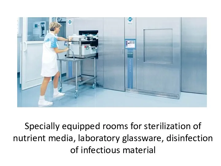
Specially equipped rooms for sterilization of nutrient media, laboratory glassware, disinfection
Specially equipped rooms for sterilization of nutrient media, laboratory glassware, disinfection
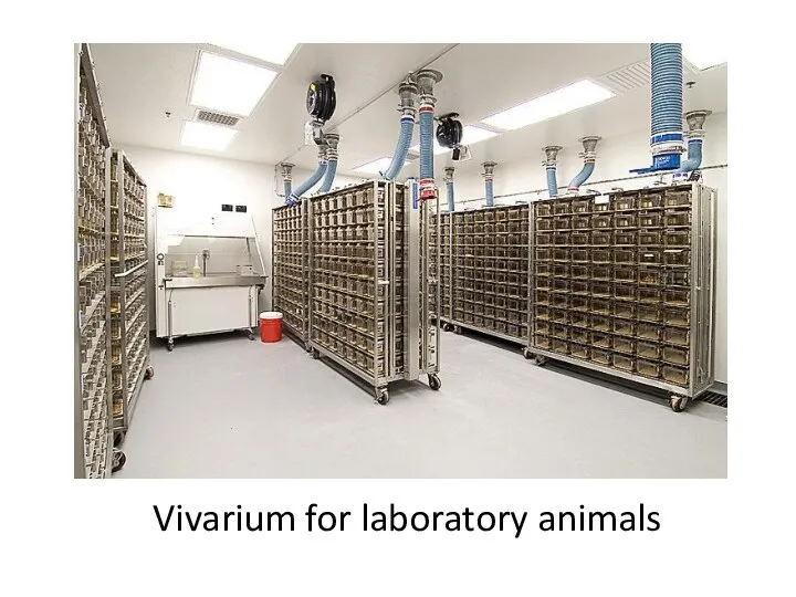
Vivarium for laboratory animals
Vivarium for laboratory animals
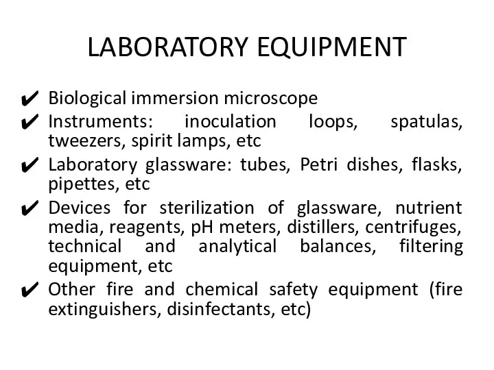
LABORATORY EQUIPMENT
Biological immersion microscope
Instruments: inoculation loops, spatulas, tweezers, spirit lamps, etc
Laboratory
LABORATORY EQUIPMENT
Biological immersion microscope
Instruments: inoculation loops, spatulas, tweezers, spirit lamps, etc
Laboratory
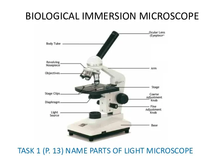
BIOLOGICAL IMMERSION MICROSCOPE
TASK 1 (P. 13) NAME PARTS OF LIGHT MICROSCOPE
BIOLOGICAL IMMERSION MICROSCOPE
TASK 1 (P. 13) NAME PARTS OF LIGHT MICROSCOPE
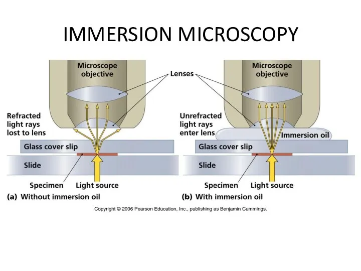
IMMERSION MICROSCOPY
IMMERSION MICROSCOPY

IMMERSION MICROSCOPY
TASK 2 (P. 13) DRAW WAY OF LIGHT IN IMMERSION
IMMERSION MICROSCOPY
TASK 2 (P. 13) DRAW WAY OF LIGHT IN IMMERSION
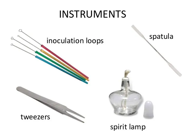
INSTRUMENTS
inoculation loops
spatula
tweezers
spirit lamp
INSTRUMENTS
inoculation loops
spatula
tweezers
spirit lamp
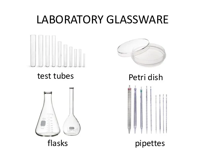
LABORATORY GLASSWARE
test tubes
Petri dish
flasks
pipettes
LABORATORY GLASSWARE
test tubes
Petri dish
flasks
pipettes
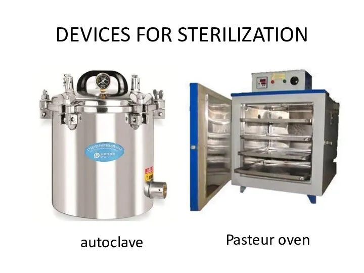
DEVICES FOR STERILIZATION
autoclave
Pasteur oven
DEVICES FOR STERILIZATION
autoclave
Pasteur oven
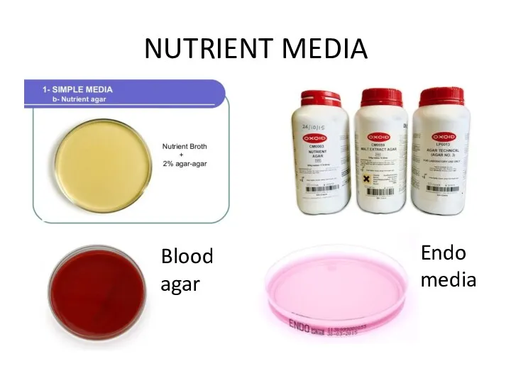
NUTRIENT MEDIA
Blood
agar
Endo
media
NUTRIENT MEDIA
Blood
agar
Endo
media
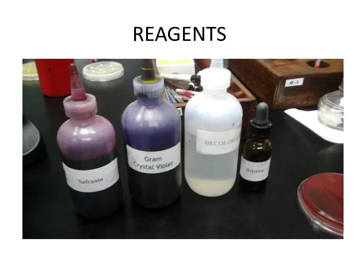
REAGENTS
REAGENTS
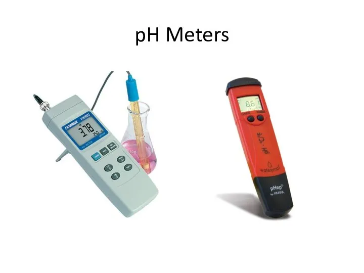
pH Meters
pH Meters
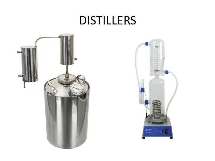
DISTILLERS
DISTILLERS
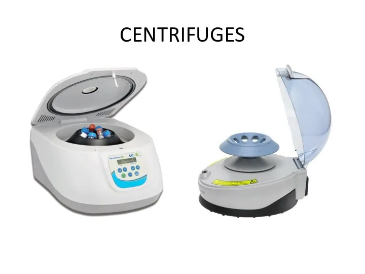
CENTRIFUGES
CENTRIFUGES
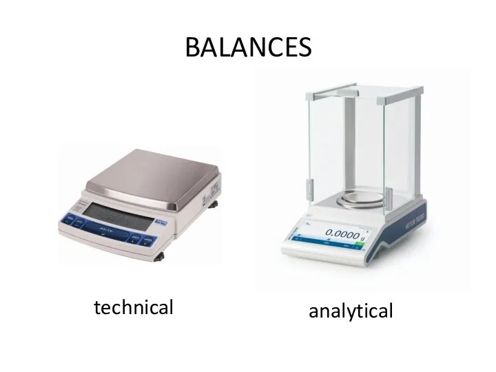
BALANCES
technical
analytical
BALANCES
technical
analytical
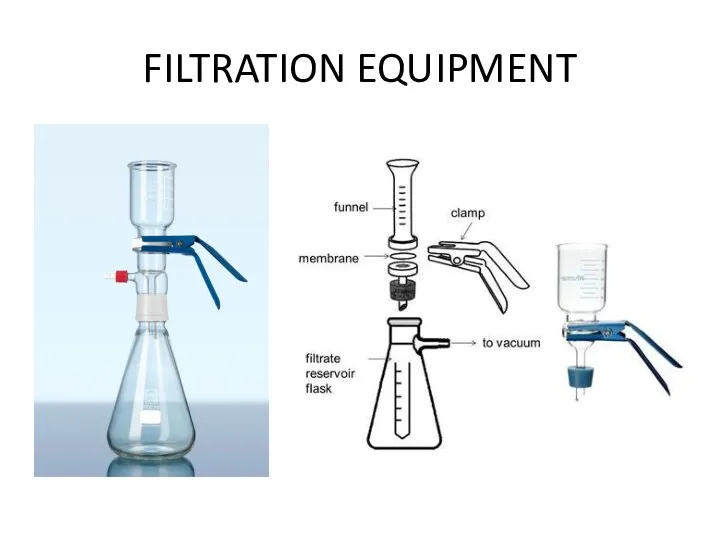
FILTRATION EQUIPMENT
FILTRATION EQUIPMENT
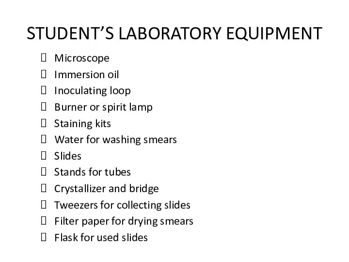
STUDENT’S LABORATORY EQUIPMENT
Microscope
Immersion oil
Inoculating loop
Burner or spirit lamp
Staining kits
Water for washing
STUDENT’S LABORATORY EQUIPMENT
Microscope
Immersion oil
Inoculating loop
Burner or spirit lamp
Staining kits
Water for washing
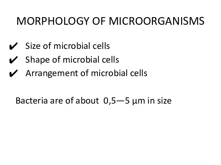
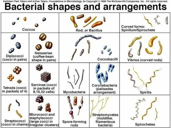
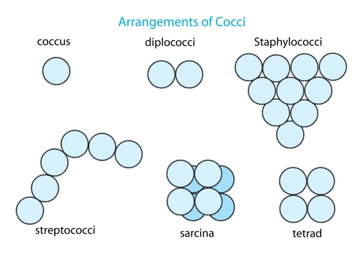
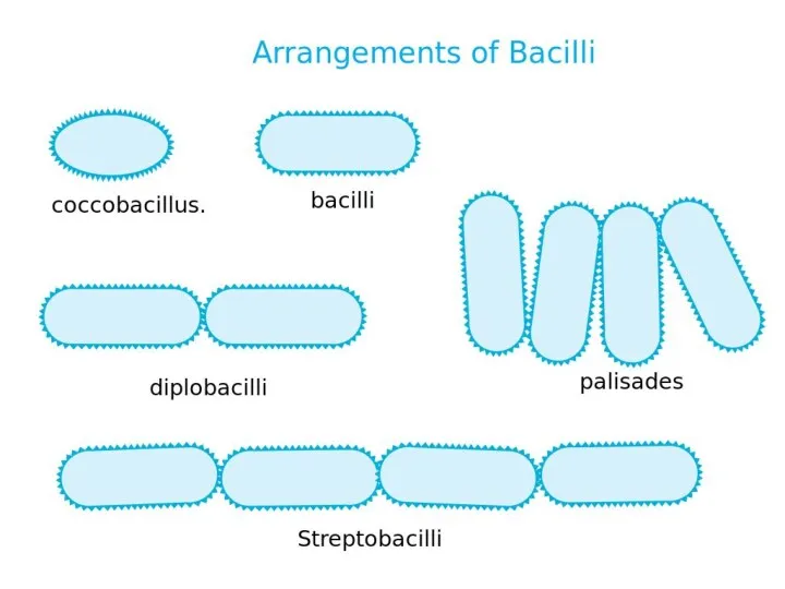
MORPHOLOGY OF MICROORGANISMS
Size of microbial cells
Shape of microbial cells
Arrangement of microbial
MORPHOLOGY OF MICROORGANISMS
Size of microbial cells
Shape of microbial cells
Arrangement of microbial
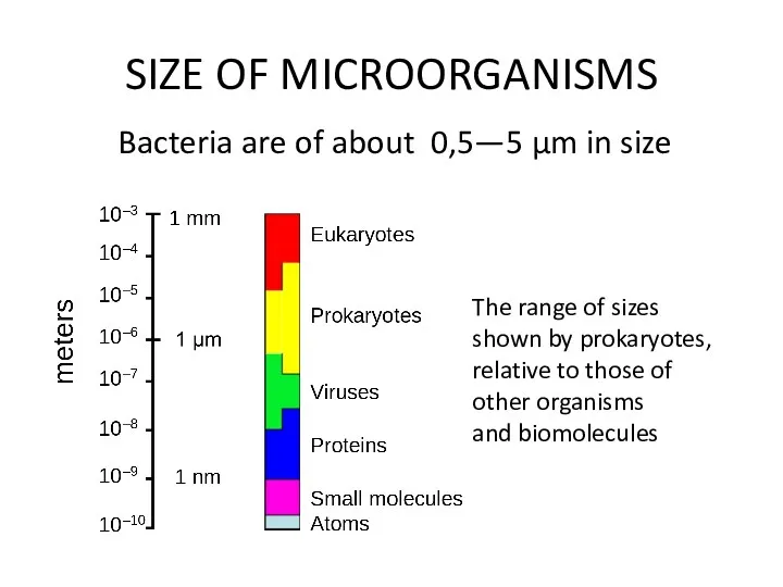
SIZE OF MICROORGANISMS
Bacteria are of about 0,5—5 µm in size
The range
SIZE OF MICROORGANISMS
Bacteria are of about 0,5—5 µm in size
The range
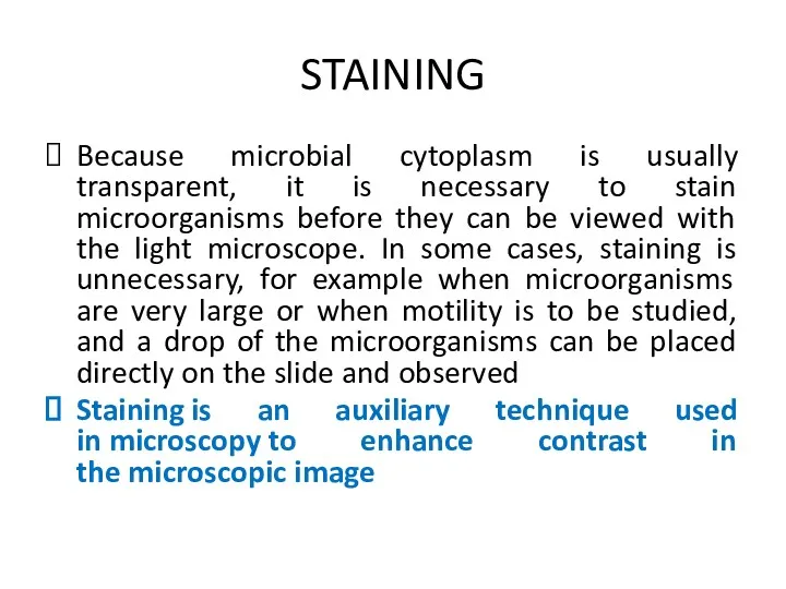
STAINING
Because microbial cytoplasm is usually transparent, it is necessary to stain
STAINING
Because microbial cytoplasm is usually transparent, it is necessary to stain
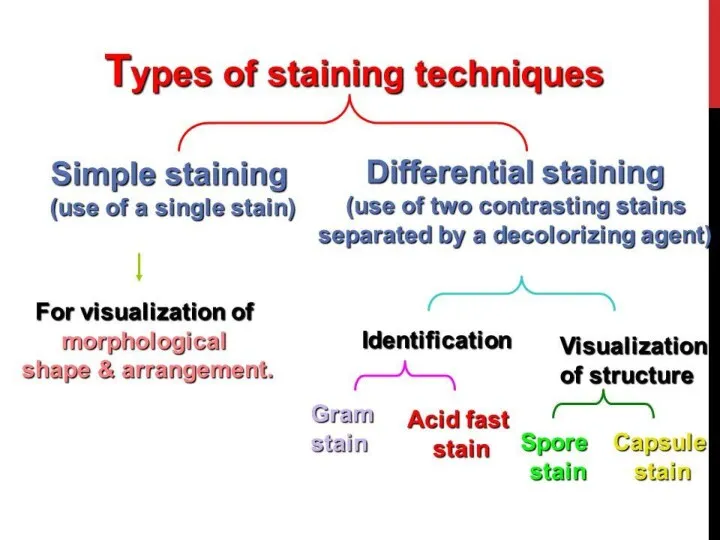
STAINING
Simple stain techniques
Staining can be performed with basic dyes such as
STAINING
Simple stain techniques
Staining can be performed with basic dyes such as
IMMERSION MICROSCOPY PROCEDURE
1. Work well seated.
2. Lift up the condenser to
IMMERSION MICROSCOPY PROCEDURE
1. Work well seated.
2. Lift up the condenser to
Escherichia coli
(simple stain by fuchsine)
Escherichia coli
(scanning electron microscope)
Escherichia coli
(simple stain by fuchsine)
Escherichia coli
(scanning electron microscope)
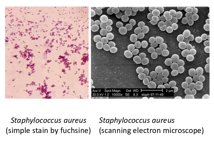
Staphylococcus aureus
(simple stain by fuchsine)
Staphylococcus aureus
(scanning electron microscope)
Staphylococcus aureus
(simple stain by fuchsine)
Staphylococcus aureus
(scanning electron microscope)
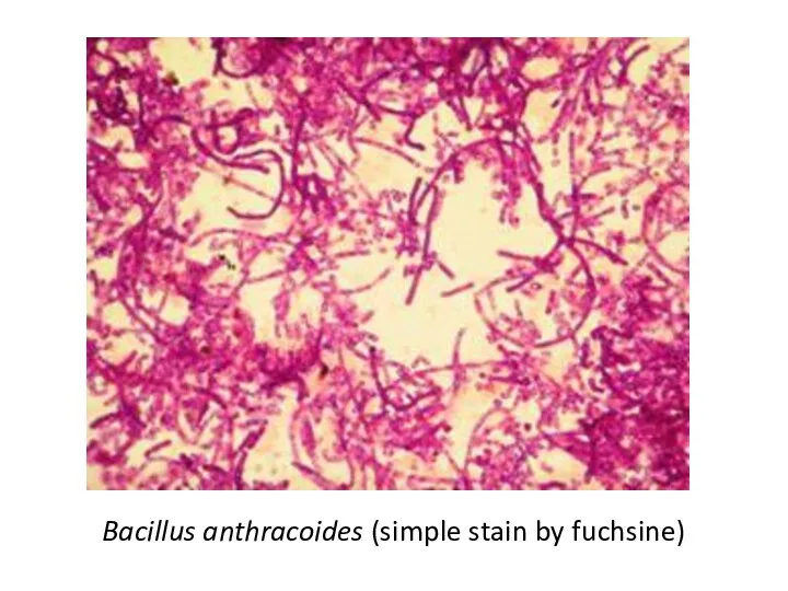
Bacillus anthracoides (simple stain by fuchsine)
Bacillus anthracoides (simple stain by fuchsine)
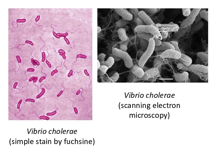
Vibrio cholerae
(simple stain by fuchsine)
Vibrio cholerae
(scanning electron microscopy)
Vibrio cholerae
(simple stain by fuchsine)
Vibrio cholerae
(scanning electron microscopy)
TASK 3
DESCRIBE THE MORPHOLOGY OF YOUR PREPARATION
Species Escherichia coli
Size small
Cell form
TASK 3
DESCRIBE THE MORPHOLOGY OF YOUR PREPARATION
Species Escherichia coli
Size small
Cell form
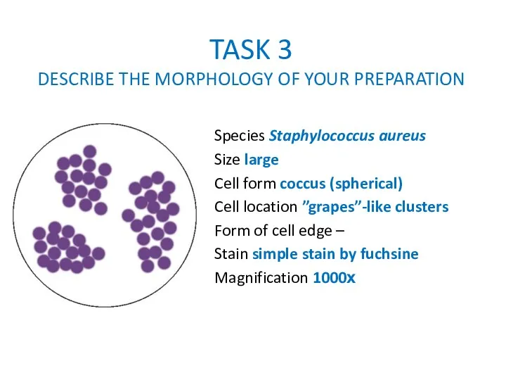
TASK 3
DESCRIBE THE MORPHOLOGY OF YOUR PREPARATION
Species Staphylococcus aureus
Size large
Cell form
TASK 3
DESCRIBE THE MORPHOLOGY OF YOUR PREPARATION
Species Staphylococcus aureus
Size large
Cell form
TASK 3
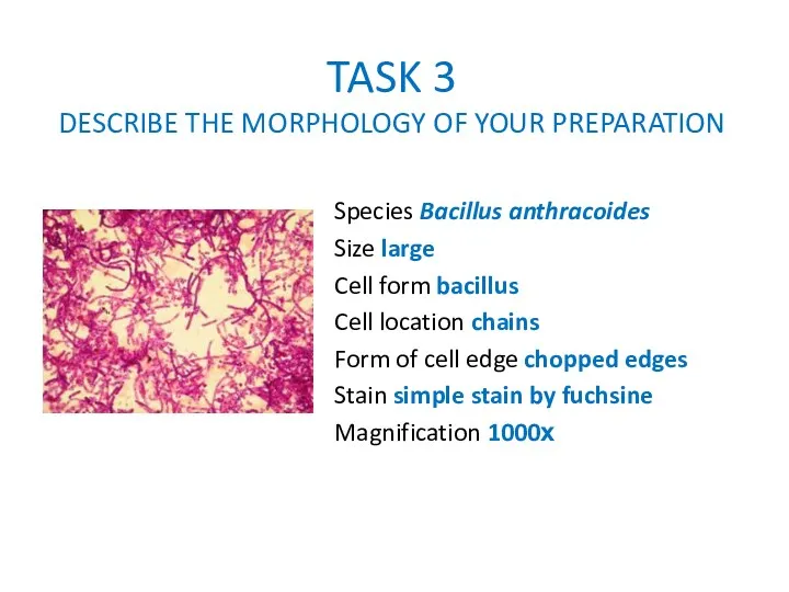
DESCRIBE THE MORPHOLOGY OF YOUR PREPARATION
Species Bacillus anthracoides
Size large
Cell
TASK 3
DESCRIBE THE MORPHOLOGY OF YOUR PREPARATION
Species Bacillus anthracoides
Size large
Cell
TASK 3
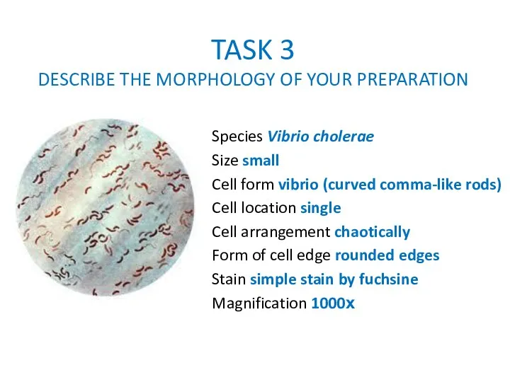
DESCRIBE THE MORPHOLOGY OF YOUR PREPARATION
Species Vibrio cholerae
Size small
Cell
TASK 3
DESCRIBE THE MORPHOLOGY OF YOUR PREPARATION
Species Vibrio cholerae
Size small
Cell
 Устройство увеличительных приборов
 Дидактическая игра Корзина грецких орехов
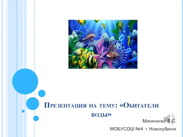 Обитатели воды
 Ферменты: строение, свойства, функции
 Селекция кошек
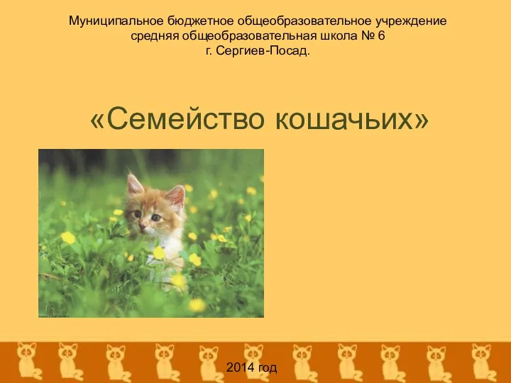 Семейство кошачьих
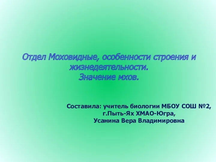 Отдел Моховидные, особенности строения и жизнедеятельности. Значение мхов
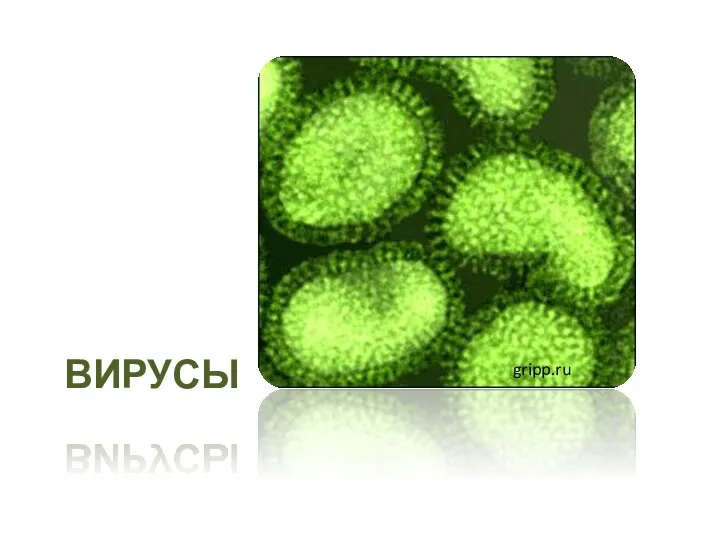 Вирусы. Классификация вирусов
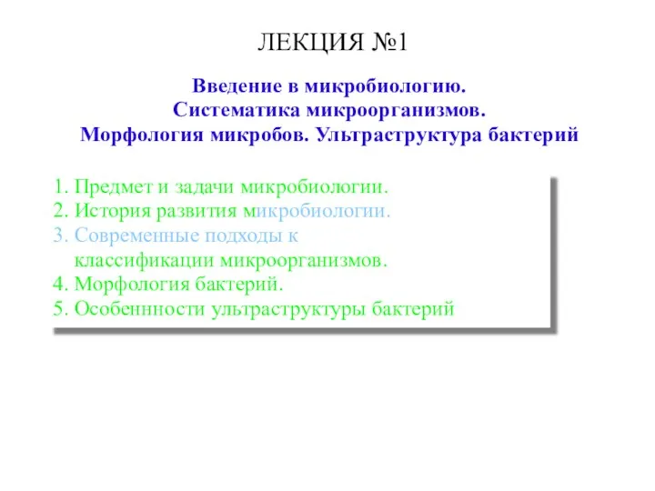 Введение в микробиологию. Систематика микроорганизмов. Морфология микробов. Ультраструктура бактерий
 Внеклассное мероприятие Красная книга Оренбургской области
 Практико-ориентированные задания на уроках биологии, как этап подготовки к итоговой аттестации
 Композиция и сюжет в изобразительном и декоративно-прикладном искусстве
 Образование половых клеток. Мейоз
 Значение растений в природе и жизни человека
 Красная Книга России
 Генетика людини (11 клас)
 Поле и его обитатели
 Индивидуальное развитие организмов (онтогенез)
 Класс птицы как высокоорганизованная и специализированная группа. Морфофизиологические особенности, биология, распространение
 Жизненные формы растений. 6 класс
 Человек – предмет изучения анатомии и физиологии
 Топографічна анатомія шиї
 презентация к уроку по теме Кровь и кровообращение. 8 класс
 Углеводный обмен
 Мәдени өсімдіктер
 Презентация к внеурочному мероприятию по биологии Что?Где?Когда?
 Презентация по теме: Особенности высшей нервной деятельности человека.
 Анатомия ЦНС. Задний мозг. Варолиев мост