- Physiology of Bacteria

Содержание
- 2. Microbial Metabolism The primary function of all living cells is to grow and reproduce Growth &
- 3. Microbial Metabolism The metabolic process that involves the degradation of chemical components is called catabolism The
- 4. Most matabolic processes in the cell would take forever if it were not for enzymes. Enzymes
- 5. Classification of enzymes Oxidoreductases are involved in electron ( hydrogen) transfer reactions. Transferases transfer specific groups
- 6. Classification of enzymes Lyases remove chemical groups from substrates, forming double bonds, or add chemical groups
- 7. Classification of enzymes Enzymes synthesized by the cell remain within the cell to carry out specific
- 8. Classification of enzymes Pathogenicity enzymes - are enzymes that damage cells and tissues. Coagulase –enables the
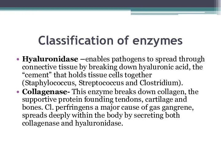
- 9. Classification of enzymes Hyaluronidase –enables pathogens to spread through connective tissue by breaking down hyaluronic acid,
- 10. Classification of enzymes Hemolysin- enzyme that cause damage to the host’s red blood cells. In the
- 11. Classification of enzymes Hyaluronidase –enables pathogens to spread through connective tissue by breaking down hyaluronic acid,
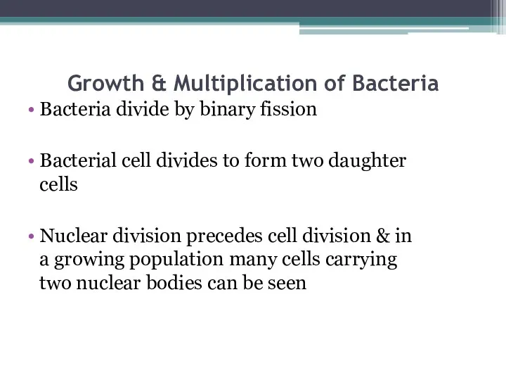
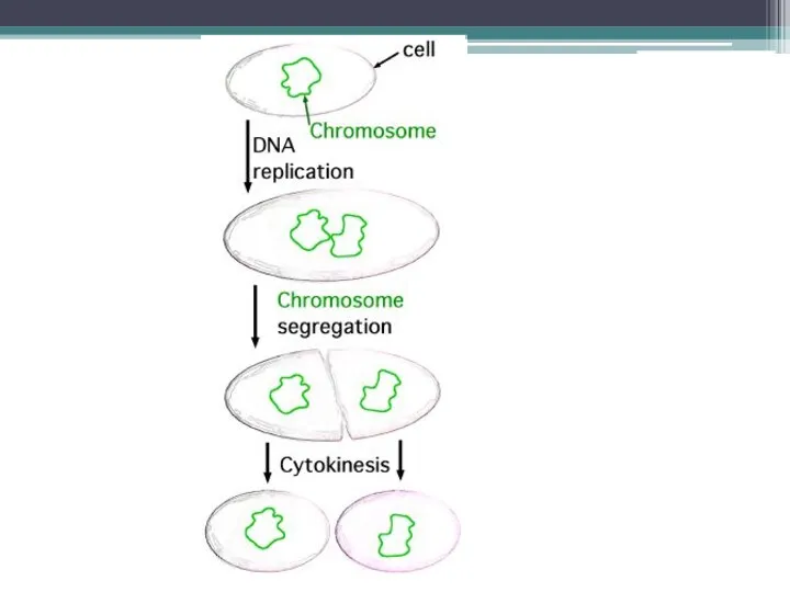
- 12. Growth & Multiplication of Bacteria Bacteria divide by binary fission Bacterial cell divides to form two
- 14. The interval of time between two cell division, or the time required for a bacterium to
- 15. Growth & Multiplication of Bacteria Bacteria divide by binary fission Bacterial cell divides to form two
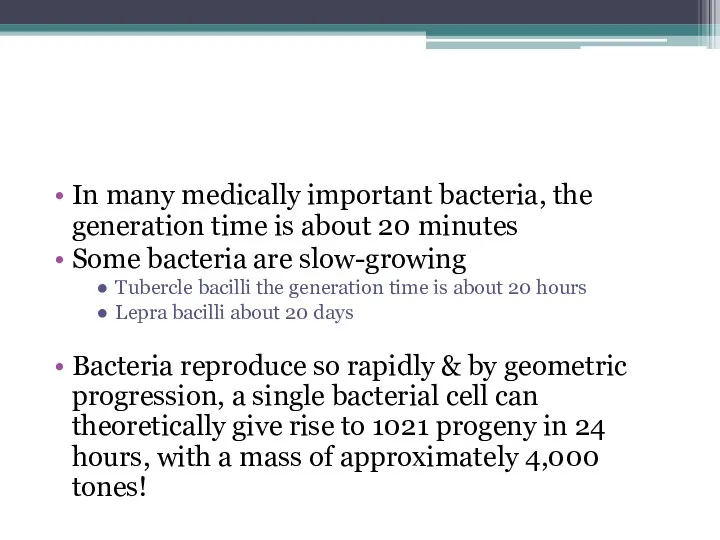
- 17. In many medically important bacteria, the generation time is about 20 minutes Some bacteria are slow-growing
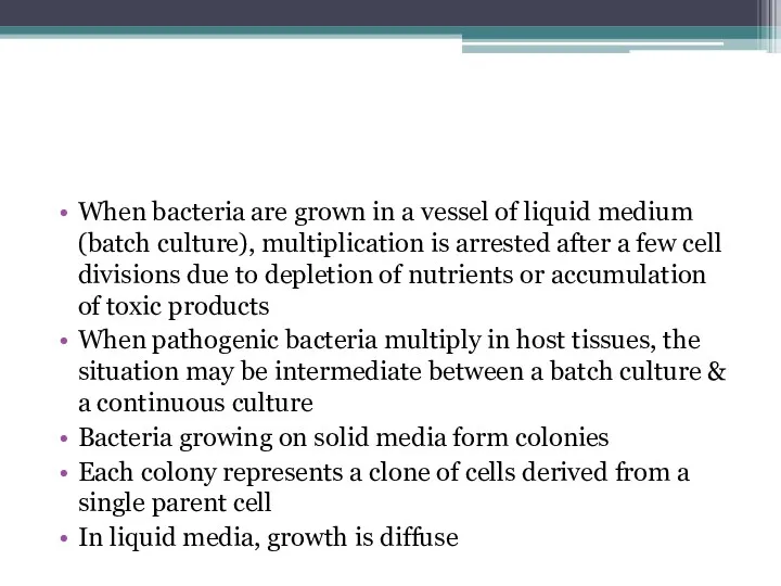
- 18. When bacteria are grown in a vessel of liquid medium (batch culture), multiplication is arrested after
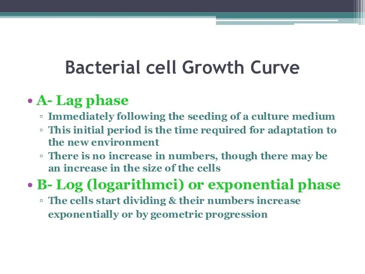
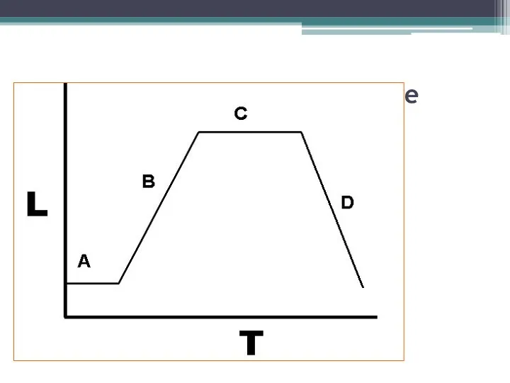
- 19. Bacterial cell Growth Curve A- Lag phase Immediately following the seeding of a culture medium This
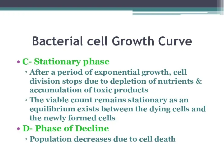
- 20. Bacterial cell Growth Curve C- Stationary phase After a period of exponential growth, cell division stops
- 21. Bacterial cell Growth Curve
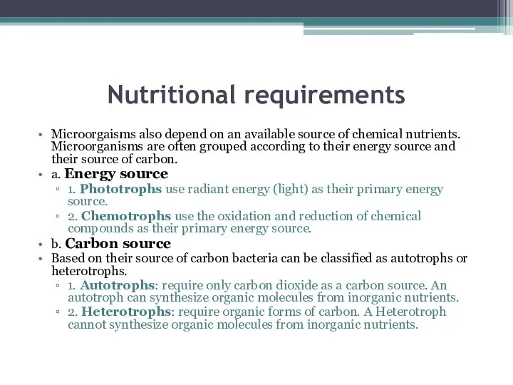
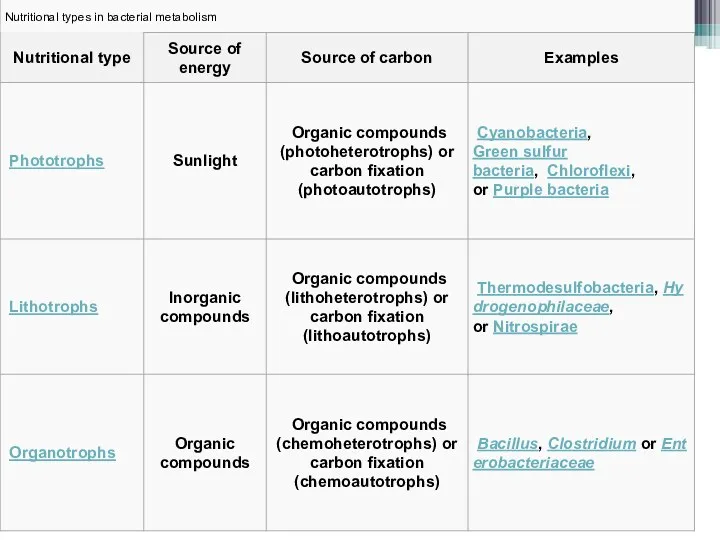
- 22. Nutritional requirements Microorgaisms also depend on an available source of chemical nutrients. Microorganisms are often grouped
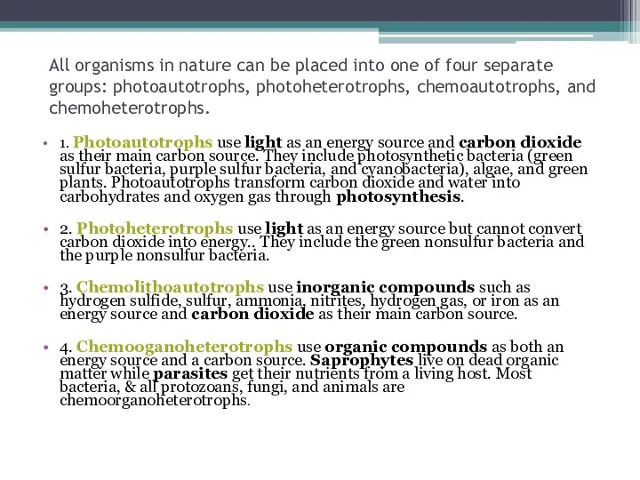
- 24. All organisms in nature can be placed into one of four separate groups: photoautotrophs, photoheterotrophs, chemoautotrophs,
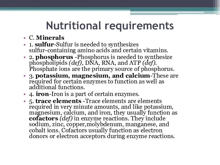
- 25. Nutritional requirements C. Minerals 1. sulfur-Sulfur is needed to synthesizes sulfur-containing amino acids and certain vitamins.
- 26. Nutritional requirements D. Water E. Growth factors Growth factors are organic compounds such as amino acids
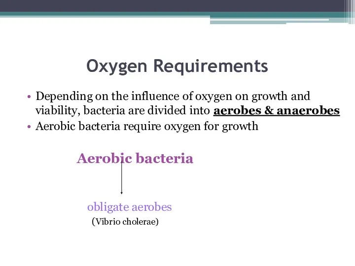
- 27. Oxygen Requirements Depending on the influence of oxygen on growth and viability, bacteria are divided into
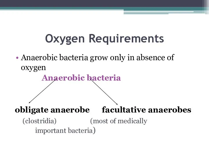
- 28. Oxygen Requirements Anaerobic bacteria grow only in absence of oxygen Anaerobic bacteria obligate anaerobe facultative anaerobes
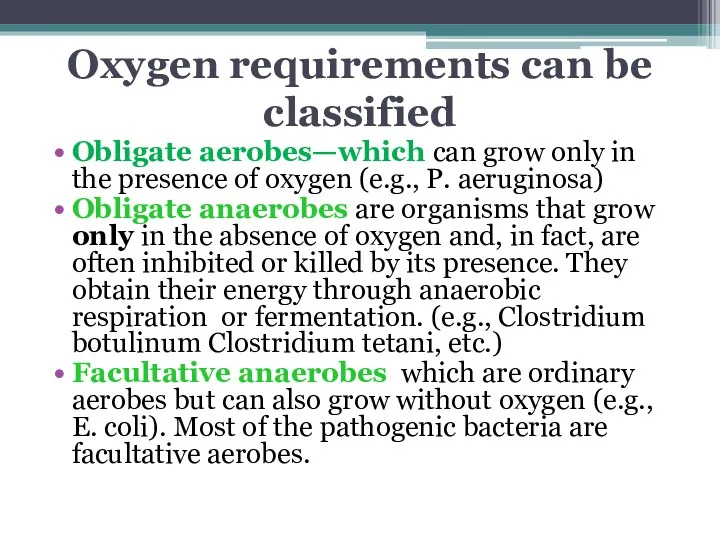
- 29. Oxygen requirements can be classified Obligate aerobes—which can grow only in the presence of oxygen (e.g.,
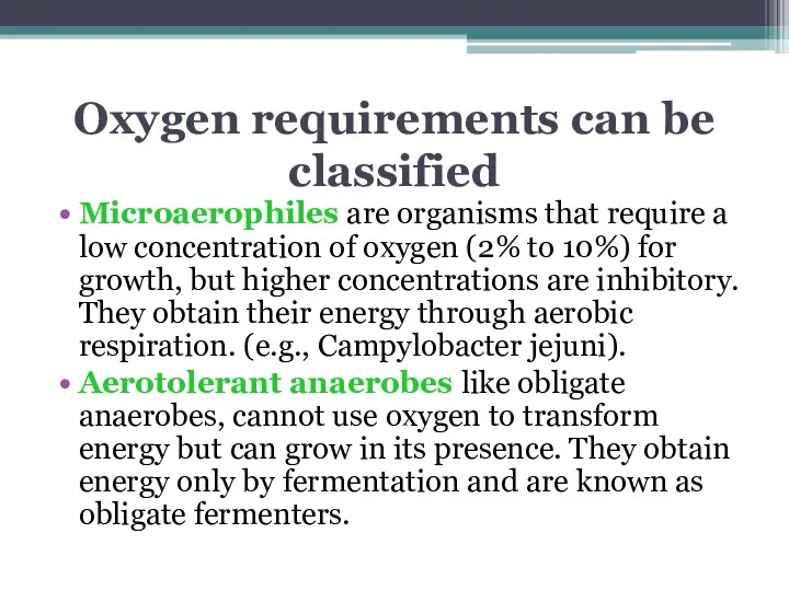
- 30. Oxygen requirements can be classified Microaerophiles are organisms that require a low concentration of oxygen (2%
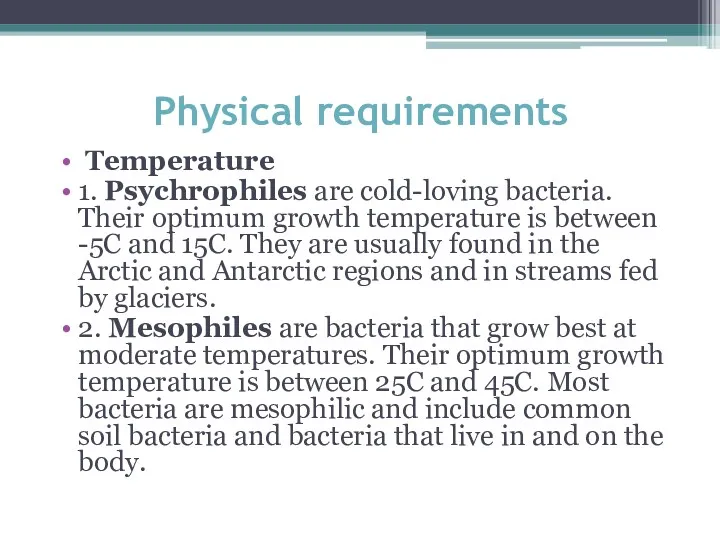
- 31. Physical requirements Temperature 1. Psychrophiles are cold-loving bacteria. Their optimum growth temperature is between -5C and
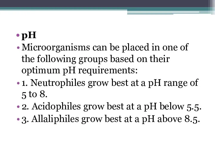
- 32. pH Microorganisms can be placed in one of the following groups based on their optimum pH
- 33. Culture Media A growth medium or culture medium is a substance in which microorganisms or cells
- 34. Types of Growth Media The most common growth media for microorganisms are nutrient broths (liquid nutrient
- 35. Types of Growth Media Nutrient media Undefined media (also known as basal or complex media) Defined
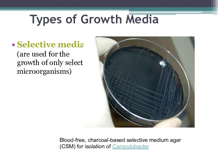
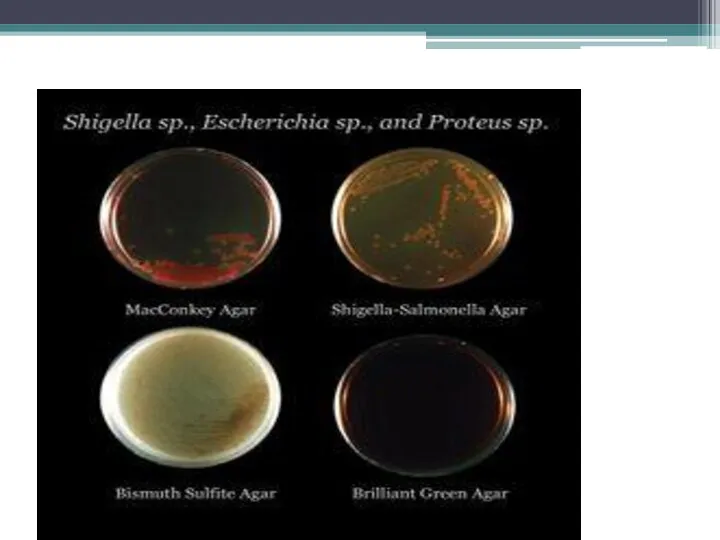
- 36. Types of Growth Media Selective media (are used for the growth of only select microorganisms) Blood-free,
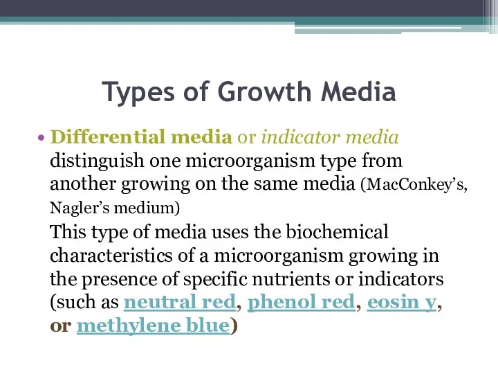
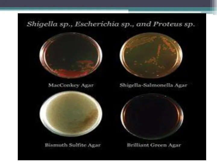
- 37. Types of Growth Media Differential media or indicator media distinguish one microorganism type from another growing
- 38. Types of Growth Media
- 39. Types of Growth Media Enriched media contain the nutrients required to support the growth of a
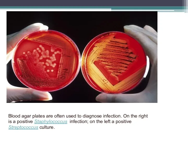
- 40. Blood agar plates are often used to diagnose infection. On the right is a positive Staphylococcus
- 41. Types of Growth Media Transport media used for the temporary storage of specimens being transported to
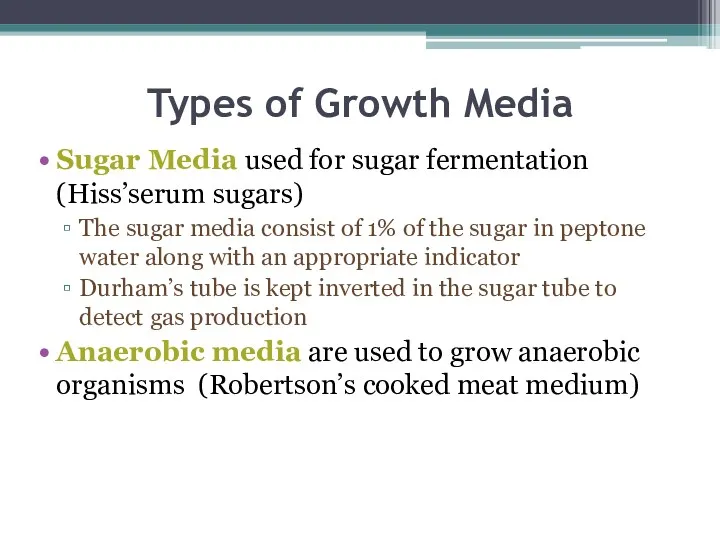
- 42. Types of Growth Media Sugar Media used for sugar fermentation (Hiss’serum sugars) The sugar media consist
- 43. Isolation of bacteria forms a very significant step in the diagnosis and management of the illness.
- 44. Common specimens include urine, faeces, wound swabs, throat swabs, vaginal swabs, sputum, and blood. Less common,
- 45. It is preferred to obtain the samples for bacteriological culture before antibiotic therapy is started. This
- 46. Specimens must be accurately labelled and accompanied by a properly completed requisition form, indicating the nature
- 47. Specimens should be transported as soon as possible to the laboratory. In case a delay is
- 48. (ii) Minimize the multiplication of bacteria (e.g. coliforms) within specimens before they reach the laboratory. In
- 49. CULTURE ON SOLID MEDIA The principal method for the detection of bacteria from clinical specimens is
- 50. Different bacteria produce different but characteristic colonies, allowing for early presumptive identification and easy identification of
- 51. Types of Growth Media
- 52. Blood agar plates are often used to diagnose infection. On the right is a positive Staphylococcus
- 53. Method of inoculating the solid culture media For obtaining the isolated colonies streaking method is used,
- 54. In this method single bacterial cells get isolated by the streaking, and when the plate is
- 55. Colony Morphology of Bacteria Bacteria grow on solid media as colonies. A colony is defined as
- 57. Form of the bacterial colony: – The form refers to the shape of the colony. These
- 58. Margin of bacterial colony: The margin or edge of a colony may be an important characteristic
- 59. Size of the bacterial colony: The size of the colony can be a useful characteristic for
- 60. Appearance of the colony surface: Bacterial colonies are frequently shiny and smooth in appearance. Other surface
- 61. Color of the colonies (pigmentation): Some bacteria produce pigment when they grow in the medium e.g.,
- 63. Скачать презентацию
Microbial Metabolism
The primary function of all living cells is to grow
Microbial Metabolism
The primary function of all living cells is to grow
Microbial Metabolism
The metabolic process that involves the degradation of chemical components
Microbial Metabolism
The metabolic process that involves the degradation of chemical components
Most matabolic processes in the cell would take forever if it
Most matabolic processes in the cell would take forever if it
Classification of enzymes
Oxidoreductases are involved in electron ( hydrogen) transfer reactions.
Transferases
Classification of enzymes
Oxidoreductases are involved in electron ( hydrogen) transfer reactions.
Transferases
Classification of enzymes
Lyases remove chemical groups from substrates, forming double bonds,
Classification of enzymes
Lyases remove chemical groups from substrates, forming double bonds,
Classification of enzymes
Enzymes synthesized by the cell remain within the cell
Classification of enzymes
Enzymes synthesized by the cell remain within the cell
Classification of enzymes
Pathogenicity enzymes - are enzymes that damage cells and
Classification of enzymes
Pathogenicity enzymes - are enzymes that damage cells and

Classification of enzymes
Hyaluronidase –enables pathogens to spread through connective tissue by
Classification of enzymes
Hyaluronidase –enables pathogens to spread through connective tissue by
Classification of enzymes
Hemolysin- enzyme that cause damage to the host’s red
Classification of enzymes
Hemolysin- enzyme that cause damage to the host’s red
Classification of enzymes
Hyaluronidase –enables pathogens to spread through connective tissue by
Classification of enzymes
Hyaluronidase –enables pathogens to spread through connective tissue by
Growth & Multiplication of Bacteria
Bacteria divide by binary fission
Bacterial cell divides
Growth & Multiplication of Bacteria
Bacteria divide by binary fission
Bacterial cell divides
The interval of time between two cell division, or the time

Growth & Multiplication of Bacteria
Bacteria divide by binary fission
Bacterial cell divides
Growth & Multiplication of Bacteria
Bacteria divide by binary fission
Bacterial cell divides

In many medically important bacteria, the generation time is about 20
In many medically important bacteria, the generation time is about 20
When bacteria are grown in a vessel of liquid medium (batch
When bacteria are grown in a vessel of liquid medium (batch
Bacterial cell Growth Curve
A- Lag phase
Immediately following the seeding of
Bacterial cell Growth Curve
A- Lag phase
Immediately following the seeding of
Bacterial cell Growth Curve
C- Stationary phase
After a period of exponential growth,
Bacterial cell Growth Curve
C- Stationary phase
After a period of exponential growth,
Bacterial cell Growth Curve
Bacterial cell Growth Curve
Nutritional requirements
Microorgaisms also depend on an available source of chemical nutrients.
Nutritional requirements
Microorgaisms also depend on an available source of chemical nutrients.
All organisms in nature can be placed into one of four
All organisms in nature can be placed into one of four
Nutritional requirements
C. Minerals
1. sulfur-Sulfur is needed to synthesizes sulfur-containing amino
Nutritional requirements
C. Minerals
1. sulfur-Sulfur is needed to synthesizes sulfur-containing amino
Nutritional requirements
D. Water
E. Growth factors
Growth factors are organic compounds
Nutritional requirements
D. Water
E. Growth factors
Growth factors are organic compounds
Oxygen Requirements
Depending on the influence of oxygen on growth and
Oxygen Requirements
Depending on the influence of oxygen on growth and
Oxygen Requirements
Anaerobic bacteria grow only in absence of oxygen
Anaerobic
Oxygen Requirements
Anaerobic bacteria grow only in absence of oxygen
Anaerobic
Oxygen requirements can be classified
Obligate aerobes—which can grow only in
Oxygen requirements can be classified
Obligate aerobes—which can grow only in
Oxygen requirements can be classified
Microaerophiles are organisms that require a
Oxygen requirements can be classified
Microaerophiles are organisms that require a
Physical requirements
Temperature
1. Psychrophiles are cold-loving bacteria. Their optimum growth
Physical requirements
Temperature
1. Psychrophiles are cold-loving bacteria. Their optimum growth
pH
Microorganisms can be placed in one of the following groups
pH
Microorganisms can be placed in one of the following groups
Culture Media
A growth medium or culture medium is a substance in
Culture Media
A growth medium or culture medium is a substance in
Types of Growth Media
The most common growth media for microorganisms are
Types of Growth Media
The most common growth media for microorganisms are
Types of Growth Media
Nutrient media
Undefined media (also known as basal or
Types of Growth Media
Nutrient media
Undefined media (also known as basal or
Types of Growth Media
Selective media (are used for the growth of
Types of Growth Media
Selective media (are used for the growth of
Types of Growth Media
Differential media or indicator media distinguish one microorganism
Types of Growth Media
Differential media or indicator media distinguish one microorganism
Types of Growth Media
Types of Growth Media
Types of Growth Media
Enriched media contain the nutrients required to support
Types of Growth Media
Enriched media contain the nutrients required to support

Blood agar plates are often used to diagnose infection. On the
Blood agar plates are often used to diagnose infection. On the
Types of Growth Media
Transport media used for the temporary storage of
Types of Growth Media
Transport media used for the temporary storage of
Types of Growth Media
Sugar Media used for sugar fermentation (Hiss’serum sugars)
The
Types of Growth Media
Sugar Media used for sugar fermentation (Hiss’serum sugars)
The
Isolation of bacteria forms a very significant step in the diagnosis
Isolation of bacteria forms a very significant step in the diagnosis
Common specimens include urine, faeces, wound swabs, throat swabs, vaginal swabs,
Common specimens include urine, faeces, wound swabs, throat swabs, vaginal swabs,
It is preferred to obtain the samples for bacteriological culture before
It is preferred to obtain the samples for bacteriological culture before
Specimens must be accurately labelled and accompanied by a properly completed
Specimens must be accurately labelled and accompanied by a properly completed
Specimens should be transported as soon as possible to the laboratory.
Specimens should be transported as soon as possible to the laboratory.
(ii) Minimize the multiplication of bacteria (e.g. coliforms) within specimens before
(ii) Minimize the multiplication of bacteria (e.g. coliforms) within specimens before
CULTURE ON SOLID MEDIA
The principal method for the detection of
CULTURE ON SOLID MEDIA
The principal method for the detection of
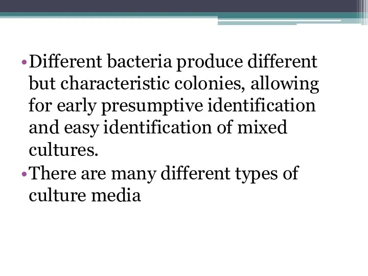
Different bacteria produce different but characteristic colonies, allowing for early presumptive
Different bacteria produce different but characteristic colonies, allowing for early presumptive
Types of Growth Media
Types of Growth Media
Blood agar plates are often used to diagnose infection. On the
Blood agar plates are often used to diagnose infection. On the
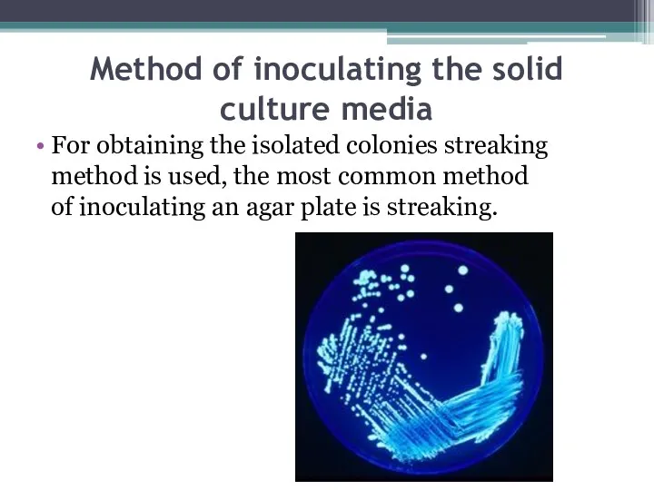
Method of inoculating the solid culture media
For obtaining the isolated colonies
Method of inoculating the solid culture media
For obtaining the isolated colonies
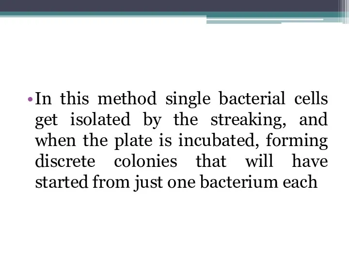
In this method single bacterial cells get isolated by the streaking,
In this method single bacterial cells get isolated by the streaking,
Colony Morphology of Bacteria
Bacteria grow on solid media as colonies. A
Colony Morphology of Bacteria
Bacteria grow on solid media as colonies. A
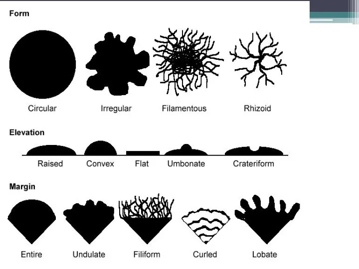
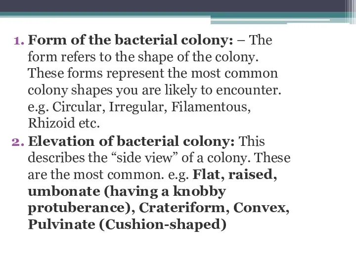
Form of the bacterial colony: – The form refers to the shape
Form of the bacterial colony: – The form refers to the shape
Margin of bacterial colony: The margin or edge of a colony may
Margin of bacterial colony: The margin or edge of a colony may
Size of the bacterial colony: The size of the colony can be
Size of the bacterial colony: The size of the colony can be
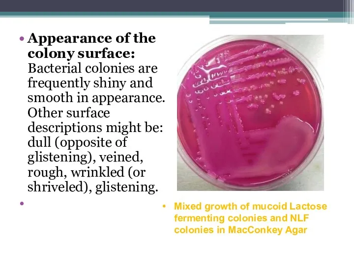
Appearance of the colony surface: Bacterial colonies are frequently shiny and
Appearance of the colony surface: Bacterial colonies are frequently shiny and
 Животный организм и особенности
 Chicory (cichorium intybus)
 Презентация к уроку окружающего мира 4 класс Мир глазами эколога.
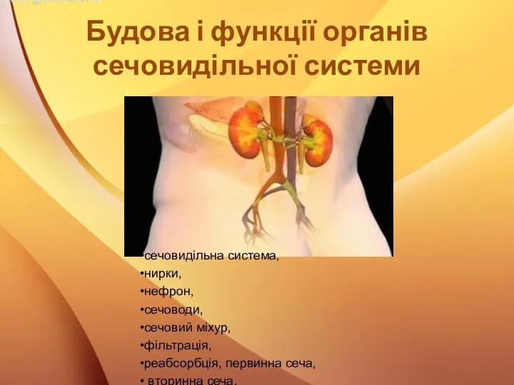 Будова і функції органів сечовидільної системи
 Жизненные формы растений. Растительные ткани
 Есту анализаторы
 Новые открытия в биотехнологии за 10 лет
 Двигательная деятельность человека
 Растения и животные разных материков
 Protein denatu-ration
 Морфологическое, систематическое и экологическое разнообразие насекомых
 История изучения клетки. Клеточная теория
 Сучасні критерії виду
 Жылқы –малдың патшасы
 Свиньи. Породы свиней
 Акулы - морской хищник
 Высшие растения мхи. Общая характеристика моховидных
 Особенности двудольных растений. Обобщение игра. 7 класс
 Дендрофлора лесов мира
 Цитологические основы законов Менделя
 Разнообразие, распространение и значение растений
 Птицы Ненецкого Автономного округа
 Комнатные растения. Фиалка
 Высшие споровые растения
 Растения в домашней аптечке
 Многообразие водных биогеоценозов
 Нервная система, общие сведения. Физиология и анатомия спинного мозга. Лекция № 21
 Биоинженерия. Полимеразная цепная реакция