- Sensory systems

Содержание
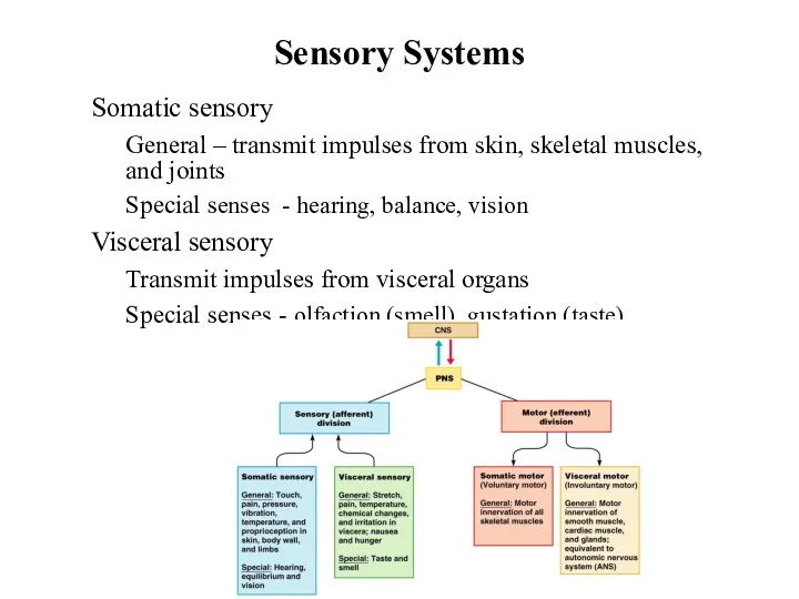
- 2. Sensory Systems Somatic sensory General – transmit impulses from skin, skeletal muscles, and joints Special senses
- 3. Stimulus - energy source Internal External Receptors Sense organs - structures specialized to respond to stimuli
- 4. Sensory Pathways Stimulus as physical energy ? sensory receptor acts as a transducer Stimulus > threshold
- 5. Classification by Function (Stimuli) Mechanoreceptors – respond to touch, pressure, vibration, stretch, and itch Thermoreceptors –
- 6. Classification by Location Exteroceptors – sensitive to stimuli arising from outside the body Located at or
- 7. Classification by Structure
- 8. General somatic – include touch, pain, vibration, pressure, temperature Proprioceptive – detect stretch in tendons and
- 9. Somatic Receptors Divided into two groups Free or Unencapsulated nerve endings Encapsulated nerve endings - consist
- 10. Free Nerve Endings Abundant in epithelia and underlying connective tissue Nociceptors - respond to pain Thermoreceptors
- 11. Encapsulated Nerve Endings Meissner’s corpuscles Spiraling nerve ending surrounded by Schwann cells Occur in the dermal
- 12. Encapsulated Nerve Endings - Proprioceptors Monitor stretch in locomotory organs Three types of proprioceptors Muscle spindles
- 13. Muscle Spindle & Golgi Tendon Organ
- 14. Special Senses Figure 10-4: Sensory pathways Taste, smell, sight, hearing, and balance Localized – confined to
- 15. Anatomy of the Eyeball Function of the eyeball Protect and support the photoreceptors Gather, focus, and
- 16. The Fibrous Layer Most external layer of the eyeball Cornea Anterior one-sixth of the fibrous tunic
- 17. The Vascular Layer Middle layer consists of choroid, ciliary body, and iris Iris and Pupil Composed
- 18. The Vascular Layer Choroid - vascular layer in the wall of the eye. Dark brown (pigmented)
- 19. The Inner Layer (Retina) Retina is the innermost layer of the eye lining the posterior cavity
- 20. Photoreceptors Two main types Rod cells More sensitive to light Allow vision in dim light In
- 21. Regional Specializations of the Retina Ora serrata retinae Neural layer ends at the posterior margin of
- 22. The Lens A thick, transparent, biconvex disc Held in place by its ciliary zonule Lens epithelium
- 23. The Eye as an Optical Device Structures in the eye bend light rays Light rays converge
- 24. Internal Chambers and Fluids Figure 16.8
- 25. Internal Chambers and Fluids Anterior segment Divided into anterior and posterior chambers Anterior chamber – between
- 26. Internal Chambers and Fluids The lens and ciliary zonules divide the eye Posterior segment (cavity) Filled
- 27. Accessory Structures of the Eye Eyebrows – coarse hairs on the superciliary arches Eyelids (palpebrae) Separated
- 28. Accessory Structures of the Eye Lacrimal apparatus – keeps the surface of the eye moist Lacrimal
- 29. Extrinsic Eye Muscles Figure 16.6a, b Six muscles that control movement of the eye Originate in
- 30. Visual Pathways to the Cerebral Cortex Pathway begins at the retina Light activates photoreceptors Photoreceptors signal
- 31. Optic nerve Optic chiasm Optic tract Thalamus Visual cortex Other pathways include the midbrain and diencephalon
- 32. The Ear: Hearing and Equilibrium The ear – receptor organ for hearing and equilibrium Composed of
- 33. The Outer (External) Ear Auricle (pinna) - helps direct sounds External acoustic meatus Lined with skin
- 34. The Middle Ear The tympanic cavity A small, air-filled space Located within the petrous portion of
- 35. Figure 16.17 The Middle Ear Ear ossicles – smallest bones in the body Malleus – attaches
- 36. The Inner (Internal) Ear Inner ear – also called the labyrinth Bony labyrinth – a cavity
- 37. The Membranous Labyrinth Figure 16.18 Membranous labyrinth - series of membrane-walled sacs and ducts Fit within
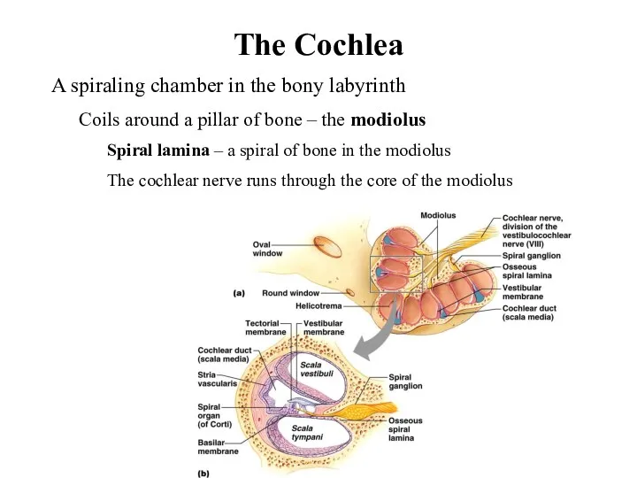
- 38. The Cochlea A spiraling chamber in the bony labyrinth Coils around a pillar of bone –
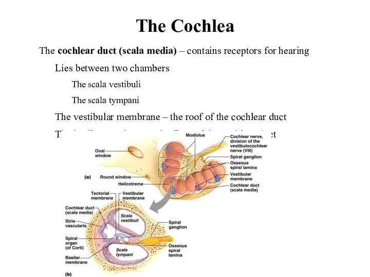
- 39. The Cochlea The cochlear duct (scala media) – contains receptors for hearing Lies between two chambers
- 40. The Cochlea The cochlear duct (scala media) – contains receptors for hearing Organ of Corti –
- 41. The Role of the Cochlea in Hearing Figure 16.20
- 42. Auditory Pathway from the Organ of Corti The ascending auditory pathway Transmits information from cochlear receptors
- 43. The Vestibule Utricle and saccule – suspended in perilymph Two egg-shaped parts of the membranous labyrinth
- 44. Anatomy and Function of the Maculae Figure 16.21b
- 45. The Semicircular Canals Lie posterior and lateral to the vestibule Anterior and posterior semicircular canals lie
- 46. The Semicircular Canals Semicircular duct – snakes through each semicircular canal Membranous ampulla – located within
- 47. Structure and Function of the Crista Ampullaris Figure 16.22b
- 48. The Chemical Senses: Taste and Smell Taste – gustation Smell – olfaction Receptors – classified as
- 49. Taste – Gustation Taste receptors Occur in taste buds Most are found on the surface of
- 50. Taste Buds Collection of 50 –100 epithelial cells Contain three major cell types (similar in all
- 51. Taste Sensation and the Gustatory Pathway Four basic qualities of taste Sweet, sour, salty, and bitter
- 52. Gustatory Pathway from Taste Buds Figure 16.2 Taste information reaches the cerebral cortex Primarily through the
- 53. Olfactory epithelium with olfactory receptors, supporting cells, basal cells Olfactory receptors are modified neurons Surfaces are
- 55. Скачать презентацию
Sensory Systems
Somatic sensory
General – transmit impulses from skin, skeletal muscles,
Sensory Systems
Somatic sensory
General – transmit impulses from skin, skeletal muscles,
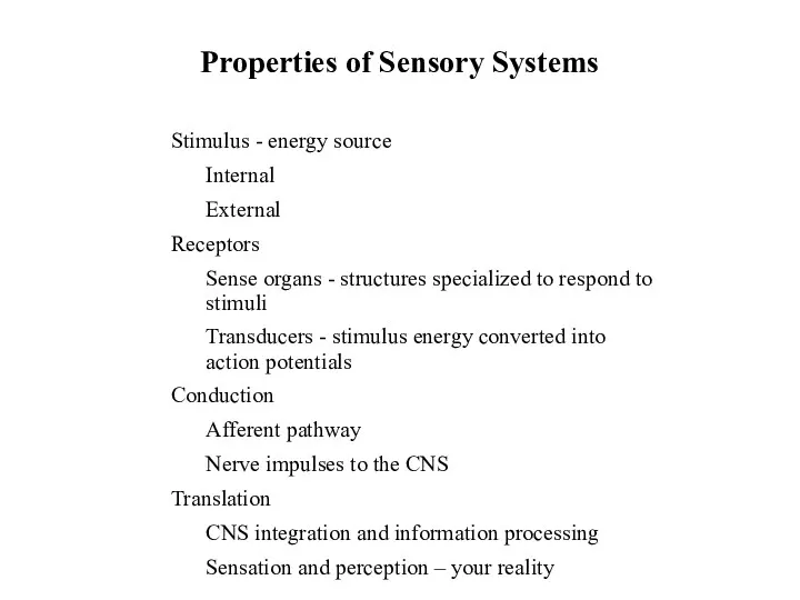
Stimulus - energy source
Internal
External
Receptors
Sense organs - structures specialized to respond
Stimulus - energy source
Internal
External
Receptors
Sense organs - structures specialized to respond
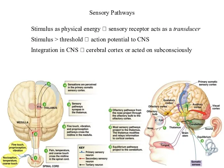
Sensory Pathways
Stimulus as physical energy ? sensory receptor acts as a
Sensory Pathways
Stimulus as physical energy ? sensory receptor acts as a
Classification by Function (Stimuli)
Mechanoreceptors – respond to touch, pressure, vibration, stretch,
Classification by Function (Stimuli)
Mechanoreceptors – respond to touch, pressure, vibration, stretch,
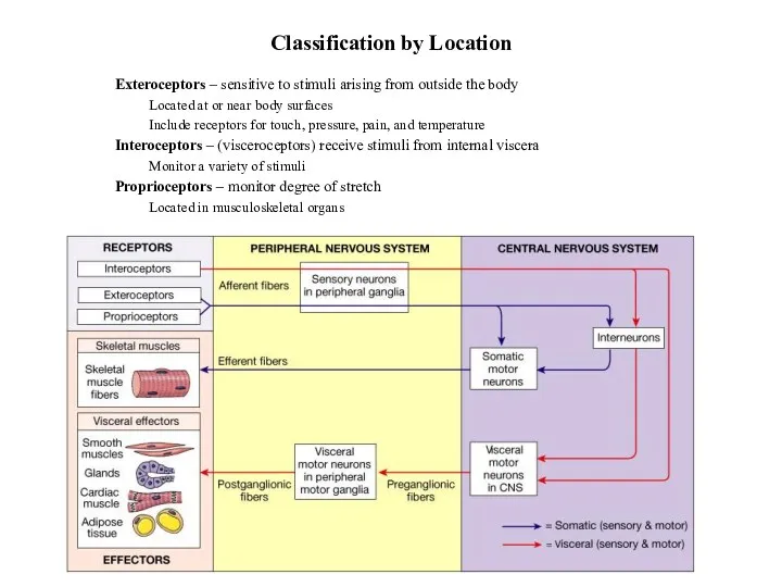
Classification by Location
Exteroceptors – sensitive to stimuli arising from outside the
Classification by Location
Exteroceptors – sensitive to stimuli arising from outside the
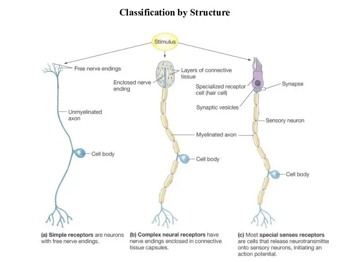
Classification by Structure
Classification by Structure
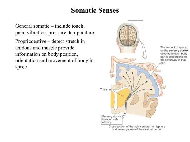
General somatic – include touch, pain, vibration, pressure, temperature
Proprioceptive – detect
General somatic – include touch, pain, vibration, pressure, temperature
Proprioceptive – detect
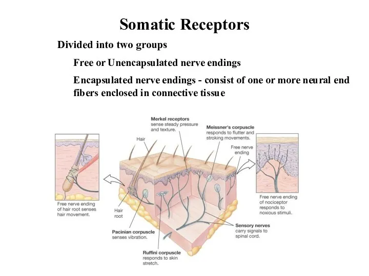
Somatic Receptors
Divided into two groups
Free or Unencapsulated nerve endings
Encapsulated nerve endings
Somatic Receptors
Divided into two groups
Free or Unencapsulated nerve endings
Encapsulated nerve endings
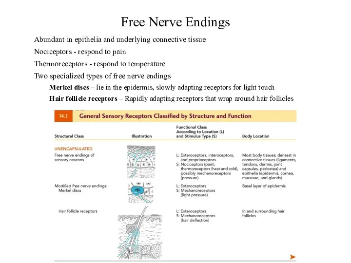
Free Nerve Endings
Abundant in epithelia and underlying connective tissue
Nociceptors - respond
Free Nerve Endings
Abundant in epithelia and underlying connective tissue
Nociceptors - respond
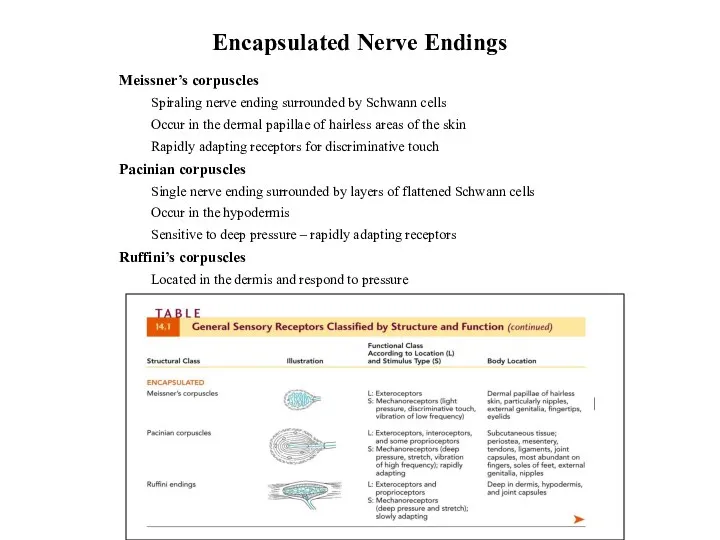
Encapsulated Nerve Endings
Meissner’s corpuscles
Spiraling nerve ending surrounded by Schwann cells
Occur
Encapsulated Nerve Endings
Meissner’s corpuscles
Spiraling nerve ending surrounded by Schwann cells
Occur
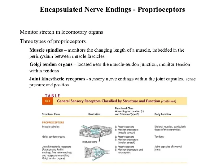
Encapsulated Nerve Endings - Proprioceptors
Monitor stretch in locomotory organs
Three types
Encapsulated Nerve Endings - Proprioceptors
Monitor stretch in locomotory organs
Three types
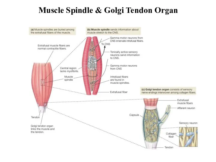
Muscle Spindle & Golgi Tendon Organ
Muscle Spindle & Golgi Tendon Organ
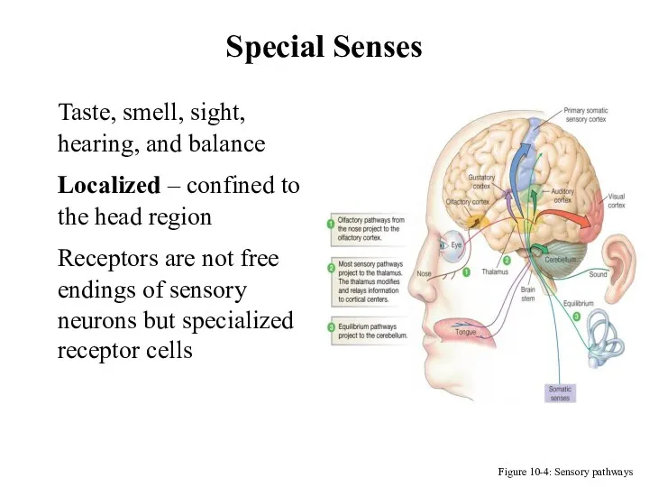
Special Senses
Figure 10-4: Sensory pathways
Taste, smell, sight, hearing, and balance
Localized –
Special Senses
Figure 10-4: Sensory pathways
Taste, smell, sight, hearing, and balance
Localized –
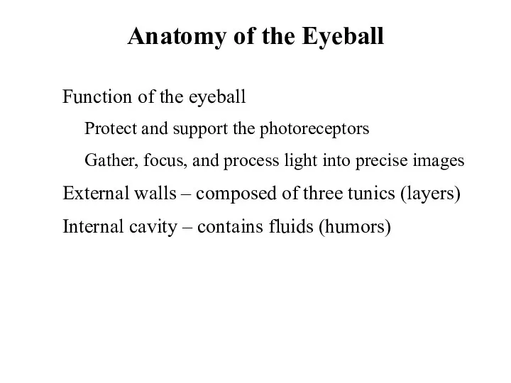
Anatomy of the Eyeball
Function of the eyeball
Protect and support the photoreceptors
Gather,
Anatomy of the Eyeball
Function of the eyeball
Protect and support the photoreceptors
Gather,
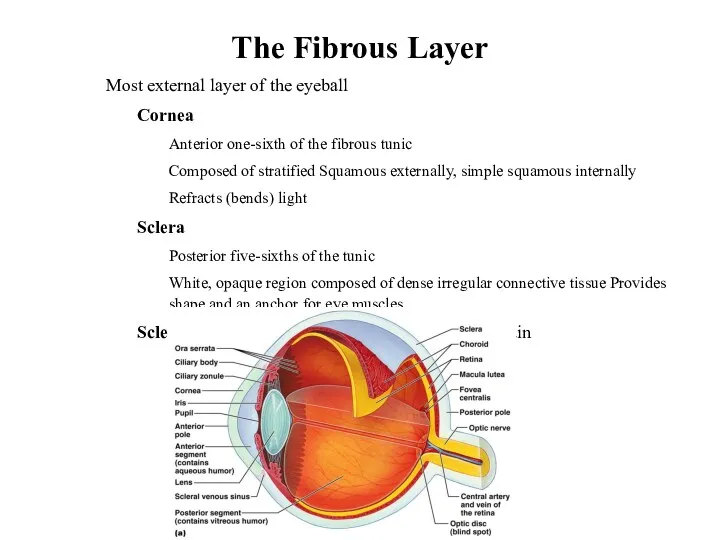
The Fibrous Layer
Most external layer of the eyeball
Cornea
Anterior one-sixth of the
The Fibrous Layer
Most external layer of the eyeball
Cornea
Anterior one-sixth of the
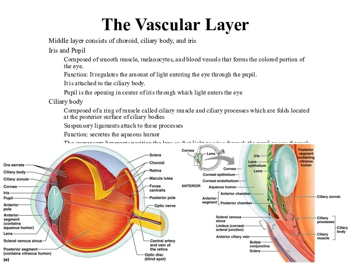
The Vascular Layer
Middle layer consists of choroid, ciliary body, and iris
The Vascular Layer
Middle layer consists of choroid, ciliary body, and iris
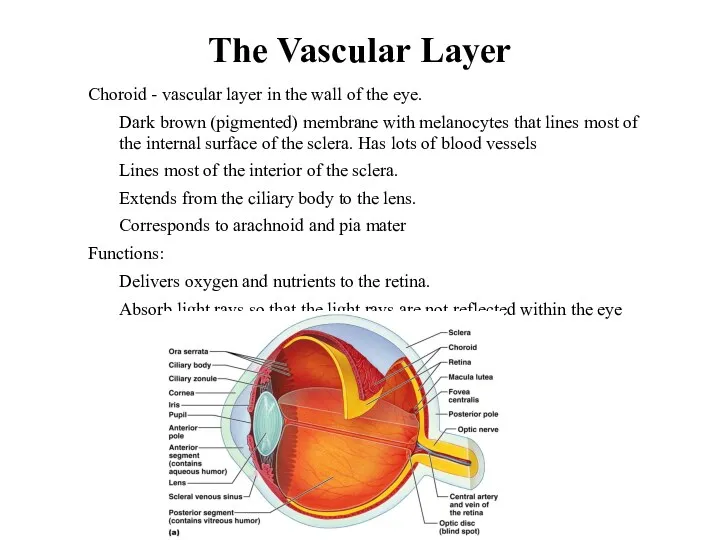
The Vascular Layer
Choroid - vascular layer in the wall of the
The Vascular Layer
Choroid - vascular layer in the wall of the
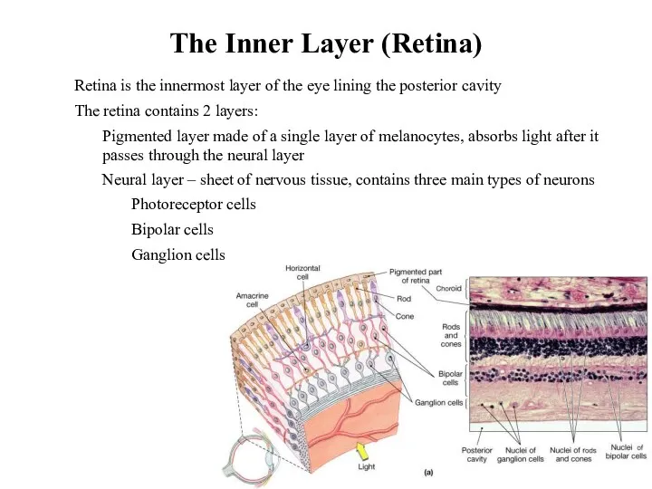
The Inner Layer (Retina)
Retina is the innermost layer of the eye
The Inner Layer (Retina)
Retina is the innermost layer of the eye
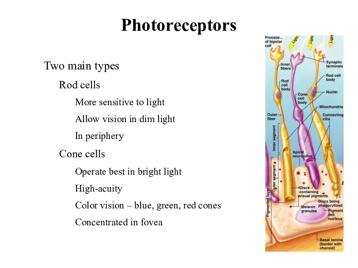
Photoreceptors
Two main types
Rod cells
More sensitive to light
Allow vision in
Photoreceptors
Two main types
Rod cells
More sensitive to light
Allow vision in
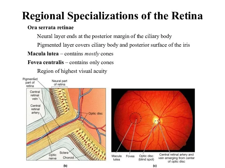
Regional Specializations of the Retina
Ora serrata retinae
Neural layer ends at
Regional Specializations of the Retina
Ora serrata retinae
Neural layer ends at
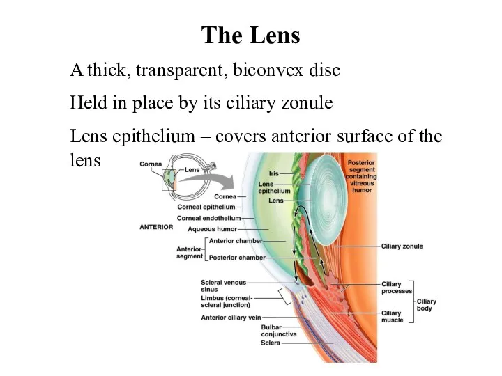
The Lens
A thick, transparent, biconvex disc
Held in place by its ciliary
The Lens
A thick, transparent, biconvex disc
Held in place by its ciliary
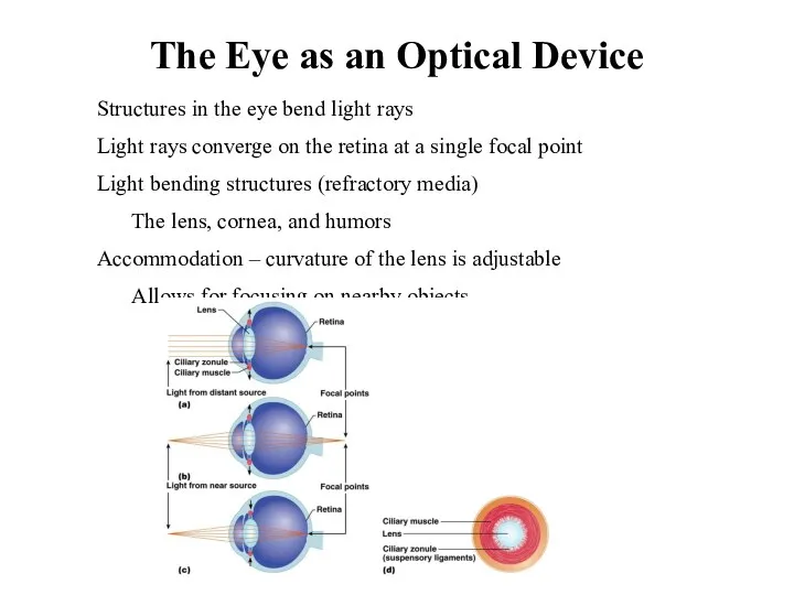
The Eye as an Optical Device
Structures in the eye bend light
The Eye as an Optical Device
Structures in the eye bend light
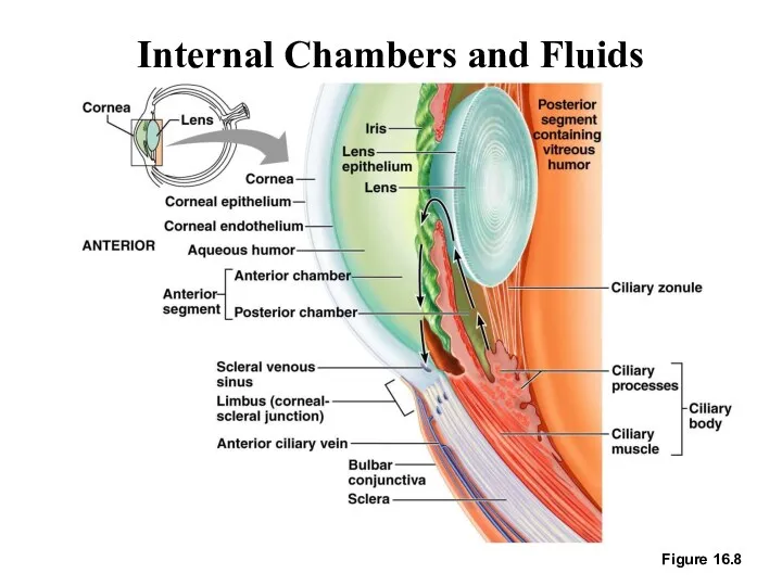
Internal Chambers and Fluids
Figure 16.8
Internal Chambers and Fluids
Figure 16.8
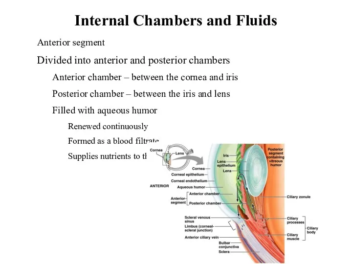
Internal Chambers and Fluids
Anterior segment
Divided into anterior and posterior chambers
Anterior chamber
Internal Chambers and Fluids
Anterior segment
Divided into anterior and posterior chambers
Anterior chamber
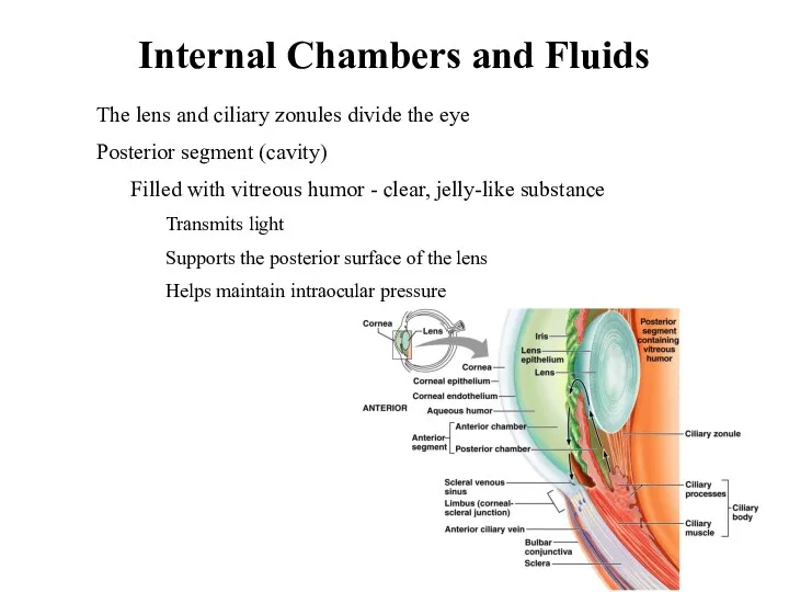
Internal Chambers and Fluids
The lens and ciliary zonules divide the eye
Internal Chambers and Fluids
The lens and ciliary zonules divide the eye
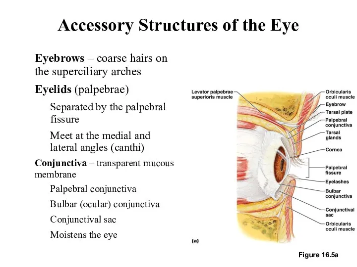
Accessory Structures of the Eye
Eyebrows – coarse hairs on the superciliary
Accessory Structures of the Eye
Eyebrows – coarse hairs on the superciliary
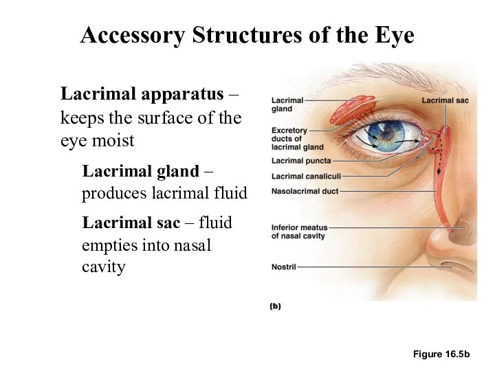
Accessory Structures of the Eye
Lacrimal apparatus – keeps the surface of
Accessory Structures of the Eye
Lacrimal apparatus – keeps the surface of
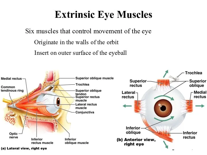
Extrinsic Eye Muscles
Figure 16.6a, b
Six muscles that control movement of the
Extrinsic Eye Muscles
Figure 16.6a, b
Six muscles that control movement of the
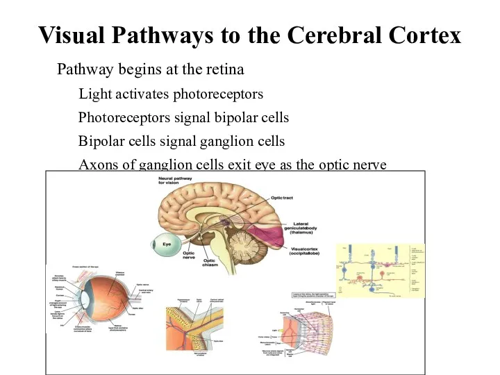
Visual Pathways to the Cerebral Cortex
Pathway begins at the retina
Light activates
Visual Pathways to the Cerebral Cortex
Pathway begins at the retina
Light activates
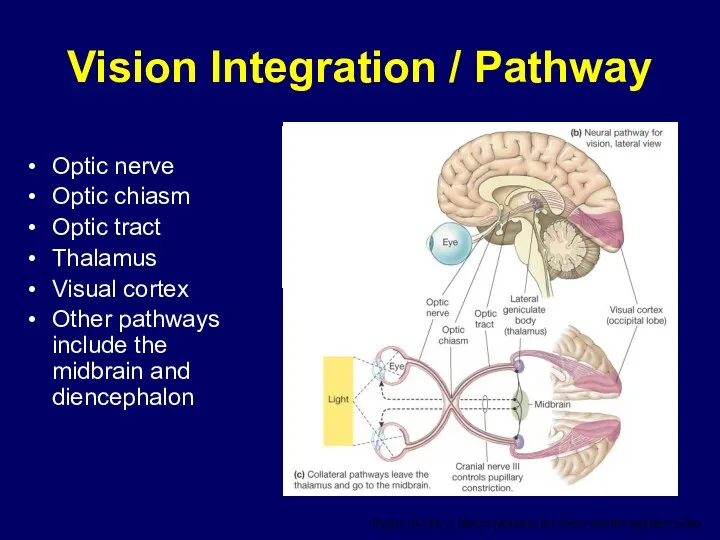
Optic nerve
Optic chiasm
Optic tract
Thalamus
Visual cortex
Other pathways include the midbrain and
Optic nerve
Optic chiasm
Optic tract
Thalamus
Visual cortex
Other pathways include the midbrain and
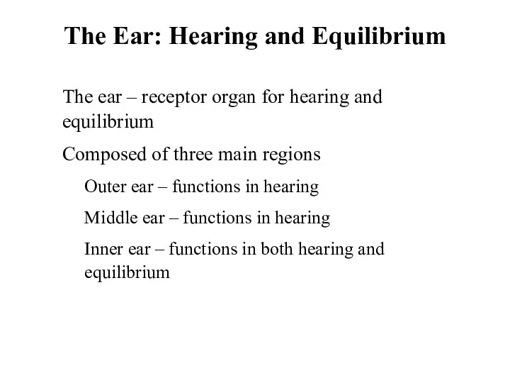
The Ear: Hearing and Equilibrium
The ear – receptor organ for hearing
The Ear: Hearing and Equilibrium
The ear – receptor organ for hearing
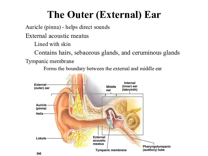
The Outer (External) Ear
Auricle (pinna) - helps direct sounds
External acoustic meatus
Lined
The Outer (External) Ear
Auricle (pinna) - helps direct sounds
External acoustic meatus
Lined
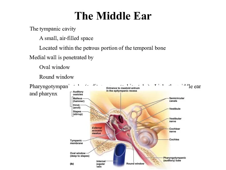
The Middle Ear
The tympanic cavity
A small, air-filled space
Located within
The Middle Ear
The tympanic cavity
A small, air-filled space
Located within
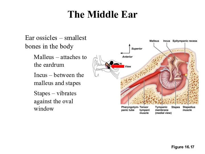
Figure 16.17
The Middle Ear
Ear ossicles – smallest bones in the body
Malleus
Figure 16.17
The Middle Ear
Ear ossicles – smallest bones in the body
Malleus
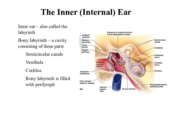
The Inner (Internal) Ear
Inner ear – also called the labyrinth
Bony labyrinth
The Inner (Internal) Ear
Inner ear – also called the labyrinth
Bony labyrinth
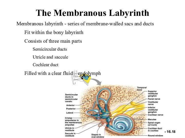
The Membranous Labyrinth
Figure 16.18
Membranous labyrinth - series of membrane-walled sacs and
The Membranous Labyrinth
Figure 16.18
Membranous labyrinth - series of membrane-walled sacs and
The Cochlea
A spiraling chamber in the bony labyrinth
Coils around a pillar
The Cochlea
A spiraling chamber in the bony labyrinth
Coils around a pillar
The Cochlea
The cochlear duct (scala media) – contains receptors for hearing
Lies
The Cochlea
The cochlear duct (scala media) – contains receptors for hearing
Lies

The Cochlea
The cochlear duct (scala media) – contains receptors for hearing
Organ
The Cochlea
The cochlear duct (scala media) – contains receptors for hearing
Organ
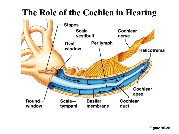
The Role of the Cochlea in Hearing
Figure 16.20
The Role of the Cochlea in Hearing
Figure 16.20
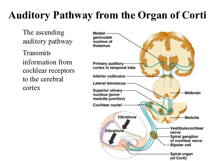
Auditory Pathway from the Organ of Corti
The ascending auditory pathway
Transmits
Auditory Pathway from the Organ of Corti
The ascending auditory pathway
Transmits
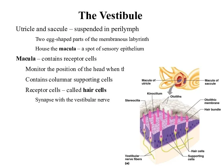
The Vestibule
Utricle and saccule – suspended in perilymph
Two egg-shaped parts
The Vestibule
Utricle and saccule – suspended in perilymph
Two egg-shaped parts
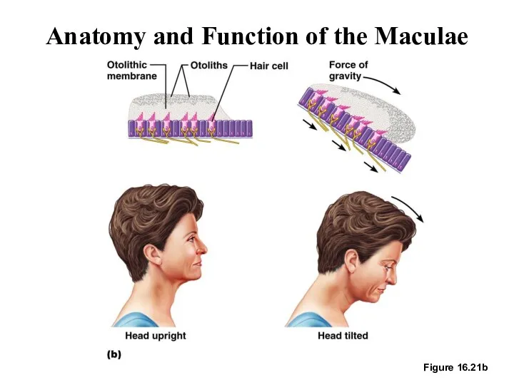
Anatomy and Function of the Maculae
Figure 16.21b
Anatomy and Function of the Maculae
Figure 16.21b
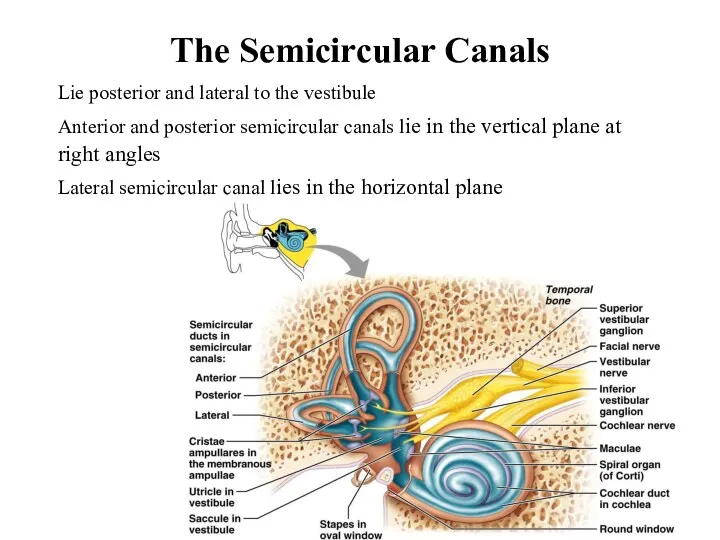
The Semicircular Canals
Lie posterior and lateral to the vestibule
Anterior and posterior
The Semicircular Canals
Lie posterior and lateral to the vestibule
Anterior and posterior
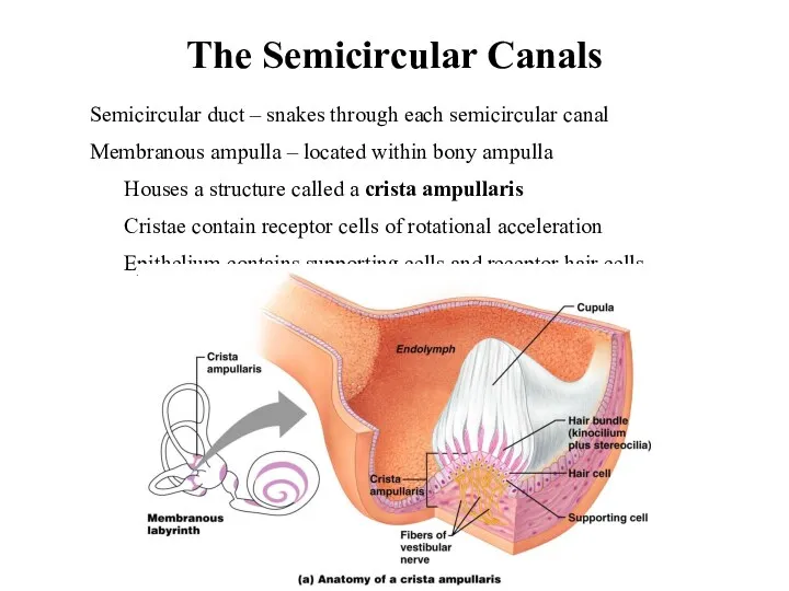
The Semicircular Canals
Semicircular duct – snakes through each semicircular canal
Membranous ampulla
The Semicircular Canals
Semicircular duct – snakes through each semicircular canal
Membranous ampulla
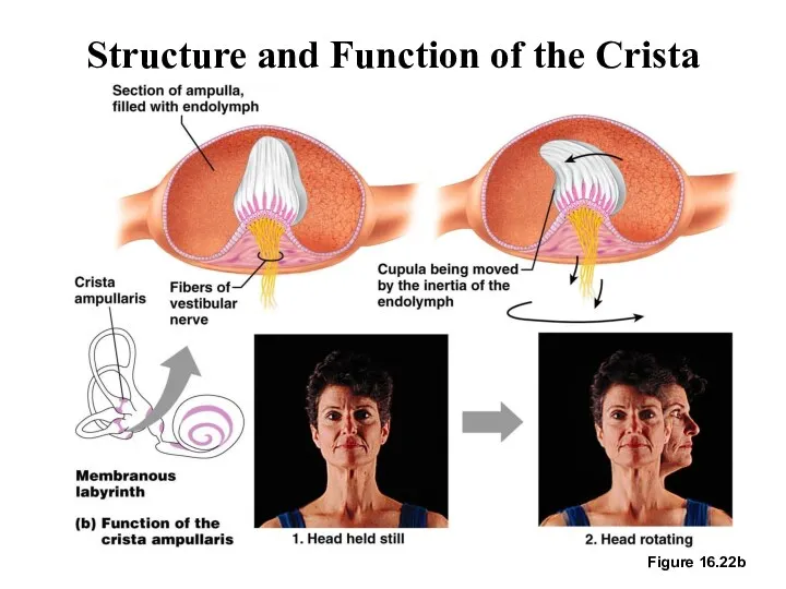
Structure and Function of the Crista Ampullaris
Figure 16.22b
Structure and Function of the Crista Ampullaris
Figure 16.22b
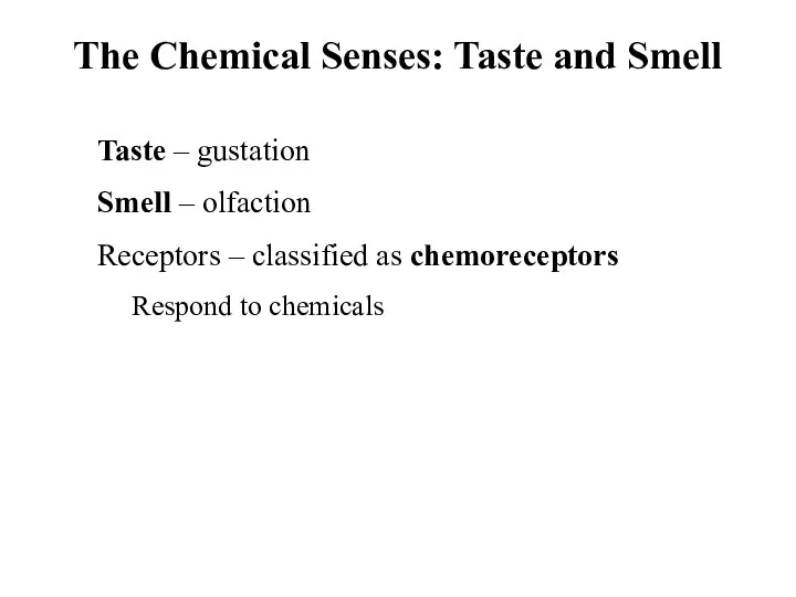
The Chemical Senses: Taste and Smell
Taste – gustation
Smell – olfaction
Receptors
The Chemical Senses: Taste and Smell
Taste – gustation
Smell – olfaction
Receptors
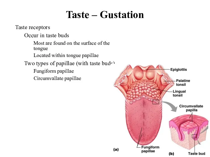
Taste – Gustation
Taste receptors
Occur in taste buds
Most are found on the
Taste – Gustation
Taste receptors
Occur in taste buds
Most are found on the
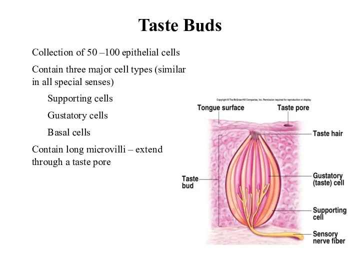
Taste Buds
Collection of 50 –100 epithelial cells
Contain three major cell types
Taste Buds
Collection of 50 –100 epithelial cells
Contain three major cell types
Taste Sensation and the Gustatory Pathway
Four basic qualities of taste
Sweet, sour,
Taste Sensation and the Gustatory Pathway
Four basic qualities of taste
Sweet, sour,
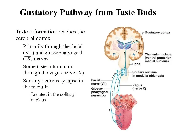
Gustatory Pathway from Taste Buds
Figure 16.2
Taste information reaches the cerebral cortex
Primarily
Gustatory Pathway from Taste Buds
Figure 16.2
Taste information reaches the cerebral cortex
Primarily
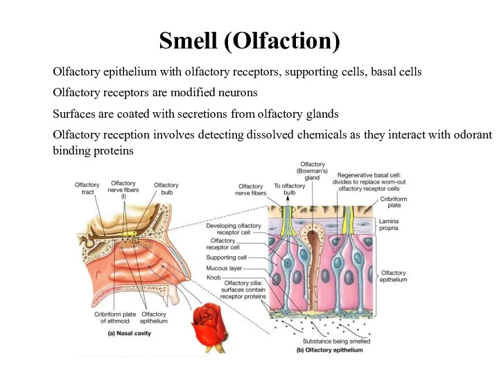
Olfactory epithelium with olfactory receptors, supporting cells, basal cells
Olfactory receptors are
Olfactory epithelium with olfactory receptors, supporting cells, basal cells
Olfactory receptors are
 Красная книга Белгородчины
 Значение дыхания, газообмен, типы дыхания. Дыхание растений
 Природные зоны Южной Америки
 Головной мозг
 Органоиды клетки
 Строение глаза
 Посевные качества семян
 Класс рыбы: хрящевые, костные
 Классификация животных. Основные систематические группы. Влияние человека на животных
 Путешествие в царство Природы
 Хромосомний імпринтинг. Однобатьківська дисомія. Фенокопії
 Подготовка учащихся к ЕГЭ по биологии
 Клетка. Основные положения клеточной теории. Органоиды клетки
 Способы размножения растений
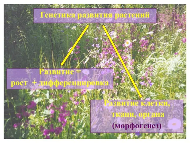 Генетика развития растений
 Потенциал покоя и потенциал действия клетки
 Гнилостные бактерии
 Физиология сердечно-сосудистой системы. Занятие № 26
 Неклеточные формы жизни. Вирусы и бактериофаги.
 Фасциолез. Профилактика
 Воздействие человека и его деятельности на животных
 Вирусы
 Отдел Настоящие грибы
 Анатомо-физиологические особенности полости рта, глотки, пищевода, желудка, кишечника
 Тундровый волк
 Ознакомление с фитонцидными растениями и выявление возможности их использования в интерьере. Практическая работа
 Cell structure. Cell Theory
 Обмен веществ и энергии в организме