- The Molecular Basis of Inheritance

Содержание
- 2. Figure 16.1 Overview: Life’s Operating Instructions In 1953, James Watson and Francis Crick shook the world
- 3. DNA, the substance of inheritance Is the most celebrated molecule of our time Hereditary information Is
- 4. Concept 16.1: DNA is the genetic material Early in the 20th century The identification of the
- 5. The Search for the Genetic Material: Scientific Inquiry The role of DNA in heredity Was first
- 6. Evidence That DNA Can Transform Bacteria Frederick Griffith was studying Streptococcus pneumoniae A bacterium that causes
- 7. Bacteria of the “S” (smooth) strain of Streptococcus pneumoniae are pathogenic because they have a capsule
- 8. Griffith called the phenomenon transformation Now defined as a change in genotype and phenotype due to
- 9. Evidence That Viral DNA Can Program Cells Additional evidence for DNA as the genetic material Came
- 10. Figure 16.3 Viruses that infect bacteria, bacteriophages Are widely used as tools by researchers in molecular
- 11. Alfred Hershey and Martha Chase Performed experiments showing that DNA is the genetic material of a
- 12. In their famous 1952 experiment, Alfred Hershey and Martha Chase used radioactive sulfur and phosphorus to
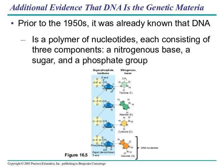
- 13. Additional Evidence That DNA Is the Genetic Materia Prior to the 1950s, it was already known
- 14. Erwin Chargaff analyzed the base composition of DNA From a number of different organisms In 1947,
- 15. Building a Structural Model of DNA: Scientific Inquiry Once most biologists were convinced that DNA was
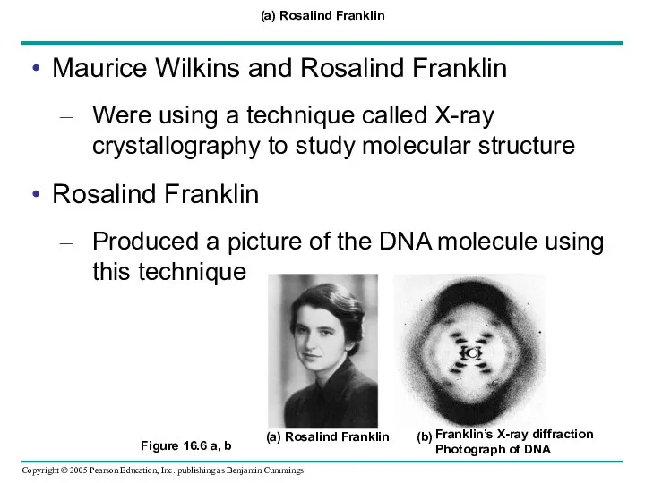
- 16. (a) Rosalind Franklin Maurice Wilkins and Rosalind Franklin Were using a technique called X-ray crystallography to
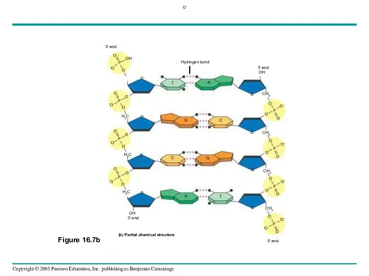
- 17. Figure 16.7a, c Watson and Crick deduced that DNA was a double helix Through observations of
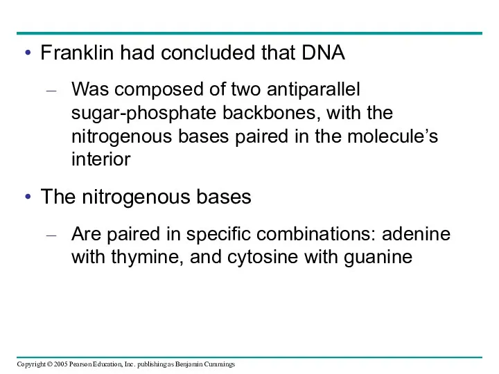
- 18. Franklin had concluded that DNA Was composed of two antiparallel sugar-phosphate backbones, with the nitrogenous bases
- 19. O
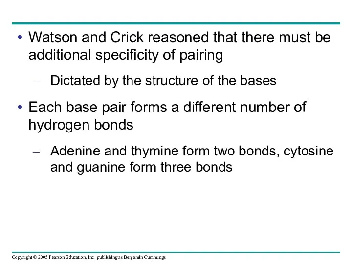
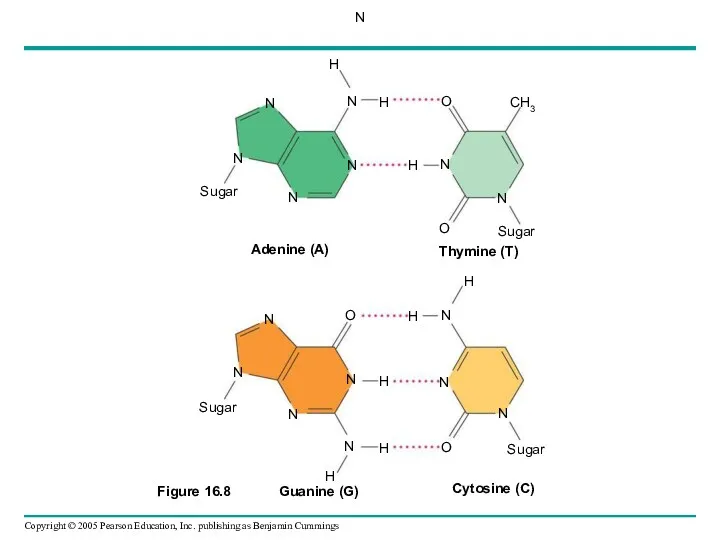
- 20. Watson and Crick reasoned that there must be additional specificity of pairing Dictated by the structure
- 21. N
- 22. Concept 16.2: Many proteins work together in DNA replication and repair The relationship between structure and
- 23. The Basic Principle: Base Pairing to a Template Strand Since the two strands of DNA are
- 24. (a) The parent molecule has two complementary strands of DNA. Each base is paired by hydrogen
- 25. Figure 16.10 a–c DNA replication is semiconservative Each of the two new daughter molecules will have
- 26. Figure 16.11 Experiments performed by Meselson and Stahl Supported the semiconservative model of DNA replication
- 27. CONCLUSION
- 28. DNA Replication: A Closer Look The copying of DNA Is remarkable in its speed and accuracy
- 29. Getting Started: Origins of Replication The replication of a DNA molecule Begins at special sites called
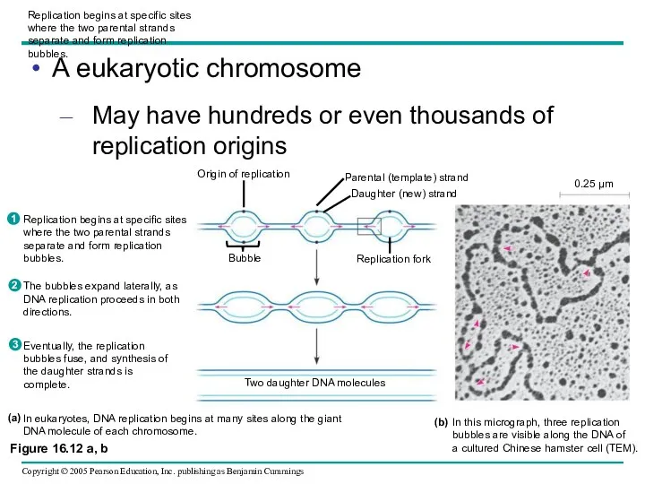
- 30. Replication begins at specific sites where the two parental strands separate and form replication bubbles. A
- 31. Elongating a New DNA Strand Elongation of new DNA at a replication fork Is catalyzed by
- 32. Antiparallel Elongation How does the antiparallel structure of the double helix affect replication?
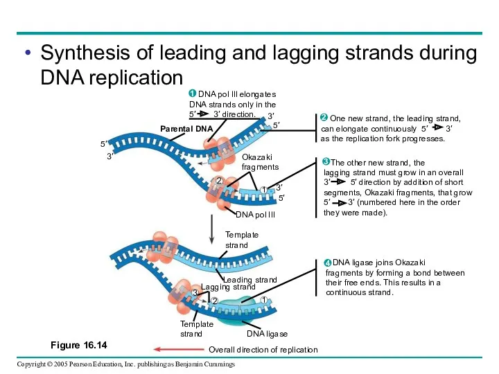
- 33. DNA polymerases add nucleotides Only to the free 3′ end of a growing strand Along one
- 34. To elongate the other new strand of DNA, the lagging strand DNA polymerase III must work
- 35. Synthesis of leading and lagging strands during DNA replication
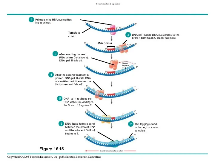
- 36. Priming DNA Synthesis DNA polymerases cannot initiate the synthesis of a polynucleotide They can only add
- 37. Only one primer is needed for synthesis of the leading strand But for synthesis of the
- 38. Overall direction of replication
- 39. Other Proteins That Assist DNA Replication Helicase, topoisomerase, single-strand binding protein Are all proteins that assist
- 40. Figure 16.16 A summary of DNA replication
- 41. The DNA Replication Machine as a Stationary Complex The various proteins that participate in DNA replication
- 42. Proofreading and Repairing DNA DNA polymerases proofread newly made DNA Replacing any incorrect nucleotides In mismatch
- 43. Figure 16.17 Nuclease DNA polymerase DNA ligase A nuclease enzyme cuts the damaged DNA strand at
- 44. Replicating the Ends of DNA Molecules The ends of eukaryotic chromosomal DNA Get shorter with each
- 45. Figure 16.19 Eukaryotic chromosomal DNA molecules Have at their ends nucleotide sequences, called telomeres, that postpone
- 47. Скачать презентацию
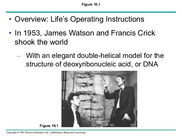
Figure 16.1
Overview: Life’s Operating Instructions
In 1953, James Watson and Francis Crick
Figure 16.1
Overview: Life’s Operating Instructions
In 1953, James Watson and Francis Crick

DNA, the substance of inheritance
Is the most celebrated molecule of our
DNA, the substance of inheritance
Is the most celebrated molecule of our
Concept 16.1: DNA is the genetic material
Early in the 20th century
The
Concept 16.1: DNA is the genetic material
Early in the 20th century
The
The Search for the Genetic Material: Scientific Inquiry
The role of DNA
The Search for the Genetic Material: Scientific Inquiry
The role of DNA
Evidence That DNA Can Transform Bacteria
Frederick Griffith was studying Streptococcus pneumoniae
A
Evidence That DNA Can Transform Bacteria
Frederick Griffith was studying Streptococcus pneumoniae
A
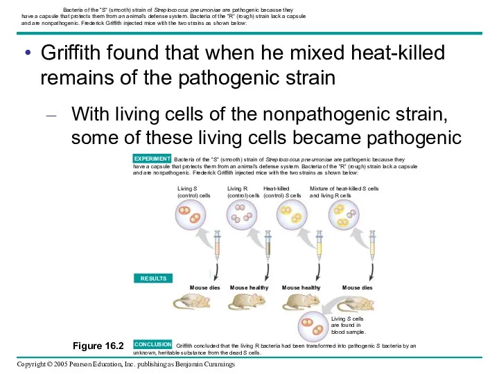
Bacteria of the “S” (smooth) strain of Streptococcus pneumoniae are
Bacteria of the “S” (smooth) strain of Streptococcus pneumoniae are
Griffith called the phenomenon transformation
Now defined as a change in genotype
Griffith called the phenomenon transformation
Now defined as a change in genotype
Evidence That Viral DNA Can Program Cells
Additional evidence for DNA as
Evidence That Viral DNA Can Program Cells
Additional evidence for DNA as
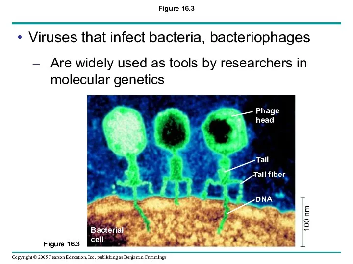
Figure 16.3
Viruses that infect bacteria, bacteriophages
Are widely used as tools by
Figure 16.3
Viruses that infect bacteria, bacteriophages
Are widely used as tools by
Alfred Hershey and Martha Chase
Performed experiments showing that DNA is the
Alfred Hershey and Martha Chase
Performed experiments showing that DNA is the
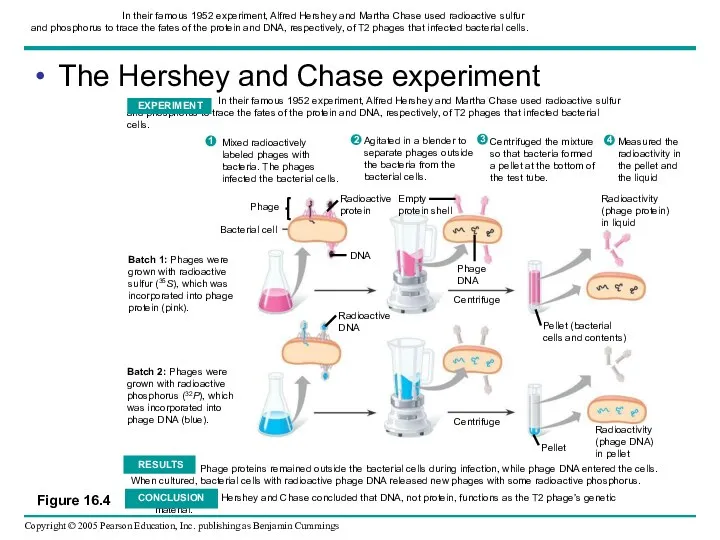
In their famous 1952 experiment, Alfred Hershey and Martha Chase
In their famous 1952 experiment, Alfred Hershey and Martha Chase
Additional Evidence That DNA Is the Genetic Materia
Prior to the 1950s,
Additional Evidence That DNA Is the Genetic Materia
Prior to the 1950s,
Erwin Chargaff analyzed the base composition of DNA
From a number of
Erwin Chargaff analyzed the base composition of DNA
From a number of
Building a Structural Model of DNA: Scientific Inquiry
Once most biologists were
Building a Structural Model of DNA: Scientific Inquiry
Once most biologists were
(a) Rosalind Franklin
Maurice Wilkins and Rosalind Franklin
Were using a technique called
(a) Rosalind Franklin
Maurice Wilkins and Rosalind Franklin
Were using a technique called
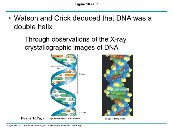
Figure 16.7a, c
Watson and Crick deduced that DNA was a double
Figure 16.7a, c
Watson and Crick deduced that DNA was a double
Franklin had concluded that DNA
Was composed of two antiparallel sugar-phosphate backbones,
Franklin had concluded that DNA
Was composed of two antiparallel sugar-phosphate backbones,
O
O
Watson and Crick reasoned that there must be additional specificity of
Watson and Crick reasoned that there must be additional specificity of
N
N
Concept 16.2: Many proteins work together in DNA replication and repair
The
Concept 16.2: Many proteins work together in DNA replication and repair
The
The Basic Principle: Base Pairing to a Template Strand
Since the two
The Basic Principle: Base Pairing to a Template Strand
Since the two
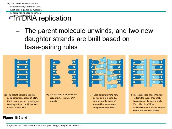
(a) The parent molecule has two
complementary strands of DNA.
Each
(a) The parent molecule has two complementary strands of DNA. Each
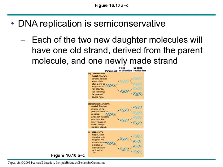
Figure 16.10 a–c
DNA replication is semiconservative
Each of the two new daughter
Figure 16.10 a–c
DNA replication is semiconservative
Each of the two new daughter
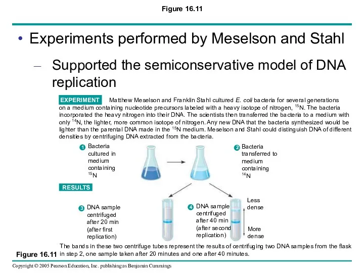
Figure 16.11
Experiments performed by Meselson and Stahl
Supported the semiconservative model of
Figure 16.11
Experiments performed by Meselson and Stahl
Supported the semiconservative model of
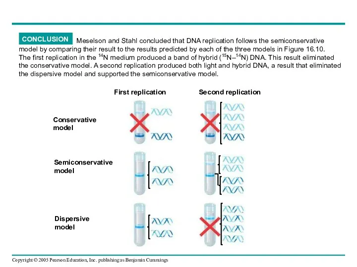
CONCLUSION
CONCLUSION
DNA Replication: A Closer Look
The copying of DNA
Is remarkable in its
DNA Replication: A Closer Look
The copying of DNA
Is remarkable in its
Getting Started: Origins of Replication
The replication of a DNA molecule
Begins at
Getting Started: Origins of Replication
The replication of a DNA molecule
Begins at
Replication begins at specific sites
where the two parental strands
separate and form
Replication begins at specific sites
where the two parental strands
separate and form
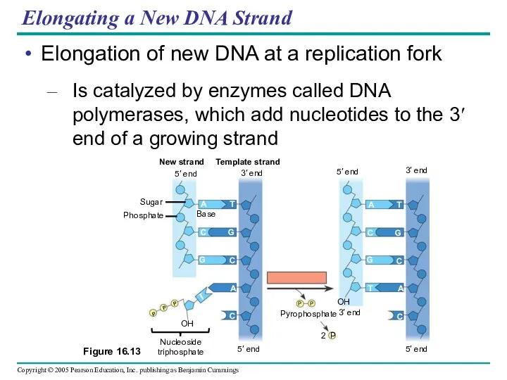
Elongating a New DNA Strand
Elongation of new DNA at a replication
Elongating a New DNA Strand
Elongation of new DNA at a replication
Antiparallel Elongation
How does the antiparallel structure of the double helix affect
Antiparallel Elongation
How does the antiparallel structure of the double helix affect
DNA polymerases add nucleotides
Only to the free 3′ end of a
DNA polymerases add nucleotides
Only to the free 3′ end of a
To elongate the other new strand of DNA, the lagging strand
DNA
To elongate the other new strand of DNA, the lagging strand
DNA
Synthesis of leading and lagging strands during DNA replication
Synthesis of leading and lagging strands during DNA replication

Priming DNA Synthesis
DNA polymerases cannot initiate the synthesis of a polynucleotide
They
Priming DNA Synthesis
DNA polymerases cannot initiate the synthesis of a polynucleotide
They
Only one primer is needed for synthesis of the leading strand
But
Only one primer is needed for synthesis of the leading strand
But
Overall direction of replication
Overall direction of replication
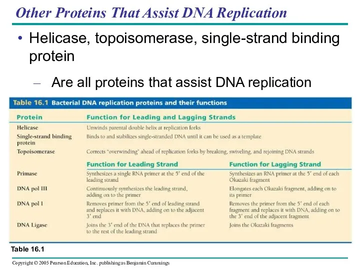
Other Proteins That Assist DNA Replication
Helicase, topoisomerase, single-strand binding protein
Are all
Other Proteins That Assist DNA Replication
Helicase, topoisomerase, single-strand binding protein
Are all
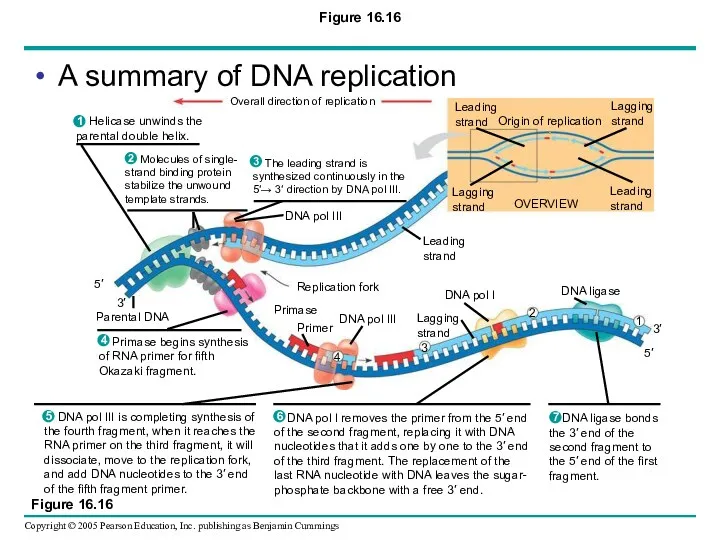
Figure 16.16
A summary of DNA replication
Figure 16.16
A summary of DNA replication
The DNA Replication Machine as a Stationary Complex
The various proteins that
The DNA Replication Machine as a Stationary Complex
The various proteins that
Proofreading and Repairing DNA
DNA polymerases proofread newly made DNA
Replacing any incorrect
Proofreading and Repairing DNA
DNA polymerases proofread newly made DNA
Replacing any incorrect
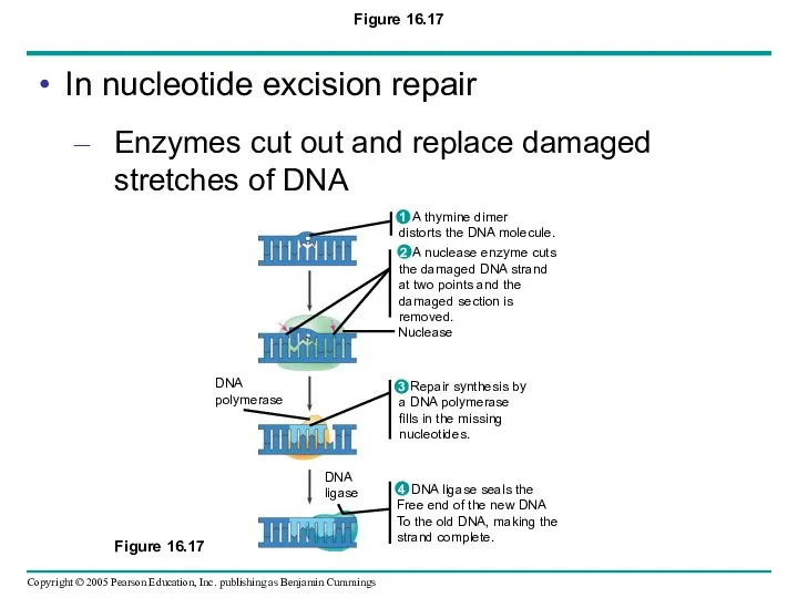
Figure 16.17
Nuclease
DNA
polymerase
DNA
ligase
A nuclease enzyme cuts
the damaged DNA strand
at two points
Figure 16.17
Nuclease
DNA
polymerase
DNA
ligase
A nuclease enzyme cuts
the damaged DNA strand
at two points
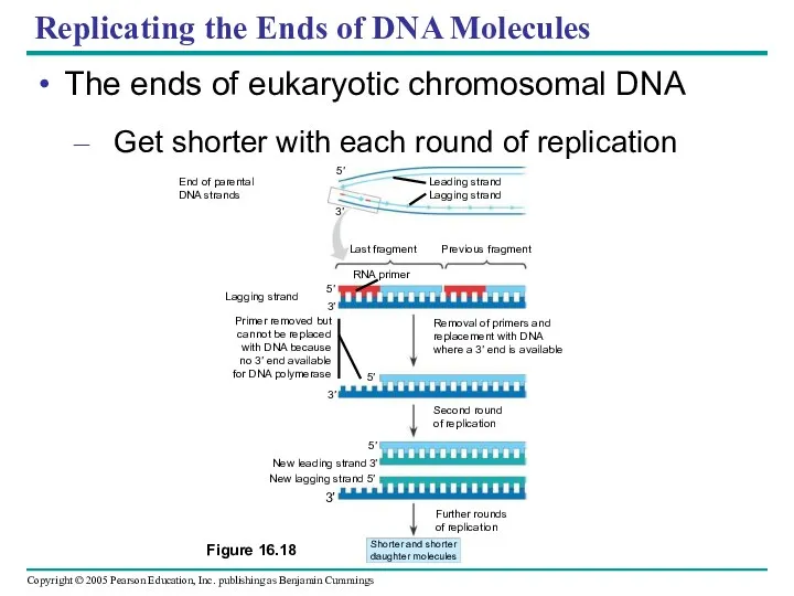
Replicating the Ends of DNA Molecules
The ends of eukaryotic chromosomal DNA
Get
Replicating the Ends of DNA Molecules
The ends of eukaryotic chromosomal DNA
Get
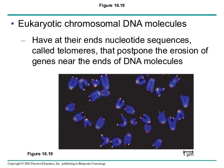
Figure 16.19
Eukaryotic chromosomal DNA molecules
Have at their ends nucleotide sequences, called
Figure 16.19
Eukaryotic chromosomal DNA molecules
Have at their ends nucleotide sequences, called
 Биотехнологическое производство сыра
 Историческое и индивидуальное в развитии
 Черенкование комнатных растений
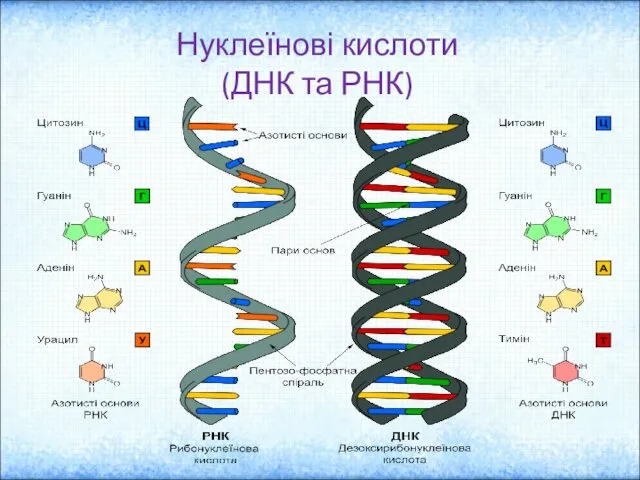 Нуклеїнові кислоти (ДНК та РНК)
 Микроэкология тела человека. Дисбактериоз и методы исследования
 Возникновение многоклеточности. Признаки многоклеточного организма Теории происхождения многоклеточных животных
 11 Интересных фактов о птицах
 Неживая природа осенью (2 класс)
 Структура бактериальной клетки
 Адаптація людини до навколишнього середовища . Фізіологічні основи загартування
 Гіпотези виникнення життя на Землі
 презентация по биологии Наследственные болезни
 Нервная система. Спинной мозг. Рефлектроная дуга
 Неживая и живая природа. (2 класс)
 Кровообращение как функциональная система. Анатомия сердца и сосудов. Характерные черты строения и функционирования сердечной мышцы
 Зоология позвоночных. Надкласс четвероногие. (Лекция 7)
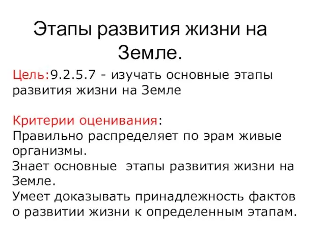 Этапы развития жизни на Земли
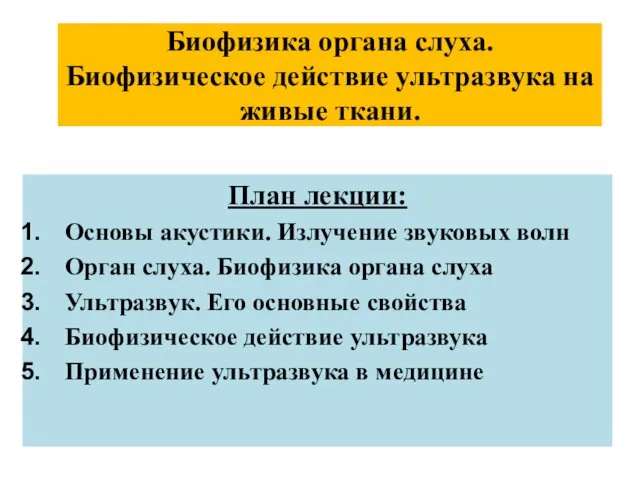 Биофизика органа слуха. Биофизическое действие ультразвука на живые ткани
 Денатурация белков
 Прокариотты және эукариотты өсімдіктер клеткасы
 СРС на тему: “Везикулярный транспорт”
 Рыбы. Систематика рыб
 Физиология микроорганизмов
 Паразитические грибы и бактерии
 Флуоресцентная микроскопия
 Клетки растений
 Ядовитые растения Самарской области
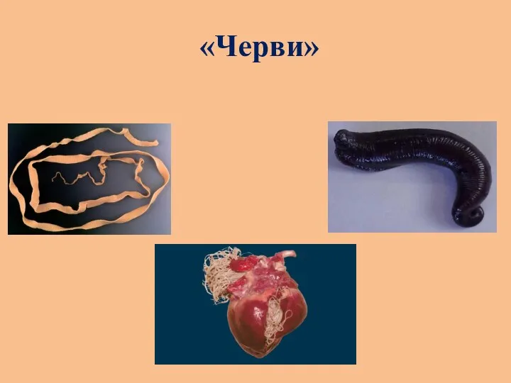 Плоские черви