- Mechanical oscillations and waves. Bioacoustics. Ultrasound

Содержание
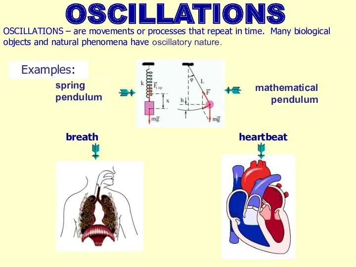
- 2. OSCILLATIONS OSCILLATIONS – are movements or processes that repeat in time. Many biological objects and natural
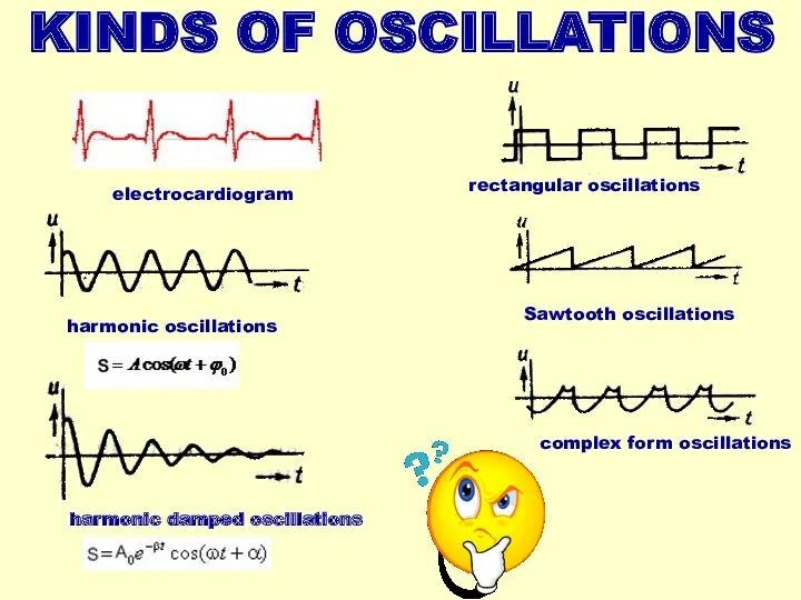
- 3. KINDS OF OSCILLATIONS complex form oscillations rectangular oscillations Sawtooth oscillations harmonic oscillations harmonic damped oscillations electrocardiogram
- 4. PARAMETERS OF OSCILLATIONS AMPLITUDE- maximal displacement from equilibrium PERIOD- time of one full oscillation cycle FREQUENCY
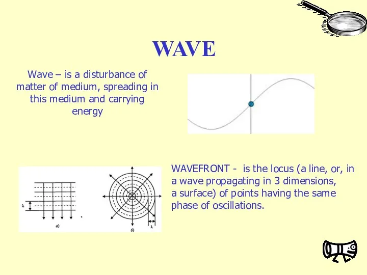
- 5. WAVE WAVEFRONT - is the locus (a line, or, in a wave propagating in 3 dimensions,
- 6. According the wavefront waves can be: spherical plane
- 7. transversal – particles of medium oscillate perpendiculary to the direction of wave spreading (electromagnetic waves, waves
- 8. RESONANCE Resonance (fr. resonance, from lat. resono - respond) - phenomenon of a sharp increase of
- 9. SOUND WAVES SOUND WAVES – are special case of elastic waves, which spread only in elastic
- 10. ν І NOISE SPECTRUM ν COMPLEX TONE SPECTRUM І SOUND CHARACTERISTICS OBJECTIVE AND SUBJECTIVE SOUND CHARACTERICTICS
- 11. Volume scale Weber–Fechner law Where L – volume; I0 – іntensity on threshold of hearing; I
- 12. Doppler effect for sound waves u – velocity of receiver relative to the medium (positive, if
- 13. Doppler effect for sound waves An animation illustrating how the Doppler effect causes a car engine
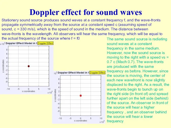
- 14. Doppler effect for sound waves Stationary sound source produces sound waves at a constant frequency f,
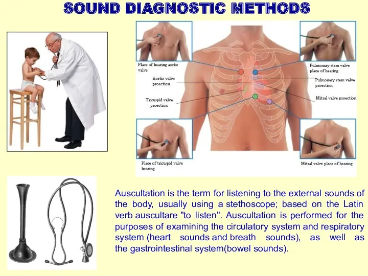
- 15. SOUND DIAGNOSTIC METHODS Auscultation is the term for listening to the external sounds of the body,
- 16. ULTRASOUND IN MEDICINE IN DIAGNOSTICS IN CURING Ultrasound physiotherapy Ultrasound surgery Mechanical impact (cavitation) Thermal impact
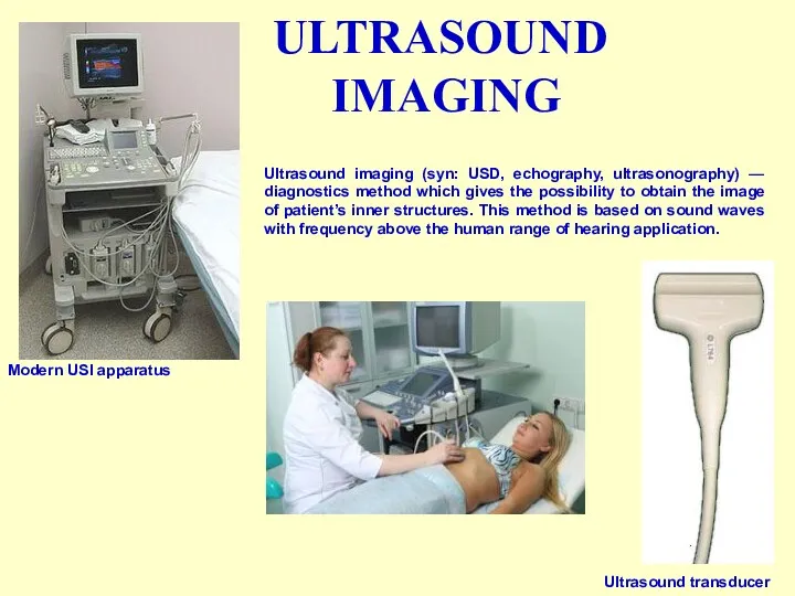
- 17. ULTRASOUND IMAGING Modern USI apparatus Ultrasound transducer Ultrasound imaging (syn: USD, echography, ultrasonography) — diagnostics method
- 18. US scanning modes A-mode or “Amplitude mode” - A single transducer scans a line through the
- 19. 3D USI 3D USI gives a three dimensional image. USI transducer gets a number of images
- 20. Dopplerography Spectral doppler common carotid artery Transcranial doppler. Color doppler mapping The method is based on
- 22. Скачать презентацию
OSCILLATIONS
OSCILLATIONS – are movements or processes that repeat in time. Many
OSCILLATIONS
OSCILLATIONS – are movements or processes that repeat in time. Many
KINDS OF OSCILLATIONS
complex form oscillations
rectangular oscillations
Sawtooth oscillations
harmonic oscillations
harmonic damped oscillations
electrocardiogram
KINDS OF OSCILLATIONS
complex form oscillations
rectangular oscillations
Sawtooth oscillations
harmonic oscillations
harmonic damped oscillations
electrocardiogram
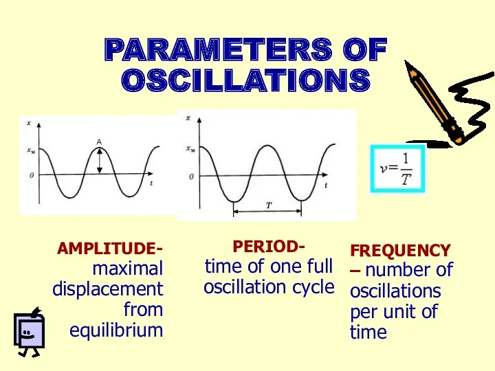
PARAMETERS OF OSCILLATIONS
AMPLITUDE-
maximal displacement from equilibrium
PERIOD-
time of one full
PARAMETERS OF OSCILLATIONS
AMPLITUDE-
maximal displacement from equilibrium
PERIOD-
time of one full
WAVE
WAVEFRONT - is the locus (a line, or, in a wave propagating in 3 dimensions, a surface)
WAVE
WAVEFRONT - is the locus (a line, or, in a wave propagating in 3 dimensions, a surface)
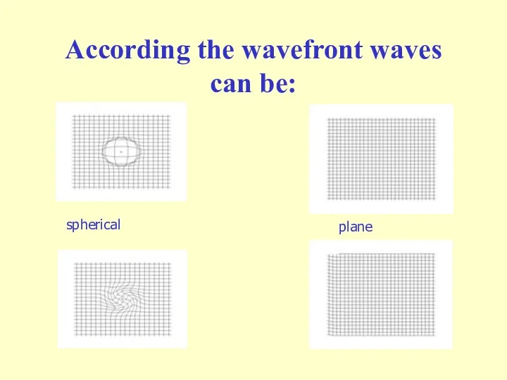
According the wavefront waves can be:
spherical
plane
According the wavefront waves can be:
spherical
plane
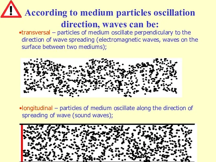
transversal – particles of medium oscillate perpendiculary to the direction of
RESONANCE
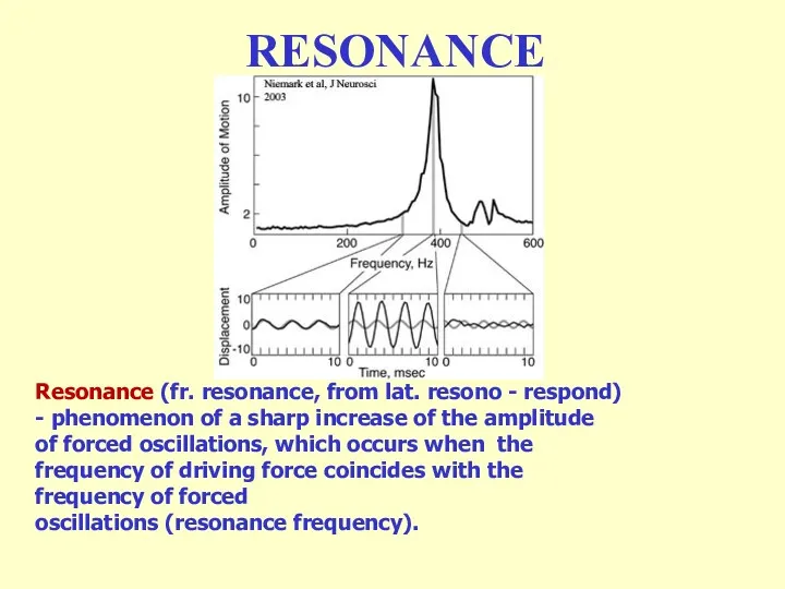
Resonance (fr. resonance, from lat. resono - respond) - phenomenon of a
RESONANCE
Resonance (fr. resonance, from lat. resono - respond) - phenomenon of a
SOUND WAVES
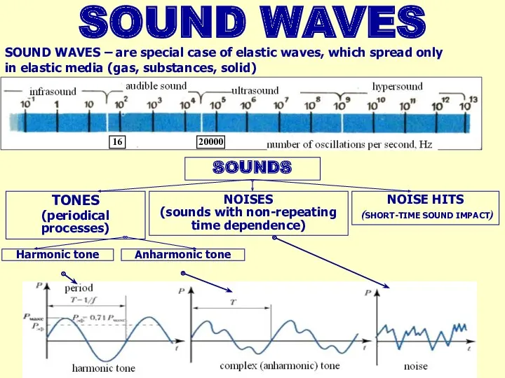
SOUND WAVES – are special case of elastic waves, which spread only
SOUND WAVES
SOUND WAVES – are special case of elastic waves, which spread only
ν
І
NOISE SPECTRUM
ν
COMPLEX TONE SPECTRUM
І
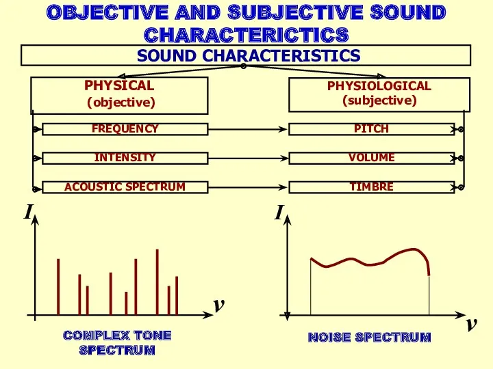
SOUND CHARACTERISTICS
OBJECTIVE AND SUBJECTIVE SOUND CHARACTERICTICS
ν
І
NOISE SPECTRUM
ν
COMPLEX TONE SPECTRUM
І
SOUND CHARACTERISTICS
OBJECTIVE AND SUBJECTIVE SOUND CHARACTERICTICS
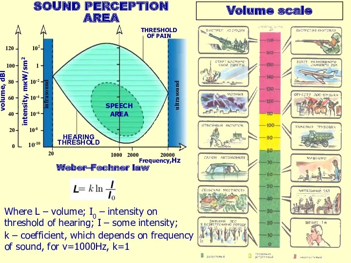
Volume scale
Weber–Fechner law
Where L – volume; I0 – іntensity on threshold
Volume scale
Weber–Fechner law
Where L – volume; I0 – іntensity on threshold
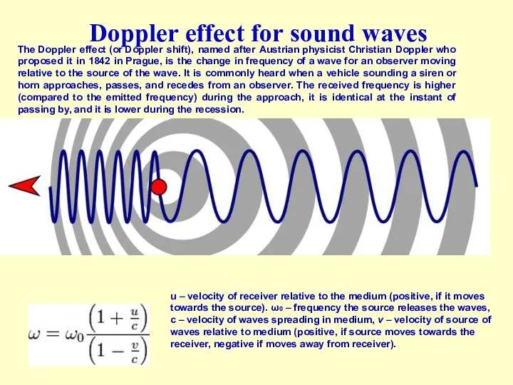
Doppler effect for sound waves
u – velocity of receiver relative to
Doppler effect for sound waves
u – velocity of receiver relative to
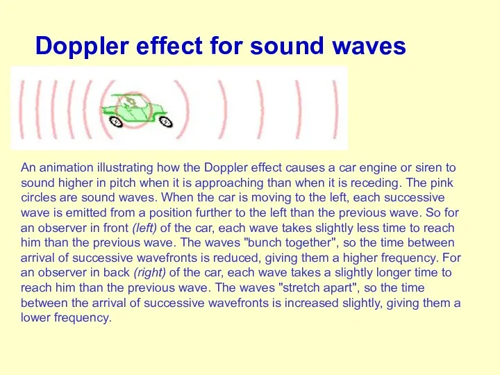
Doppler effect for sound waves
An animation illustrating how the Doppler effect
Doppler effect for sound waves
An animation illustrating how the Doppler effect
Doppler effect for sound waves
Stationary sound source produces sound waves at
Doppler effect for sound waves
Stationary sound source produces sound waves at
SOUND DIAGNOSTIC METHODS
Auscultation is the term for listening to the external sounds
SOUND DIAGNOSTIC METHODS
Auscultation is the term for listening to the external sounds
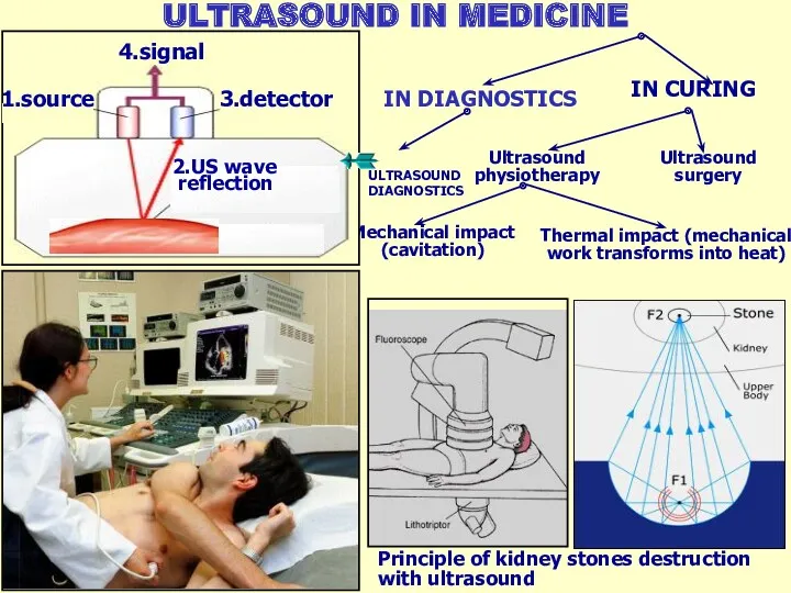
ULTRASOUND IN MEDICINE
IN DIAGNOSTICS
IN CURING
Ultrasound physiotherapy
Ultrasound surgery
Mechanical impact (cavitation)
Thermal impact (mechanical
ULTRASOUND IN MEDICINE
IN DIAGNOSTICS
IN CURING
Ultrasound physiotherapy
Ultrasound surgery
Mechanical impact (cavitation)
Thermal impact (mechanical
ULTRASOUND
IMAGING
Modern USI apparatus
Ultrasound transducer
Ultrasound imaging (syn: USD, echography, ultrasonography) —
ULTRASOUND
IMAGING
Modern USI apparatus
Ultrasound transducer
Ultrasound imaging (syn: USD, echography, ultrasonography) —
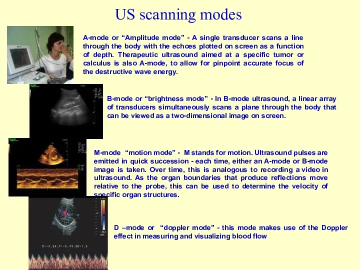
US scanning modes
A-mode or “Amplitude mode” - A single transducer scans
US scanning modes
A-mode or “Amplitude mode” - A single transducer scans
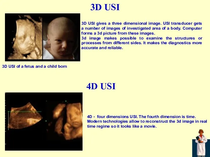
3D USI
3D USI gives a three dimensional image. USI transducer gets
3D USI
3D USI gives a three dimensional image. USI transducer gets
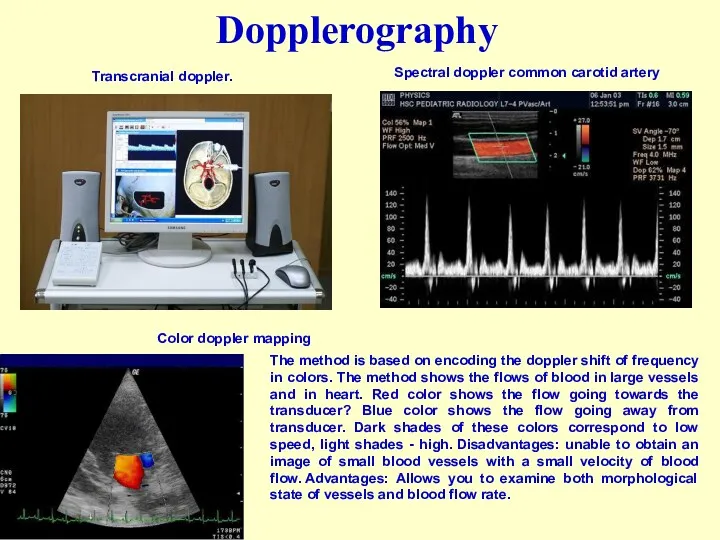
Dopplerography
Spectral doppler common carotid artery
Transcranial doppler.
Color doppler mapping
The method is based on encoding
Dopplerography
Spectral doppler common carotid artery
Transcranial doppler.
Color doppler mapping
The method is based on encoding
 Microelectronic front-end of receivers for wireless systems
 Балка на упругом основании
 Физика и музыка
 Глава 5. Пьезоэлектрический эффект и электрострикция
 Женские тропинки в космосе
 Механические свойства твердых тел
 Визуальная, квантовая физика
 Рулевое управление
 Көміртекті нанотүтікше
 Презентация к открытому уроку по физике Количество теплоты. Удельная теплоёмкость.
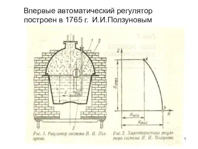 Автоматический регулятор
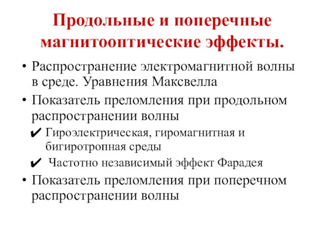 Продольные и поперечные магнитооптические эффекты
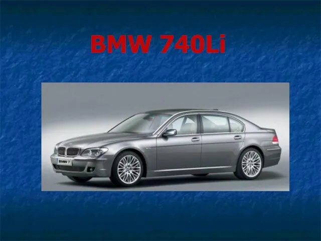 BMW 740Li
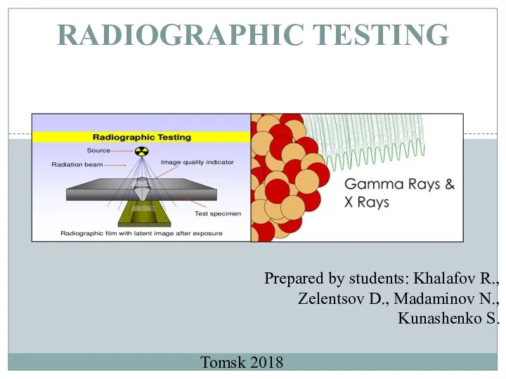 Radiographic testing
 Оптические свойства коллоидных квантовых точек
 Ядерный реактор. Преобразование внутренней энергии атомных ядер в электрическую энергиию
 Домашнее задание по курсу Детали машин №4: Проверочный расчет тихоходного вала редуктора
 Электрический ток. (Лекция 1)
 Плоские электромагнитные волны. Особенности и параметры направляемых волн
 Статичне електричне поле. (Лекція 11)
 Презентация по теме Спектры
 Условные расчеты на прочность
 Лазеры. Спонтанное и вынужденное излучение
 Презентация к уроку физики в 8 классе на тему Звуковые волны в различных средах
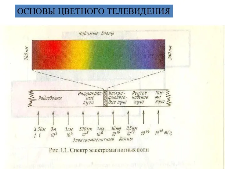 Основы цветного телевидения
 Моя будущая профессия - Автомеханик
 Жартылай өткізгіштер
 Кинематика, обобщение темы