- Microscope Measurement

Содержание
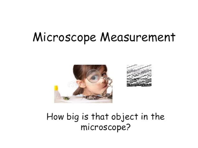
- 2. Microscope Measurement How big is that object in the microscope?
- 3. Lesson Objectives Calculate the magnification using different objective lens. Differentiate between eyepiece graticule and the stage
- 6. Light Microscope A light microscope (also, optical microscope) is an optical instrument used to make objects
- 7. Electron Microscope An electron microscope is an optical instrument that uses a beam of electrons to
- 9. Light microscope vs Electron microscope What is the difference between a light microscope and an electron
- 10. What is happening to the image as you increase the power of the objective lens?
- 11. Calculating total magnification If two lenses are always magnifying the specimen, how do you figure out
- 12. How do we find the overall magnification of a light microscope? Eyepiece Objective lens 40 100
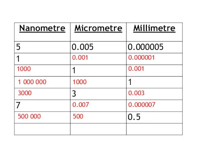
- 13. 1000 1000 1000 1000 mm Micrometre nm
- 14. 0.001 0.000001 1000 0.001 1 000 000 1000 3000 0.003 0.007 0.000007 500 000 500
- 16. The diagram below is a drawing of an organelle from a ciliated cell as seen with
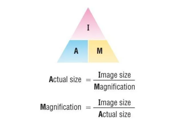
- 17. Calculating actual size: Size of the magnified image > actual size
- 18. To accurately measure the size of cellular structures we need a suitable scale:
- 19. Field of View When you look into a microscope, the “field of view” is the visible
- 20. Estimating Specimen Size The area of the slide that you see when you look through a
- 21. Ideally, we need a scale we can see directly alongside the cells we are observing:
- 22. Eye piece graticule or reticule It is a glass or plactic disc with 8 divisions etched
- 23. Stage Micrometer simply a microscope slide with a finely divided scale marked on the surface. 1
- 24. Instructions Take sample of onion cell (peel of the onion) Add a drop of water Cover
- 26. Скачать презентацию
Microscope Measurement
How big is that object in the microscope?
Microscope Measurement
How big is that object in the microscope?
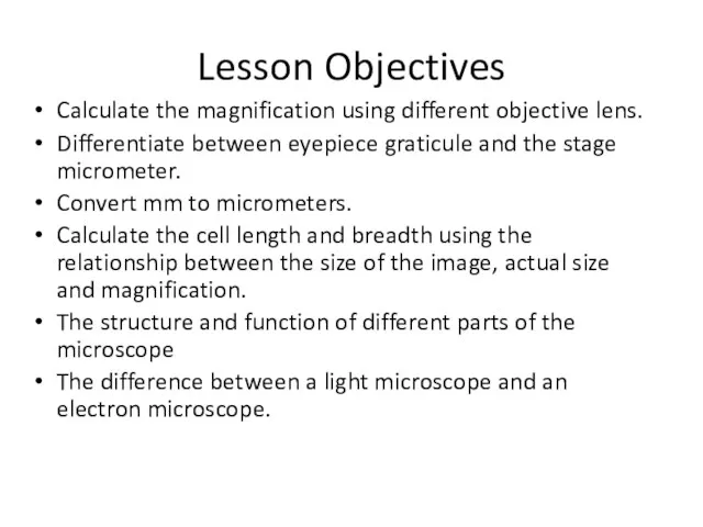
Lesson Objectives
Calculate the magnification using different objective lens.
Differentiate between eyepiece graticule
Lesson Objectives
Calculate the magnification using different objective lens.
Differentiate between eyepiece graticule
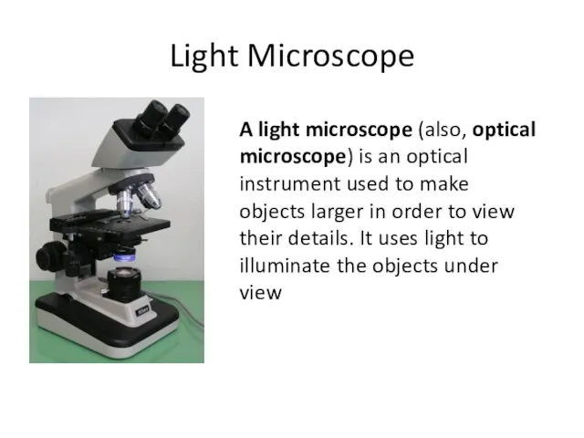
Light Microscope
A light microscope (also, optical microscope) is an optical instrument used to
Light Microscope
A light microscope (also, optical microscope) is an optical instrument used to
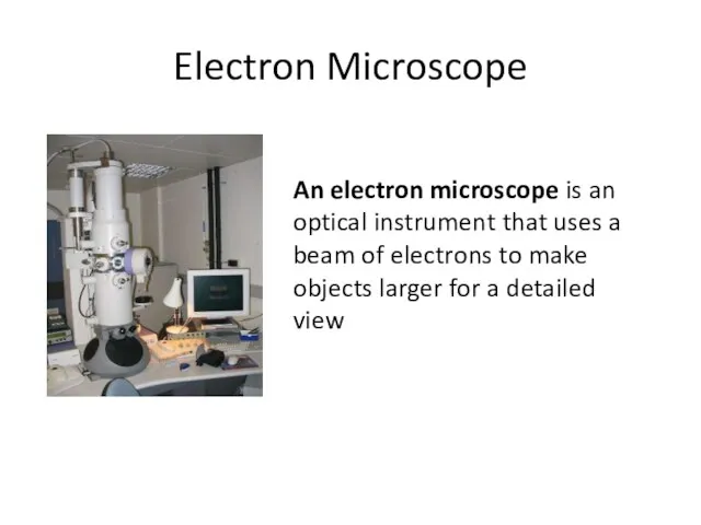
Electron Microscope
An electron microscope is an optical instrument that uses a beam
Electron Microscope
An electron microscope is an optical instrument that uses a beam
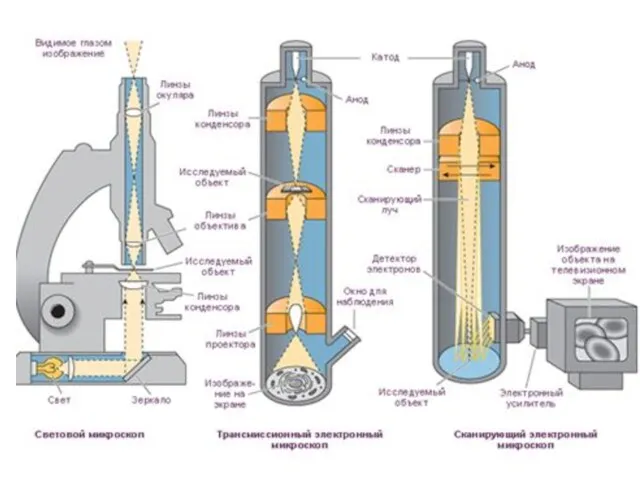
Light microscope vs Electron microscope
What is the difference between a light
Light microscope vs Electron microscope
What is the difference between a light
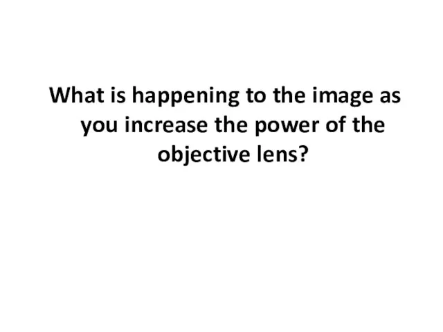
What is happening to the image as you increase the power
What is happening to the image as you increase the power
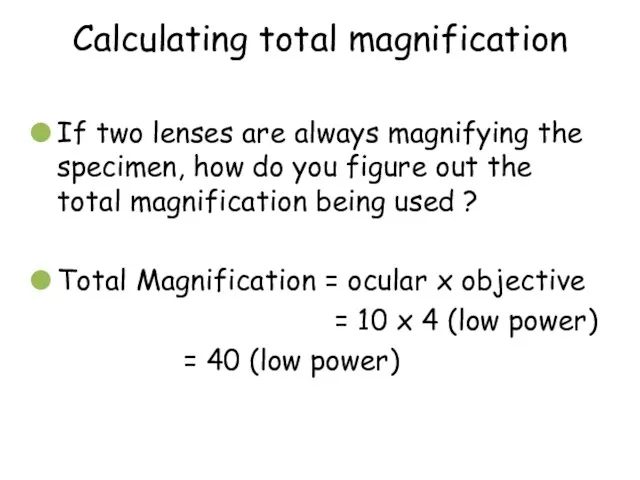
Calculating total magnification
If two lenses are always magnifying the specimen, how
Calculating total magnification
If two lenses are always magnifying the specimen, how
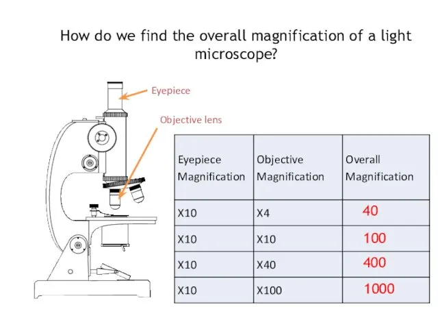
How do we find the overall magnification of a light microscope?
Eyepiece
Objective
How do we find the overall magnification of a light microscope?
Eyepiece
Objective
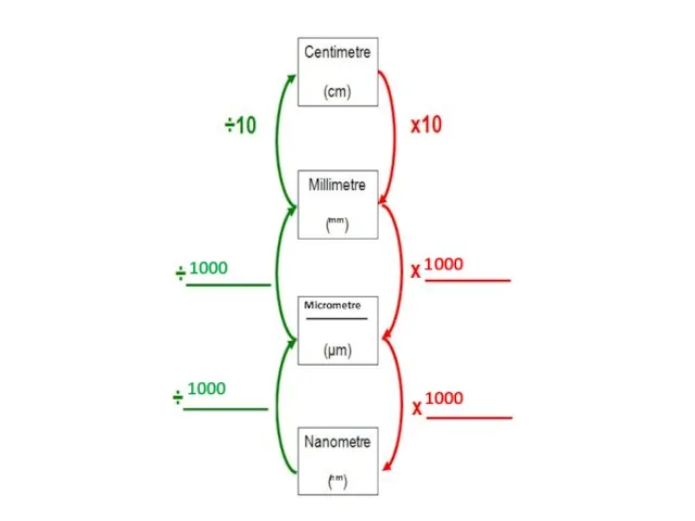
1000
1000
1000
1000
mm
Micrometre
nm
1000
1000
1000
1000
mm
Micrometre
nm
0.001
0.000001
1000
0.001
1 000 000
1000
3000
0.003
0.007
0.000007
500 000
500
0.001
0.000001
1000
0.001
1 000 000
1000
3000
0.003
0.007
0.000007
500 000
500
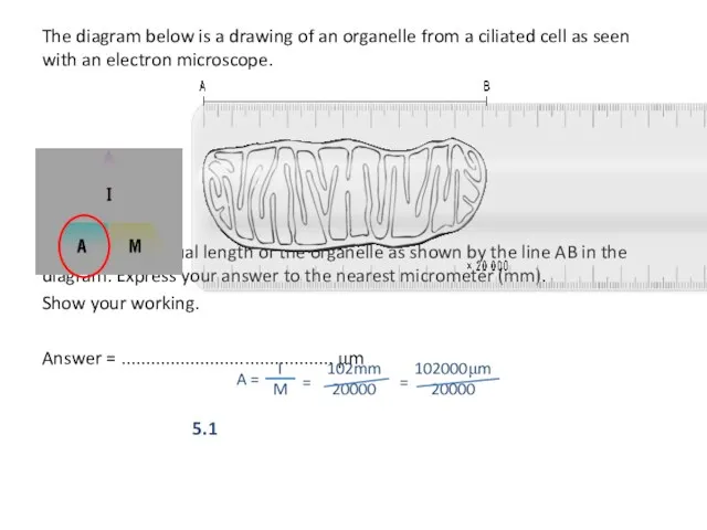
The diagram below is a drawing of an organelle from a
The diagram below is a drawing of an organelle from a
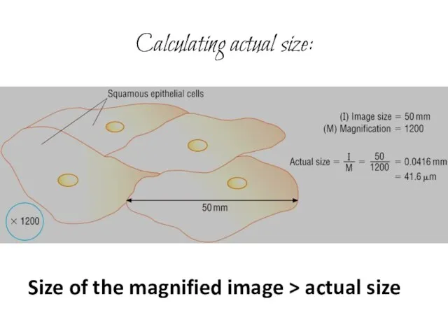
Calculating actual size:
Size of the magnified image > actual size
Calculating actual size:
Size of the magnified image > actual size
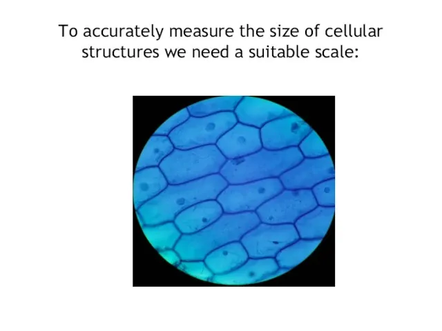
To accurately measure the size of cellular structures we need a
To accurately measure the size of cellular structures we need a
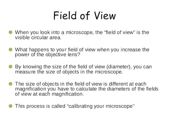
Field of View
When you look into a microscope, the “field of
Field of View
When you look into a microscope, the “field of
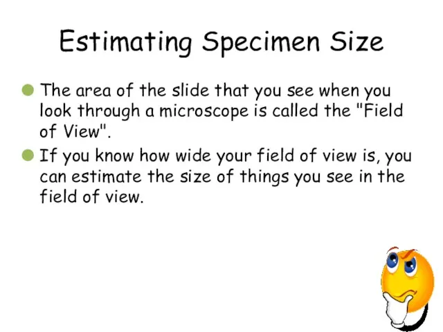
Estimating Specimen Size
The area of the slide that you see when
Estimating Specimen Size
The area of the slide that you see when
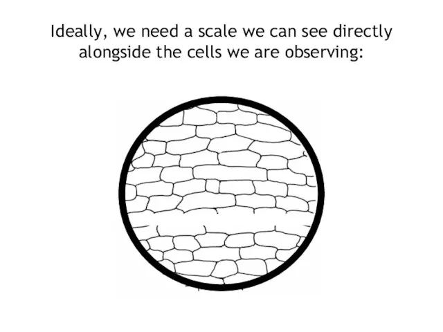
Ideally, we need a scale we can see directly alongside the
Ideally, we need a scale we can see directly alongside the
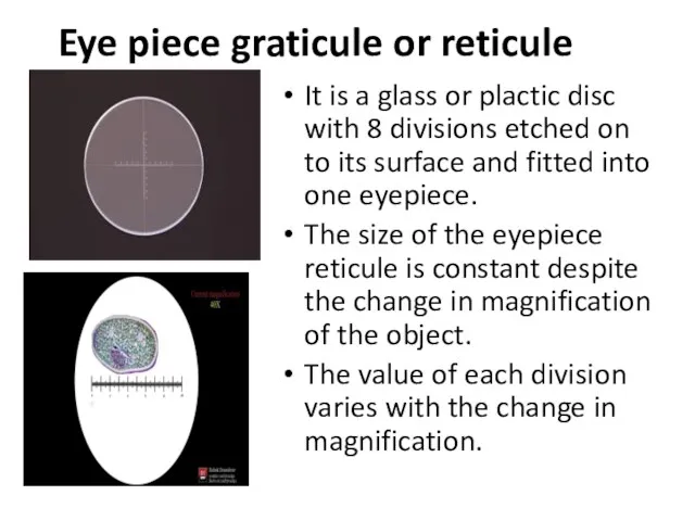
Eye piece graticule or reticule
It is a glass or plactic disc
Eye piece graticule or reticule
It is a glass or plactic disc
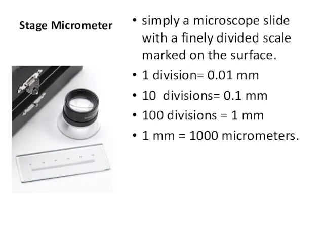
Stage Micrometer
simply a microscope slide with a finely divided scale marked
Stage Micrometer
simply a microscope slide with a finely divided scale marked
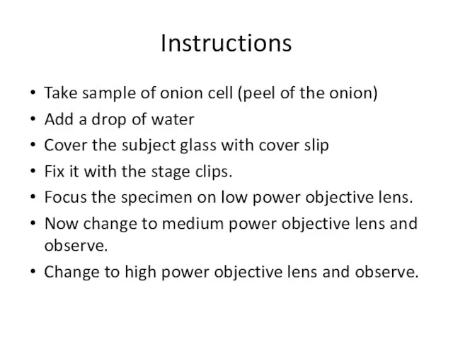
Instructions
Take sample of onion cell (peel of the onion)
Add a drop
Instructions
Take sample of onion cell (peel of the onion)
Add a drop
 Деление ядер урана. Цепные ядерные реакции
 Электростатика. Постоянный ток. (Лекция 4)
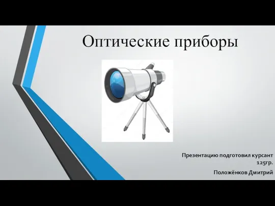 Оптические приборы
 Необычные средства связи. Викторина
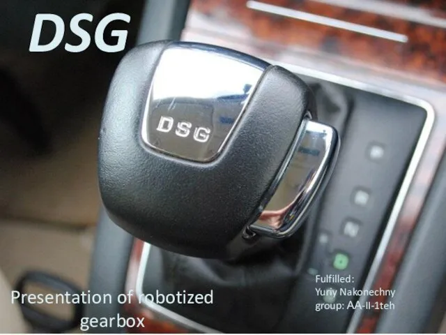 DSG. Automatic and manual modes
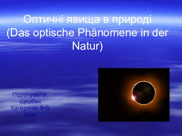 Оптичні явища у природі
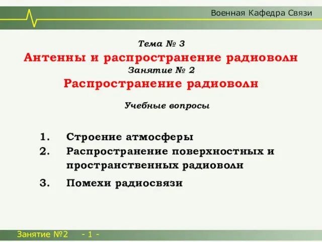 Антенны и распространение радиоволн
 Принцип дії теплових двигунів
 Презентация Тест 11 класс. Итог
 Деление атомных ядер. Цепная реакция. Термоядерный синтез
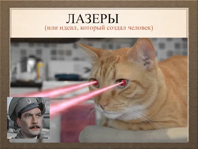 Лазеры (или идеал, который создал человек)
 Катушки со сталью в цепи синусоидального тока
 Презентация по физике на тему _Шкала электромагнитных волн_
 Турбонаддув. Турбированный бензиновый двигатель
 Магнитные преобразователи
 Механическая работа и мощность
 Ток в электролитах
 Организация работ по диагностированию, техническому обслуживанию и ремонту ЗИЛ-5301. Процесс ремонта переднего моста
 Основы теории подобия
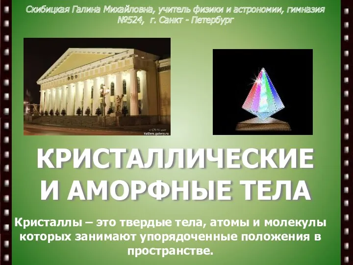 Кристаллические и аморфные тела
 Закон Кулона
 Плавление и отвердевание
 Основные положения МКТ. Физика. 10 класс
 Движение в неинерциальной системе отсчета
 Магнитное поле катушки с током. Электромагниты и их применение. 8 класс
 Акустика. Затухание волн
 Компетентностный подход в образовании.
 Передача электромагнитной энергии. Волноводы