- Cell Communication

Содержание
- 2. Overview: The Cellular Internet Cell-to-cell communication is essential for multicellular organisms Biologists have discovered some universal
- 3. Fig. 11-1
- 4. Concept 11.1: External signals are converted to responses within the cell Microbes are a window on
- 5. Evolution of Cell Signaling A signal transduction pathway is a series of steps by which a
- 6. Fig. 11-2 Receptor α factor a factor a α α a Exchange of mating factors Yeast
- 7. Pathway similarities suggest that ancestral signaling molecules evolved in prokaryotes and were modified later in eukaryotes
- 8. Fig. 11-3 Individual rod- shaped cells Spore-forming structure (fruiting body) Aggregation in process Fruiting bodies 0.5
- 9. Local and Long-Distance Signaling Cells in a multicellular organism communicate by chemical messengers Animal and plant
- 10. Fig. 11-4 Plasma membranes Gap junctions between animal cells (a) Cell junctions Plasmodesmata between plant cells
- 11. In many other cases, animal cells communicate using local regulators, messenger molecules that travel only short
- 12. Fig. 11-5 Local signaling Target cell Secreting cell Secretory vesicle Local regulator diffuses through extracellular fluid
- 13. Fig. 11-5ab Local signaling Target cell Secretory vesicle Secreting cell Local regulator diffuses through extracellular fluid
- 14. Fig. 11-5c Long-distance signaling Endocrine cell Blood vessel Hormone travels in bloodstream to target cells Target
- 15. The Three Stages of Cell Signaling: A Preview Earl W. Sutherland discovered how the hormone epinephrine
- 16. Fig. 11-6-1 Reception 1 EXTRACELLULAR FLUID Signaling molecule Plasma membrane CYTOPLASM 1 Receptor
- 17. Fig. 11-6-2 1 EXTRACELLULAR FLUID Signaling molecule Plasma membrane CYTOPLASM Transduction 2 Relay molecules in a
- 18. Fig. 11-6-3 EXTRACELLULAR FLUID Plasma membrane CYTOPLASM Receptor Signaling molecule Relay molecules in a signal transduction
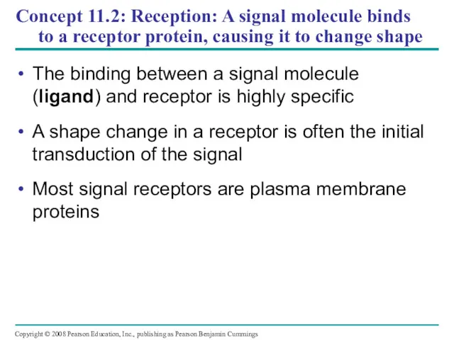
- 19. Concept 11.2: Reception: A signal molecule binds to a receptor protein, causing it to change shape
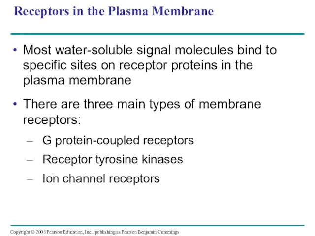
- 20. Receptors in the Plasma Membrane Most water-soluble signal molecules bind to specific sites on receptor proteins
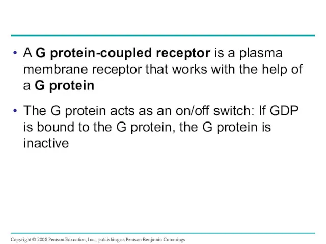
- 21. A G protein-coupled receptor is a plasma membrane receptor that works with the help of a
- 22. Fig. 11-7a Signaling-molecule binding site Segment that interacts with G proteins G protein-coupled receptor
- 23. Fig. 11-7b G protein-coupled receptor Plasma membrane Enzyme G protein (inactive) GDP CYTOPLASM Activated enzyme GTP
- 24. Receptor tyrosine kinases are membrane receptors that attach phosphates to tyrosines A receptor tyrosine kinase can
- 25. Fig. 11-7c Signaling molecule (ligand) Ligand-binding site α Helix Tyrosines Tyr Tyr Tyr Tyr Tyr Tyr
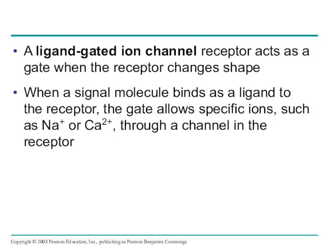
- 26. A ligand-gated ion channel receptor acts as a gate when the receptor changes shape When a
- 27. Fig. 11-7d Signaling molecule (ligand) Gate closed Ions Ligand-gated ion channel receptor Plasma membrane Gate open
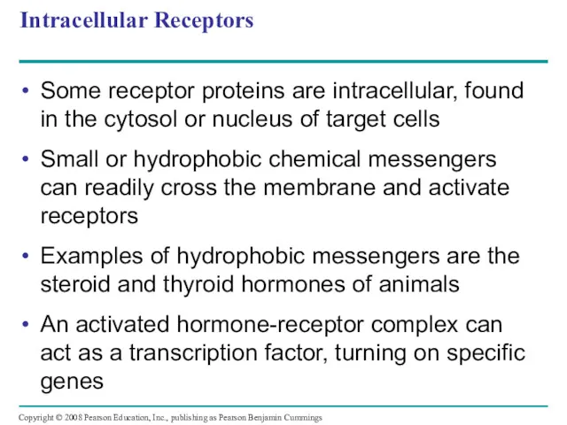
- 28. Intracellular Receptors Some receptor proteins are intracellular, found in the cytosol or nucleus of target cells
- 29. Fig. 11-8-1 Hormone (testosterone) Receptor protein Plasma membrane EXTRACELLULAR FLUID DNA NUCLEUS CYTOPLASM
- 30. Fig. 11-8-2 Receptor protein Hormone (testosterone) EXTRACELLULAR FLUID Plasma membrane Hormone- receptor complex DNA NUCLEUS CYTOPLASM
- 31. Fig. 11-8-3 Hormone (testosterone) EXTRACELLULAR FLUID Receptor protein Plasma membrane Hormone- receptor complex DNA NUCLEUS CYTOPLASM
- 32. Fig. 11-8-4 Hormone (testosterone) EXTRACELLULAR FLUID Plasma membrane Receptor protein Hormone- receptor complex DNA mRNA NUCLEUS
- 33. Fig. 11-8-5 Hormone (testosterone) EXTRACELLULAR FLUID Receptor protein Plasma membrane Hormone- receptor complex DNA mRNA NUCLEUS
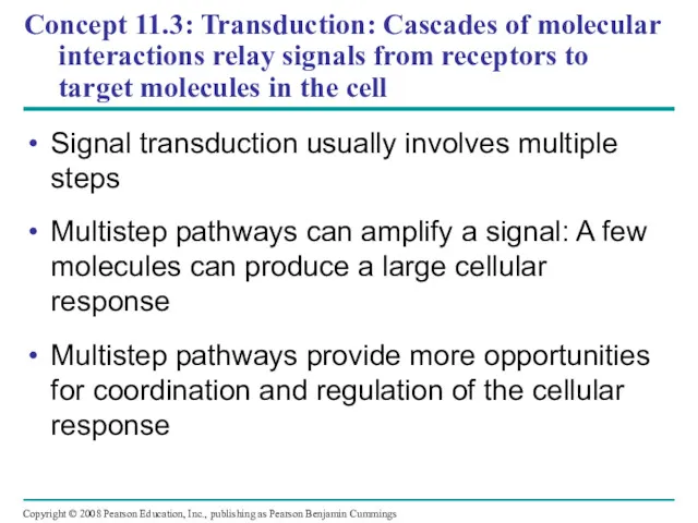
- 34. Concept 11.3: Transduction: Cascades of molecular interactions relay signals from receptors to target molecules in the
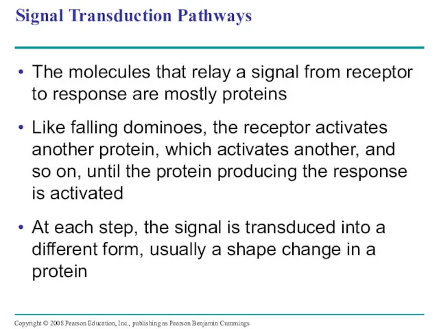
- 35. Signal Transduction Pathways The molecules that relay a signal from receptor to response are mostly proteins
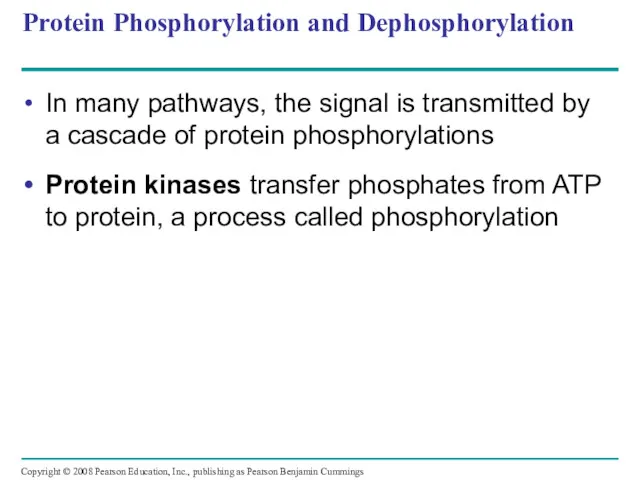
- 36. Protein Phosphorylation and Dephosphorylation In many pathways, the signal is transmitted by a cascade of protein
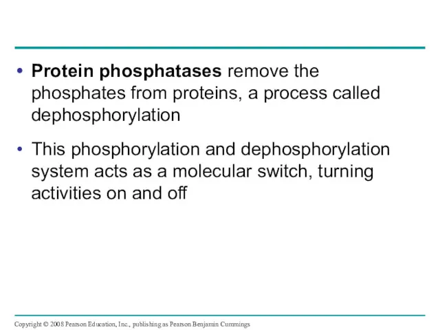
- 37. Protein phosphatases remove the phosphates from proteins, a process called dephosphorylation This phosphorylation and dephosphorylation system
- 38. Fig. 11-9 Signaling molecule Receptor Activated relay molecule Inactive protein kinase 1 Active protein kinase 1
- 39. Small Molecules and Ions as Second Messengers The extracellular signal molecule that binds to the receptor
- 40. Cyclic AMP Cyclic AMP (cAMP) is one of the most widely used second messengers Adenylyl cyclase,
- 41. Adenylyl cyclase Fig. 11-10 Pyrophosphate P P i ATP cAMP Phosphodiesterase AMP
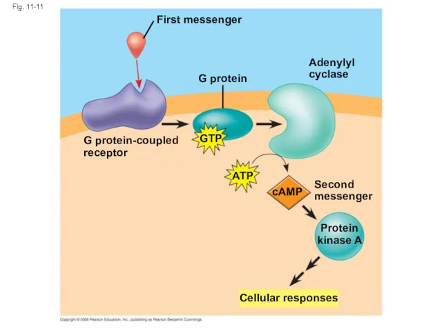
- 42. Many signal molecules trigger formation of cAMP Other components of cAMP pathways are G proteins, G
- 43. First messenger Fig. 11-11 G protein Adenylyl cyclase GTP ATP cAMP Second messenger Protein kinase A
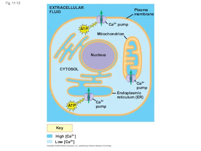
- 44. Calcium Ions and Inositol Triphosphate (IP3) Calcium ions (Ca2+) act as a second messenger in many
- 45. EXTRACELLULAR FLUID Fig. 11-12 ATP Nucleus Mitochondrion Ca2+ pump Plasma membrane CYTOSOL Ca2+ pump Endoplasmic reticulum
- 46. A signal relayed by a signal transduction pathway may trigger an increase in calcium in the
- 47. Fig. 11-13-1 EXTRA- CELLULAR FLUID Signaling molecule (first messenger) G protein GTP G protein-coupled receptor Phospholipase
- 48. Fig. 11-13-2 G protein EXTRA- CELLULAR FLUID Signaling molecule (first messenger) G protein-coupled receptor Phospholipase C
- 49. Fig. 11-13-3 G protein EXTRA- CELLULAR FLUID Signaling molecule (first messenger) G protein-coupled receptor Phospholipase C
- 50. Concept 11.4: Response: Cell signaling leads to regulation of transcription or cytoplasmic activities The cell’s response
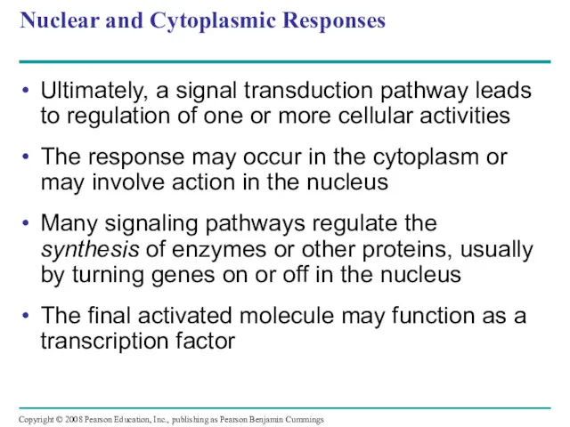
- 51. Nuclear and Cytoplasmic Responses Ultimately, a signal transduction pathway leads to regulation of one or more
- 52. Fig. 11-14 Growth factor Receptor Phosphorylation cascade Reception Transduction Active transcription factor Response P Inactive transcription
- 53. Other pathways regulate the activity of enzymes Copyright © 2008 Pearson Education, Inc., publishing as Pearson
- 54. Fig. 11-15 Reception Transduction Response Binding of epinephrine to G protein-coupled receptor (1 molecule) Inactive G
- 55. Signaling pathways can also affect the physical characteristics of a cell, for example, cell shape Copyright
- 56. Fig. 11-16 RESULTS CONCLUSION Wild-type (shmoos) ∆Fus3 ∆formin Shmoo projection forming Formin P Actin subunit P
- 57. Fig. 11-16a RESULTS Wild-type (shmoos) ∆Fus3 ∆formin
- 58. Fig. 11-16b CONCLUSION Mating factor G protein-coupled receptor GDP GTP Phosphory- lation cascade Shmoo projection forming
- 59. Fine-Tuning of the Response Multistep pathways have two important benefits: Amplifying the signal (and thus the
- 60. Signal Amplification Enzyme cascades amplify the cell’s response At each step, the number of activated products
- 61. The Specificity of Cell Signaling and Coordination of the Response Different kinds of cells have different
- 62. Fig. 11-17 Signaling molecule Receptor Relay molecules Response 1 Cell A. Pathway leads to a single
- 63. Fig. 11-17a Signaling molecule Receptor Relay molecules Response 1 Cell A. Pathway leads to a single
- 64. Fig. 11-17b Response 4 Response 5 Activation or inhibition Cell C. Cross-talk occurs between two pathways.
- 65. Signaling Efficiency: Scaffolding Proteins and Signaling Complexes Scaffolding proteins are large relay proteins to which other
- 66. Fig. 11-18 Signaling molecule Receptor Scaffolding protein Plasma membrane Three different protein kinases
- 67. Termination of the Signal Inactivation mechanisms are an essential aspect of cell signaling When signal molecules
- 68. Concept 11.5: Apoptosis (programmed cell death) integrates multiple cell-signaling pathways Apoptosis is programmed or controlled cell
- 69. Fig. 11-19 2 µm
- 70. Apoptosis in the Soil Worm Caenorhabditis elegans Apoptosis is important in shaping an organism during embryonic
- 71. Fig. 11-20 Ced-9 protein (active) inhibits Ced-4 activity Mitochondrion Receptor for death- signaling molecule Ced-4 Ced-3
- 72. Fig. 11-20a Ced-9 protein (active) inhibits Ced-4 activity Mitochondrion Ced-4 Ced-3 Receptor for death- signaling molecule
- 73. Fig. 11-20b (b) Death signal Death- signaling molecule Ced-9 (inactive) Cell forms blebs Active Ced-4 Active
- 74. Apoptotic Pathways and the Signals That Trigger Them Caspases are the main proteases (enzymes that cut
- 75. Apoptosis evolved early in animal evolution and is essential for the development and maintenance of all
- 76. Fig. 11-21 Interdigital tissue 1 mm
- 77. Fig. 11-UN1 Reception Transduction Response Receptor Relay molecules Signaling molecule Activation of cellular response 1 2
- 78. Fig. 11-UN2
- 79. You should now be able to: Describe the nature of a ligand-receptor interaction and state how
- 81. Скачать презентацию
Overview: The Cellular Internet
Cell-to-cell communication is essential for multicellular organisms
Biologists have
Overview: The Cellular Internet
Cell-to-cell communication is essential for multicellular organisms
Biologists have
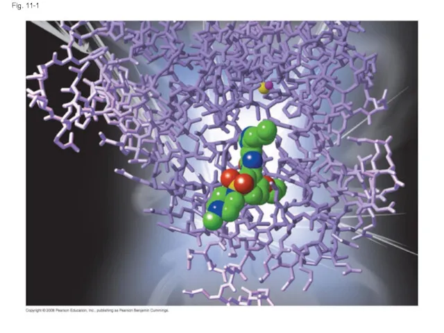
Fig. 11-1
Fig. 11-1
Concept 11.1: External signals are converted to responses within the cell
Microbes
Concept 11.1: External signals are converted to responses within the cell
Microbes
Evolution of Cell Signaling
A signal transduction pathway is a series of
Evolution of Cell Signaling
A signal transduction pathway is a series of
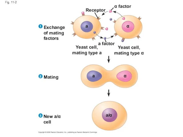
Fig. 11-2
Receptor
α factor
a factor
a
α
α
a
Exchange
of mating
factors
Yeast cell,
mating type a
Yeast cell,
mating type α
Mating
New
Fig. 11-2
Receptor
α factor
a factor
a
α
α
a
Exchange
of mating
factors
Yeast cell,
mating type a
Yeast cell,
mating type α
Mating
New
Pathway similarities suggest that ancestral signaling molecules evolved in prokaryotes and
Pathway similarities suggest that ancestral signaling molecules evolved in prokaryotes and
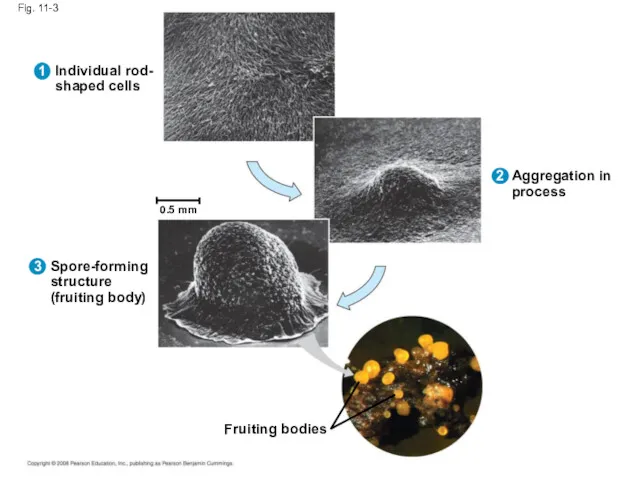
Fig. 11-3
Individual rod-
shaped cells
Spore-forming
structure
(fruiting body)
Aggregation in
process
Fruiting bodies
0.5 mm
1
3
2
Fig. 11-3
Individual rod-
shaped cells
Spore-forming
structure
(fruiting body)
Aggregation in
process
Fruiting bodies
0.5 mm
1
3
2
Local and Long-Distance Signaling
Cells in a multicellular organism communicate by chemical
Local and Long-Distance Signaling
Cells in a multicellular organism communicate by chemical
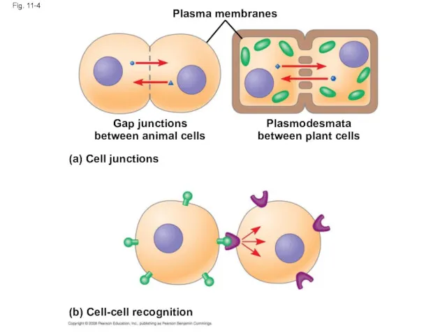
Fig. 11-4
Plasma membranes
Gap junctions
between animal cells
(a) Cell junctions
Plasmodesmata
between plant cells
(b) Cell-cell
Fig. 11-4
Plasma membranes
Gap junctions
between animal cells
(a) Cell junctions
Plasmodesmata
between plant cells
(b) Cell-cell
In many other cases, animal cells communicate using local regulators, messenger
In many other cases, animal cells communicate using local regulators, messenger
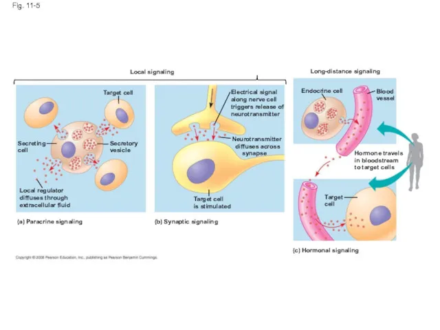
Fig. 11-5
Local signaling
Target cell
Secreting
cell
Secretory
vesicle
Local regulator
diffuses through
extracellular fluid
(a) Paracrine signaling
(b) Synaptic signaling
Target
Fig. 11-5
Local signaling
Target cell
Secreting
cell
Secretory
vesicle
Local regulator
diffuses through
extracellular fluid
(a) Paracrine signaling
(b) Synaptic signaling
Target
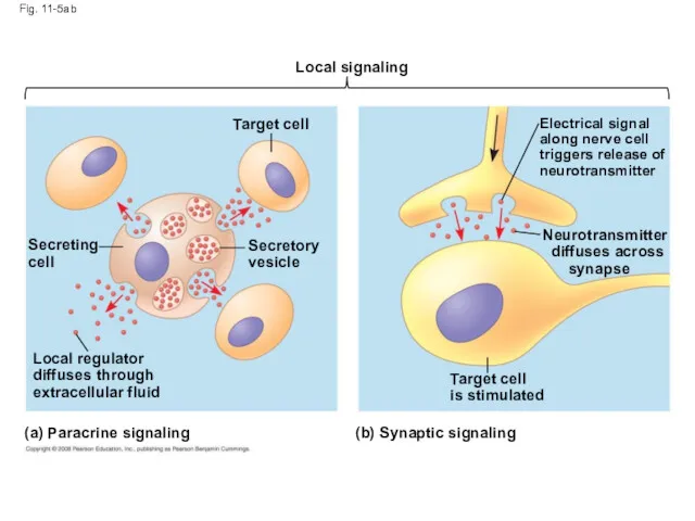
Fig. 11-5ab
Local signaling
Target cell
Secretory
vesicle
Secreting
cell
Local regulator
diffuses through
extracellular fluid
(a) Paracrine signaling
(b) Synaptic signaling
Target
Fig. 11-5ab
Local signaling
Target cell
Secretory
vesicle
Secreting
cell
Local regulator
diffuses through
extracellular fluid
(a) Paracrine signaling
(b) Synaptic signaling
Target
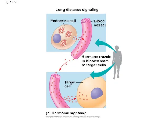
Fig. 11-5c
Long-distance signaling
Endocrine cell
Blood
vessel
Hormone travels
in bloodstream
to target cells
Target
cell
(c) Hormonal signaling
Fig. 11-5c
Long-distance signaling
Endocrine cell
Blood
vessel
Hormone travels
in bloodstream
to target cells
Target
cell
(c) Hormonal signaling
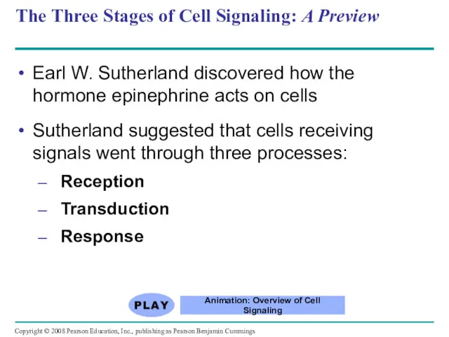
The Three Stages of Cell Signaling: A Preview
Earl W. Sutherland discovered
The Three Stages of Cell Signaling: A Preview
Earl W. Sutherland discovered
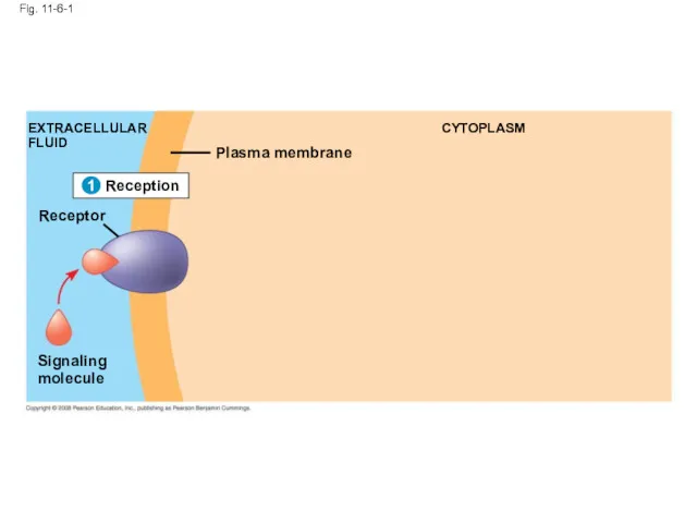
Fig. 11-6-1
Reception
1
EXTRACELLULAR
FLUID
Signaling
molecule
Plasma membrane
CYTOPLASM
1
Receptor
Fig. 11-6-1
Reception
1
EXTRACELLULAR
FLUID
Signaling
molecule
Plasma membrane
CYTOPLASM
1
Receptor
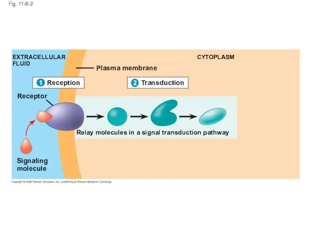
Fig. 11-6-2
1
EXTRACELLULAR
FLUID
Signaling
molecule
Plasma membrane
CYTOPLASM
Transduction
2
Relay molecules in a signal transduction pathway
Reception
1
Receptor
Fig. 11-6-2
1
EXTRACELLULAR
FLUID
Signaling
molecule
Plasma membrane
CYTOPLASM
Transduction
2
Relay molecules in a signal transduction pathway
Reception
1
Receptor
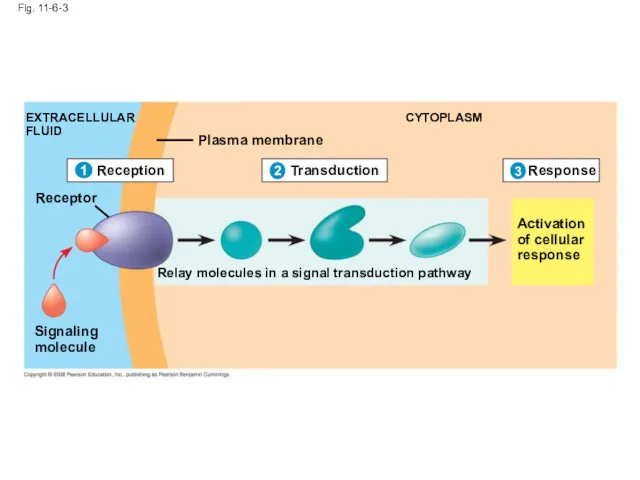
Fig. 11-6-3
EXTRACELLULAR
FLUID
Plasma membrane
CYTOPLASM
Receptor
Signaling
molecule
Relay molecules in a signal transduction pathway
Activation
of cellular
response
Transduction
Response
2
3
Reception
1
Fig. 11-6-3
EXTRACELLULAR
FLUID
Plasma membrane
CYTOPLASM
Receptor
Signaling
molecule
Relay molecules in a signal transduction pathway
Activation
of cellular
response
Transduction
Response
2
3
Reception
1
Concept 11.2: Reception: A signal molecule binds to a receptor protein,
Concept 11.2: Reception: A signal molecule binds to a receptor protein,
Receptors in the Plasma Membrane
Most water-soluble signal molecules bind to specific
Receptors in the Plasma Membrane
Most water-soluble signal molecules bind to specific
A G protein-coupled receptor is a plasma membrane receptor that works
A G protein-coupled receptor is a plasma membrane receptor that works
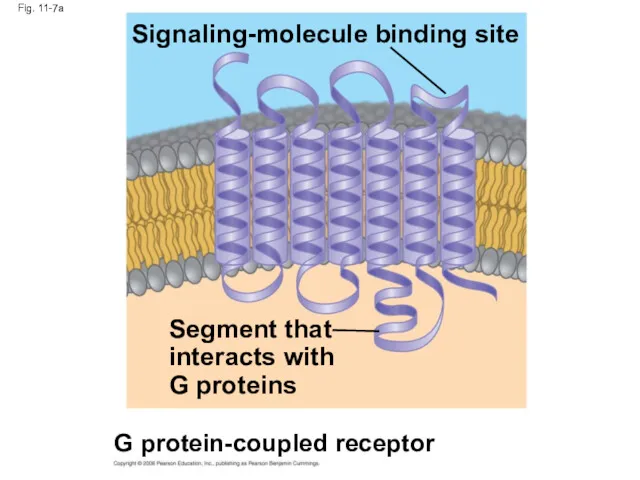
Fig. 11-7a
Signaling-molecule binding site
Segment that
interacts with
G proteins
G protein-coupled receptor
Fig. 11-7a
Signaling-molecule binding site
Segment that
interacts with
G proteins
G protein-coupled receptor
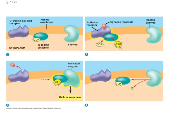
Fig. 11-7b
G protein-coupled
receptor
Plasma
membrane
Enzyme
G protein
(inactive)
GDP
CYTOPLASM
Activated
enzyme
GTP
Cellular response
GDP
P
i
Activated
receptor
GDP
GTP
Signaling molecule
Inactive
enzyme
1
2
3
4
Fig. 11-7b
G protein-coupled
receptor
Plasma
membrane
Enzyme
G protein
(inactive)
GDP
CYTOPLASM
Activated
enzyme
GTP
Cellular response
GDP
P
i
Activated
receptor
GDP
GTP
Signaling molecule
Inactive
enzyme
1
2
3
4
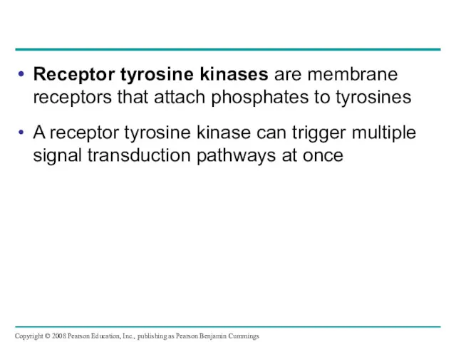
Receptor tyrosine kinases are membrane receptors that attach phosphates to tyrosines
A
Receptor tyrosine kinases are membrane receptors that attach phosphates to tyrosines
A
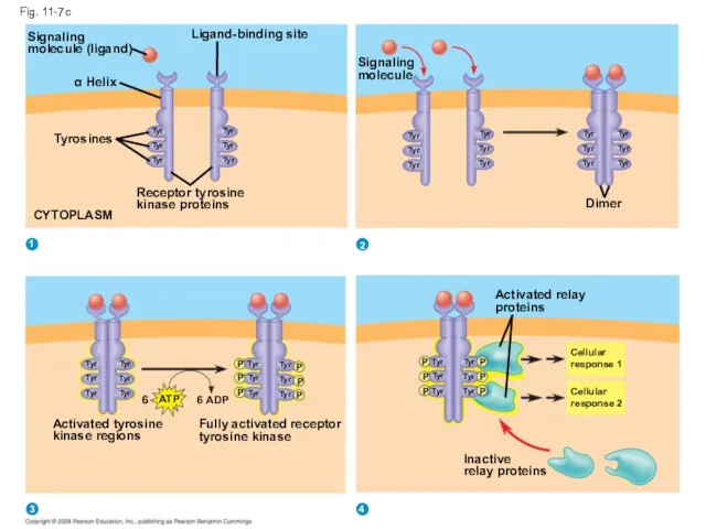
Fig. 11-7c
Signaling
molecule (ligand)
Ligand-binding site
α Helix
Tyrosines
Tyr
Tyr
Tyr
Tyr
Tyr
Tyr
Receptor tyrosine
kinase proteins
CYTOPLASM
Signaling
molecule
Tyr
Tyr
Tyr
Tyr
Tyr
Tyr
Tyr
Tyr
Tyr
Tyr
Tyr
Tyr
Dimer
Activated relay
proteins
Tyr
Tyr
Tyr
Tyr
Tyr
Tyr
P
P
P
P
P
P
Cellular
response 1
Cellular
response 2
Inactive
relay proteins
Activated
A ligand-gated ion channel receptor acts as a gate when the
A ligand-gated ion channel receptor acts as a gate when the
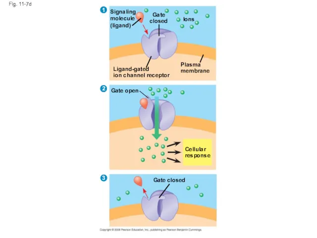
Fig. 11-7d
Signaling
molecule
(ligand)
Gate
closed
Ions
Ligand-gated
ion channel receptor
Plasma
membrane
Gate open
Cellular
response
Gate closed
3
2
1
Fig. 11-7d
Signaling
molecule
(ligand)
Gate
closed
Ions
Ligand-gated
ion channel receptor
Plasma
membrane
Gate open
Cellular
response
Gate closed
3
2
1
Intracellular Receptors
Some receptor proteins are intracellular, found in the cytosol or
Intracellular Receptors
Some receptor proteins are intracellular, found in the cytosol or
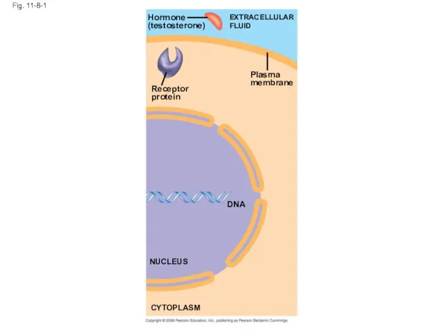
Fig. 11-8-1
Hormone
(testosterone)
Receptor
protein
Plasma
membrane
EXTRACELLULAR
FLUID
DNA
NUCLEUS
CYTOPLASM
Fig. 11-8-1
Hormone
(testosterone)
Receptor
protein
Plasma
membrane
EXTRACELLULAR
FLUID
DNA
NUCLEUS
CYTOPLASM
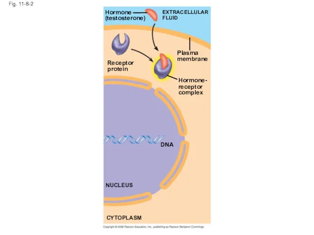
Fig. 11-8-2
Receptor
protein
Hormone
(testosterone)
EXTRACELLULAR
FLUID
Plasma
membrane
Hormone-
receptor
complex
DNA
NUCLEUS
CYTOPLASM
Fig. 11-8-2
Receptor
protein
Hormone
(testosterone)
EXTRACELLULAR
FLUID
Plasma
membrane
Hormone-
receptor
complex
DNA
NUCLEUS
CYTOPLASM

Fig. 11-8-3
Hormone
(testosterone)
EXTRACELLULAR
FLUID
Receptor
protein
Plasma
membrane
Hormone-
receptor
complex
DNA
NUCLEUS
CYTOPLASM
Fig. 11-8-3
Hormone
(testosterone)
EXTRACELLULAR
FLUID
Receptor
protein
Plasma
membrane
Hormone-
receptor
complex
DNA
NUCLEUS
CYTOPLASM
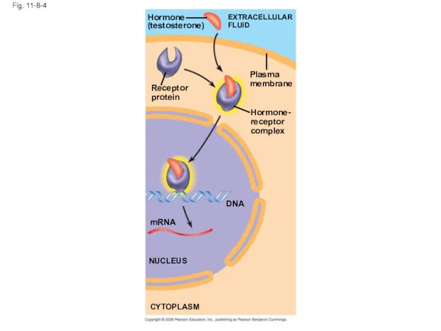
Fig. 11-8-4
Hormone
(testosterone)
EXTRACELLULAR
FLUID
Plasma
membrane
Receptor
protein
Hormone-
receptor
complex
DNA
mRNA
NUCLEUS
CYTOPLASM
Fig. 11-8-4
Hormone
(testosterone)
EXTRACELLULAR
FLUID
Plasma
membrane
Receptor
protein
Hormone-
receptor
complex
DNA
mRNA
NUCLEUS
CYTOPLASM
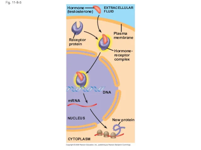
Fig. 11-8-5
Hormone
(testosterone)
EXTRACELLULAR
FLUID
Receptor
protein
Plasma
membrane
Hormone-
receptor
complex
DNA
mRNA
NUCLEUS
New protein
CYTOPLASM
Fig. 11-8-5
Hormone
(testosterone)
EXTRACELLULAR
FLUID
Receptor
protein
Plasma
membrane
Hormone-
receptor
complex
DNA
mRNA
NUCLEUS
New protein
CYTOPLASM
Concept 11.3: Transduction: Cascades of molecular interactions relay signals from receptors
Concept 11.3: Transduction: Cascades of molecular interactions relay signals from receptors
Signal Transduction Pathways
The molecules that relay a signal from receptor to
Signal Transduction Pathways
The molecules that relay a signal from receptor to
Protein Phosphorylation and Dephosphorylation
In many pathways, the signal is transmitted by
Protein Phosphorylation and Dephosphorylation
In many pathways, the signal is transmitted by
Protein phosphatases remove the phosphates from proteins, a process called dephosphorylation
This
Protein phosphatases remove the phosphates from proteins, a process called dephosphorylation
This
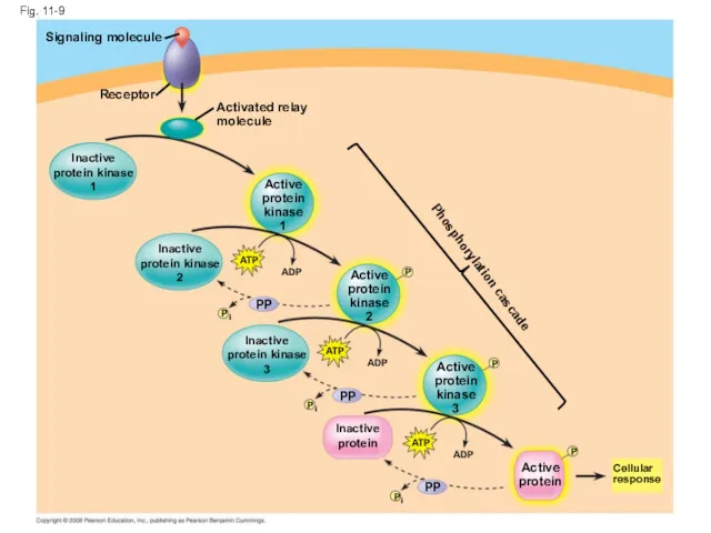
Fig. 11-9
Signaling molecule
Receptor
Activated relay
molecule
Inactive
protein kinase
1
Active
protein
kinase
1
Inactive
protein kinase
2
ATP
ADP
Active
protein
kinase
2
P
P
PP
Inactive
protein kinase
3
ATP
ADP
Active
protein
kinase
3
P
P
PP
i
ATP
ADP
P
Active
protein
PP
P
i
Inactive
protein
Cellular
response
Phosphorylation cascade
i
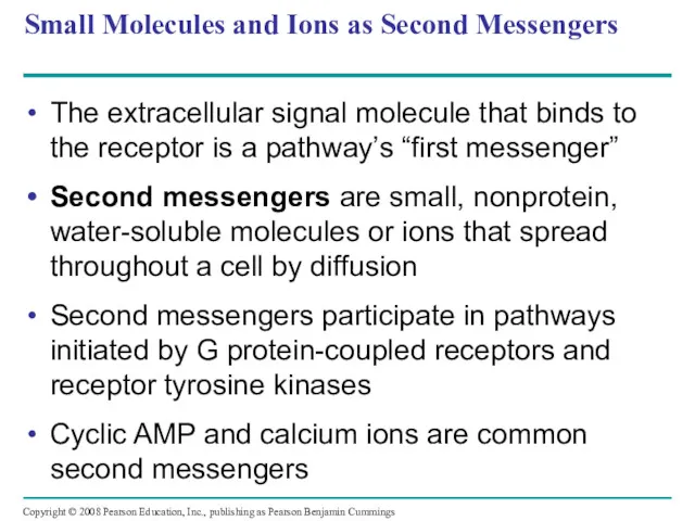
Small Molecules and Ions as Second Messengers
The extracellular signal molecule that
Small Molecules and Ions as Second Messengers
The extracellular signal molecule that
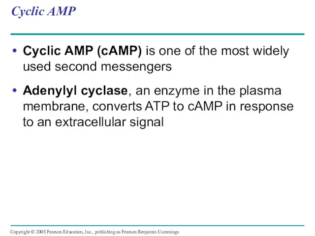
Cyclic AMP
Cyclic AMP (cAMP) is one of the most widely used
Cyclic AMP
Cyclic AMP (cAMP) is one of the most widely used
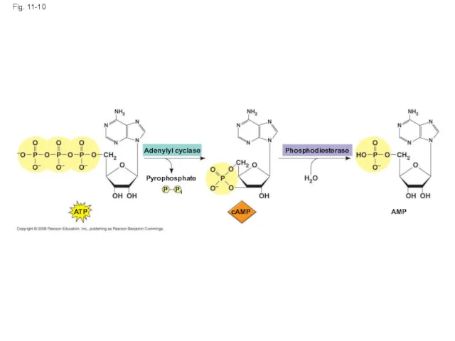
Adenylyl cyclase
Fig. 11-10
Pyrophosphate
P
P
i
ATP
cAMP
Phosphodiesterase
AMP
Adenylyl cyclase
Fig. 11-10
Pyrophosphate
P
P
i
ATP
cAMP
Phosphodiesterase
AMP
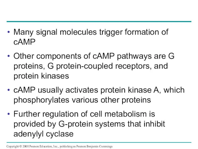
Many signal molecules trigger formation of cAMP
Other components of cAMP pathways
Many signal molecules trigger formation of cAMP
Other components of cAMP pathways
First messenger
Fig. 11-11
G protein
Adenylyl
cyclase
GTP
ATP
cAMP
Second
messenger
Protein
kinase A
G protein-coupled
receptor
Cellular responses
First messenger
Fig. 11-11
G protein
Adenylyl
cyclase
GTP
ATP
cAMP
Second
messenger
Protein
kinase A
G protein-coupled
receptor
Cellular responses
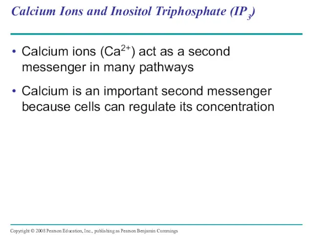
Calcium Ions and Inositol Triphosphate (IP3)
Calcium ions (Ca2+) act as a
Calcium Ions and Inositol Triphosphate (IP3)
Calcium ions (Ca2+) act as a
EXTRACELLULAR
FLUID
Fig. 11-12
ATP
Nucleus
Mitochondrion
Ca2+ pump
Plasma
membrane
CYTOSOL
Ca2+
pump
Endoplasmic
reticulum (ER)
Ca2+
pump
ATP
Key
High [Ca2+]
Low [Ca2+]
EXTRACELLULAR
FLUID
Fig. 11-12
ATP
Nucleus
Mitochondrion
Ca2+ pump
Plasma
membrane
CYTOSOL
Ca2+
pump
Endoplasmic
reticulum (ER)
Ca2+
pump
ATP
Key
High [Ca2+]
Low [Ca2+]
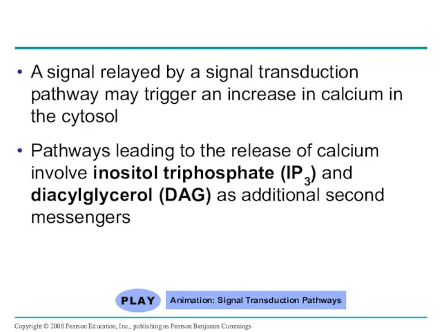
A signal relayed by a signal transduction pathway may trigger an
A signal relayed by a signal transduction pathway may trigger an
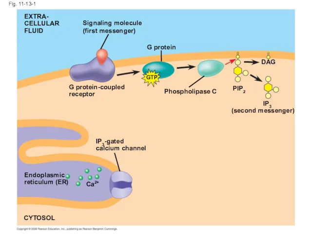
Fig. 11-13-1
EXTRA-
CELLULAR
FLUID
Signaling molecule
(first messenger)
G protein
GTP
G protein-coupled
receptor
Phospholipase C
PIP2
IP3
DAG
(second messenger)
IP3-gated
calcium channel
Endoplasmic
reticulum (ER)
Ca2+
CYTOSOL
Fig. 11-13-1
EXTRA-
CELLULAR
FLUID
Signaling molecule
(first messenger)
G protein
GTP
G protein-coupled
receptor
Phospholipase C
PIP2
IP3
DAG
(second messenger)
IP3-gated
calcium channel
Endoplasmic
reticulum (ER)
Ca2+
CYTOSOL
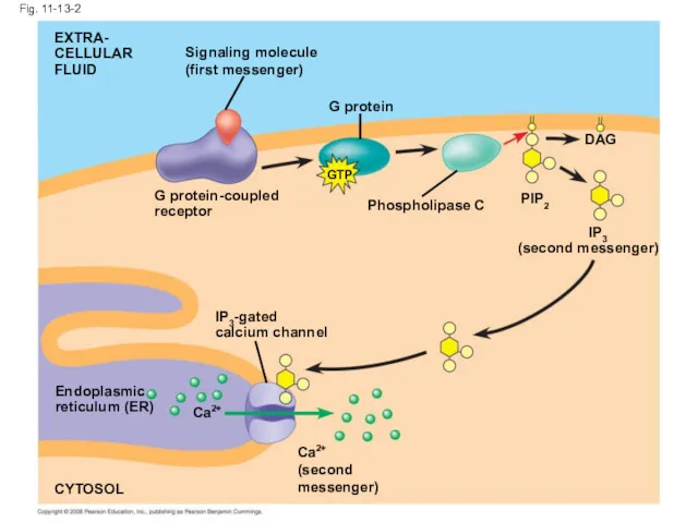
Fig. 11-13-2
G protein
EXTRA-
CELLULAR
FLUID
Signaling molecule
(first messenger)
G protein-coupled
receptor
Phospholipase C
PIP2
DAG
IP3
(second messenger)
IP3-gated
calcium channel
Endoplasmic
reticulum (ER)
Ca2+
CYTOSOL
Ca2+
(second
messenger)
GTP
Fig. 11-13-2
G protein
EXTRA-
CELLULAR
FLUID
Signaling molecule
(first messenger)
G protein-coupled
receptor
Phospholipase C
PIP2
DAG
IP3
(second messenger)
IP3-gated
calcium channel
Endoplasmic
reticulum (ER)
Ca2+
CYTOSOL
Ca2+
(second
messenger)
GTP
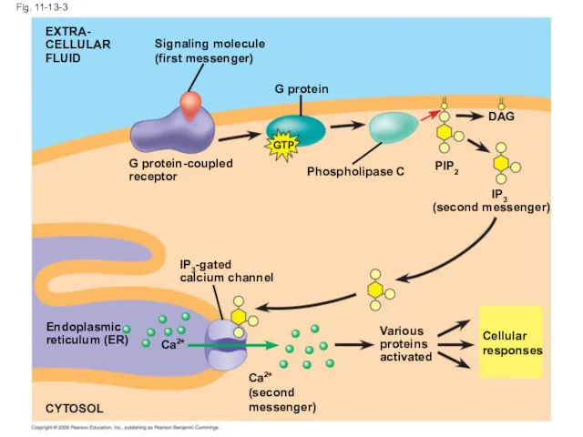
Fig. 11-13-3
G protein
EXTRA-
CELLULAR
FLUID
Signaling molecule
(first messenger)
G protein-coupled
receptor
Phospholipase C
PIP2
DAG
IP3
(second messenger)
IP3-gated
calcium channel
Endoplasmic
reticulum (ER)
Ca2+
CYTOSOL
Various
proteins
activated
Cellular
responses
Ca2+
(second
messenger)
GTP
Fig. 11-13-3
G protein
EXTRA-
CELLULAR
FLUID
Signaling molecule
(first messenger)
G protein-coupled
receptor
Phospholipase C
PIP2
DAG
IP3
(second messenger)
IP3-gated
calcium channel
Endoplasmic
reticulum (ER)
Ca2+
CYTOSOL
Various
proteins
activated
Cellular
responses
Ca2+
(second
messenger)
GTP
Concept 11.4: Response: Cell signaling leads to regulation of transcription or
Concept 11.4: Response: Cell signaling leads to regulation of transcription or
Nuclear and Cytoplasmic Responses
Ultimately, a signal transduction pathway leads to regulation
Nuclear and Cytoplasmic Responses
Ultimately, a signal transduction pathway leads to regulation
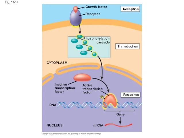
Fig. 11-14
Growth factor
Receptor
Phosphorylation
cascade
Reception
Transduction
Active
transcription
factor
Response
P
Inactive
transcription
factor
CYTOPLASM
DNA
NUCLEUS
mRNA
Gene
Fig. 11-14
Growth factor
Receptor
Phosphorylation
cascade
Reception
Transduction
Active
transcription
factor
Response
P
Inactive
transcription
factor
CYTOPLASM
DNA
NUCLEUS
mRNA
Gene
Other pathways regulate the activity of enzymes
Copyright © 2008 Pearson Education,
Other pathways regulate the activity of enzymes
Copyright © 2008 Pearson Education,
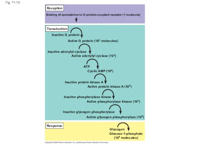
Fig. 11-15
Reception
Transduction
Response
Binding of epinephrine to G protein-coupled receptor (1 molecule)
Inactive G
Fig. 11-15
Reception
Transduction
Response
Binding of epinephrine to G protein-coupled receptor (1 molecule)
Inactive G
Signaling pathways can also affect the physical characteristics of a cell,
Signaling pathways can also affect the physical characteristics of a cell,
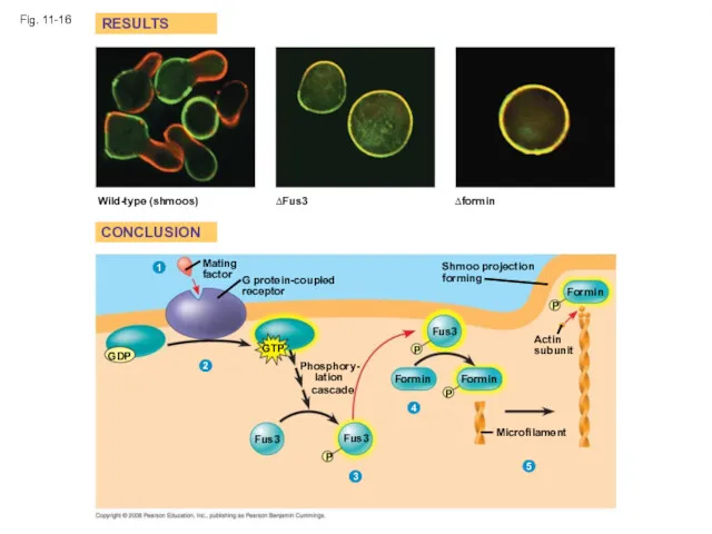
Fig. 11-16
RESULTS
CONCLUSION
Wild-type (shmoos)
∆Fus3
∆formin
Shmoo projection forming
Formin
P
Actin
subunit
P
P
Formin
Formin
Fus3
Phosphory-
lation
cascade
GTP
G protein-coupled
receptor
Mating
factor
GDP
Fus3
Fus3
P
Microfilament
1
2
3
4
5
Fig. 11-16
RESULTS
CONCLUSION
Wild-type (shmoos)
∆Fus3
∆formin
Shmoo projection forming
Formin
P
Actin
subunit
P
P
Formin
Formin
Fus3
Phosphory-
lation
cascade
GTP
G protein-coupled
receptor
Mating
factor
GDP
Fus3
Fus3
P
Microfilament
1
2
3
4
5
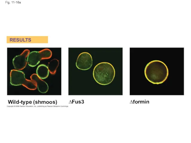
Fig. 11-16a
RESULTS
Wild-type (shmoos)
∆Fus3
∆formin
Fig. 11-16a
RESULTS
Wild-type (shmoos)
∆Fus3
∆formin
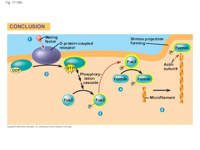
Fig. 11-16b
CONCLUSION
Mating
factor
G protein-coupled
receptor
GDP
GTP
Phosphory-
lation
cascade
Shmoo projection
forming
Fus3
Fus3
Fus3
Formin
Formin
P
P
P
Formin
P
Actin
subunit
Microfilament
1
2
3
4
5
Fig. 11-16b
CONCLUSION
Mating
factor
G protein-coupled
receptor
GDP
GTP
Phosphory-
lation
cascade
Shmoo projection
forming
Fus3
Fus3
Fus3
Formin
Formin
P
P
P
Formin
P
Actin
subunit
Microfilament
1
2
3
4
5
Fine-Tuning of the Response
Multistep pathways have two important benefits:
Amplifying the signal
Fine-Tuning of the Response
Multistep pathways have two important benefits:
Amplifying the signal
Signal Amplification
Enzyme cascades amplify the cell’s response
At each step, the number
Signal Amplification
Enzyme cascades amplify the cell’s response
At each step, the number
The Specificity of Cell Signaling and Coordination of the Response
Different kinds
The Specificity of Cell Signaling and Coordination of the Response
Different kinds
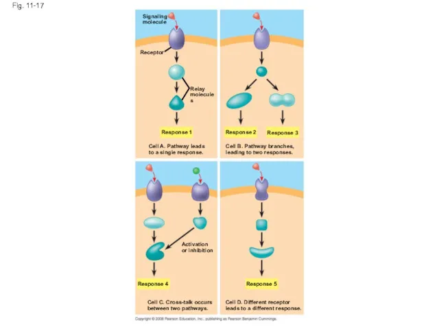
Fig. 11-17
Signaling
molecule
Receptor
Relay
molecules
Response 1
Cell A. Pathway leads
to a single response.
Response 2
Response 3
Cell
Fig. 11-17
Signaling
molecule
Receptor
Relay
molecules
Response 1
Cell A. Pathway leads
to a single response.
Response 2
Response 3
Cell

Fig. 11-17a
Signaling
molecule
Receptor
Relay
molecules
Response 1
Cell A. Pathway leads
to a single response.
Cell B. Pathway
Fig. 11-17a
Signaling
molecule
Receptor
Relay
molecules
Response 1
Cell A. Pathway leads
to a single response.
Cell B. Pathway
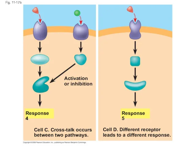
Fig. 11-17b
Response 4
Response 5
Activation
or inhibition
Cell C. Cross-talk occurs
between two pathways.
Cell D.
Fig. 11-17b
Response 4
Response 5
Activation
or inhibition
Cell C. Cross-talk occurs
between two pathways.
Cell D.
Signaling Efficiency: Scaffolding Proteins and Signaling Complexes
Scaffolding proteins are large relay
Signaling Efficiency: Scaffolding Proteins and Signaling Complexes
Scaffolding proteins are large relay
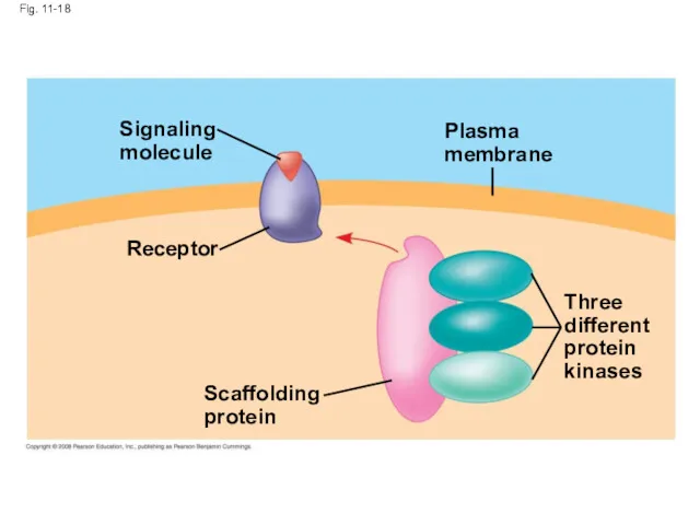
Fig. 11-18
Signaling
molecule
Receptor
Scaffolding
protein
Plasma
membrane
Three
different
protein
kinases
Fig. 11-18
Signaling
molecule
Receptor
Scaffolding
protein
Plasma
membrane
Three
different
protein
kinases
Termination of the Signal
Inactivation mechanisms are an essential aspect of cell
Termination of the Signal
Inactivation mechanisms are an essential aspect of cell
Concept 11.5: Apoptosis (programmed cell death) integrates multiple cell-signaling pathways
Apoptosis is
Concept 11.5: Apoptosis (programmed cell death) integrates multiple cell-signaling pathways
Apoptosis is

Fig. 11-19
2 µm
Fig. 11-19
2 µm
Apoptosis in the Soil Worm Caenorhabditis elegans
Apoptosis is important in shaping
Apoptosis in the Soil Worm Caenorhabditis elegans
Apoptosis is important in shaping
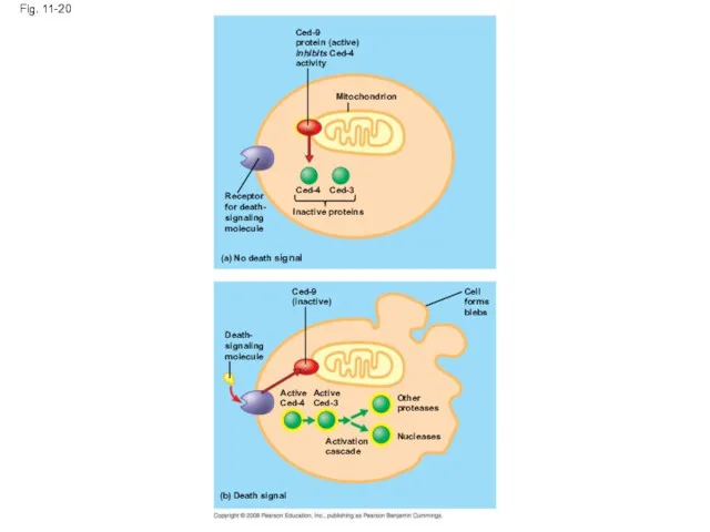
Fig. 11-20
Ced-9
protein (active)
inhibits Ced-4
activity
Mitochondrion
Receptor
for death-
signaling
molecule
Ced-4
Ced-3
Inactive proteins
(a) No death signal
Ced-9
(inactive)
Cell
forms
blebs
Death-
signaling
molecule
Other
proteases
Active
Ced-4
Active
Ced-3
Nucleases
Activation
cascade
(b) Death signal
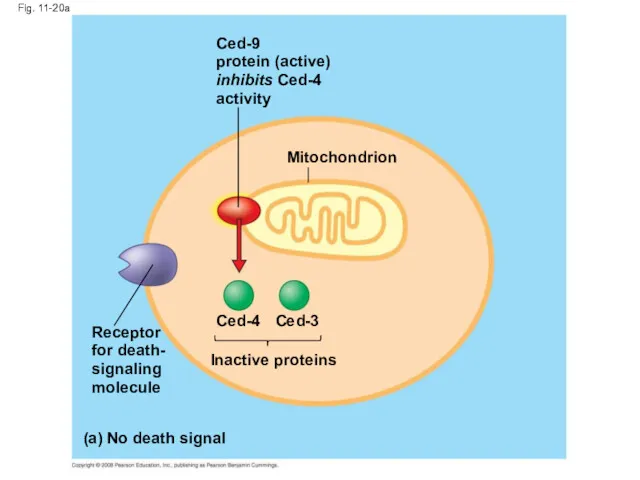
Fig. 11-20a
Ced-9
protein (active)
inhibits Ced-4
activity
Mitochondrion
Ced-4
Ced-3
Receptor
for death-
signaling
molecule
Inactive proteins
(a) No death signal
Fig. 11-20a
Ced-9
protein (active)
inhibits Ced-4
activity
Mitochondrion
Ced-4
Ced-3
Receptor
for death-
signaling
molecule
Inactive proteins
(a) No death signal
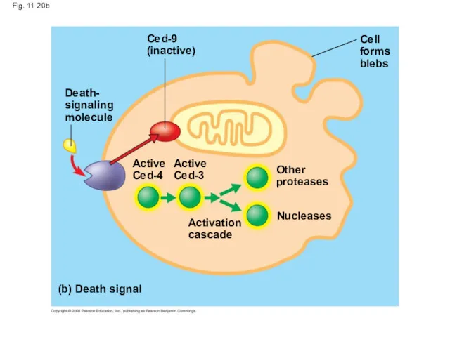
Fig. 11-20b
(b) Death signal
Death-
signaling
molecule
Ced-9
(inactive)
Cell
forms
blebs
Active
Ced-4
Active
Ced-3
Activation
cascade
Other
proteases
Nucleases
Fig. 11-20b
(b) Death signal
Death-
signaling
molecule
Ced-9
(inactive)
Cell
forms
blebs
Active
Ced-4
Active
Ced-3
Activation
cascade
Other
proteases
Nucleases
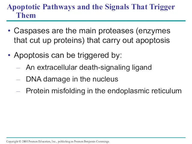
Apoptotic Pathways and the Signals That Trigger Them
Caspases are the main
Apoptotic Pathways and the Signals That Trigger Them
Caspases are the main
Apoptosis evolved early in animal evolution and is essential for the
Apoptosis evolved early in animal evolution and is essential for the
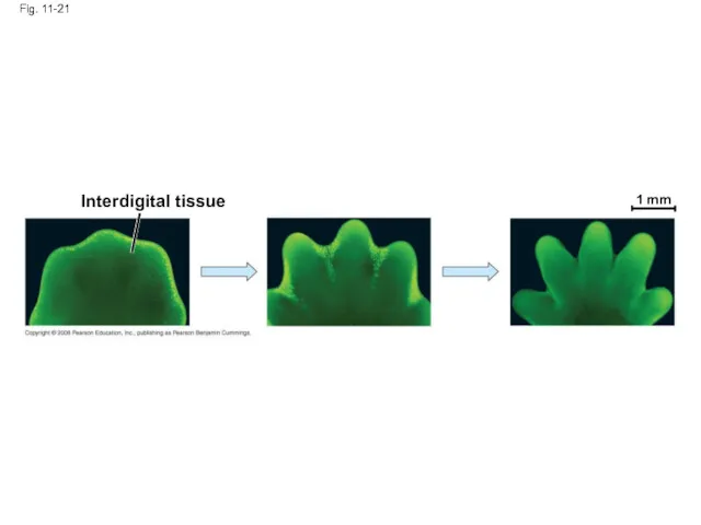
Fig. 11-21
Interdigital tissue
1 mm
Fig. 11-21
Interdigital tissue
1 mm
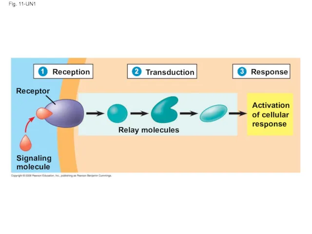
Fig. 11-UN1
Reception
Transduction
Response
Receptor
Relay molecules
Signaling
molecule
Activation
of cellular
response
1
2
3
Fig. 11-UN1
Reception
Transduction
Response
Receptor
Relay molecules
Signaling
molecule
Activation
of cellular
response
1
2
3
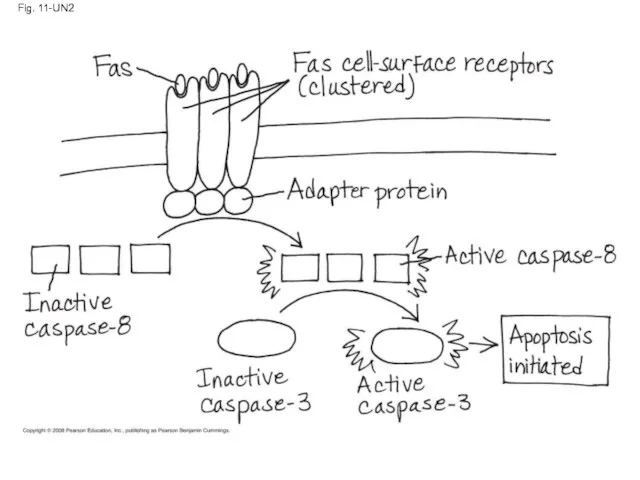
Fig. 11-UN2
Fig. 11-UN2
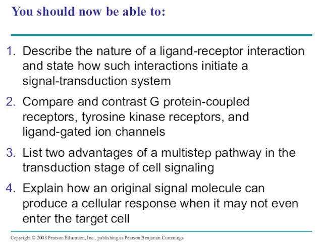
You should now be able to:
Describe the nature of a ligand-receptor
You should now be able to:
Describe the nature of a ligand-receptor
 Текстовая информация
 Установка и настройка Apache и PHP
 Презентация Состав объектов, 7 класс
 Фактологические жанры PR-текстов
 Алгоритмы. Свойства и формы представления
 Инструкция по запуску дистанционных курсов
 Эффективное использование инструментов разработки веб-страниц
 Основные понятия и определения информатики
 Перезапуск официального сайта: этапы и предложения
 Пользователи Instagram
 Ғаламтор, әлеуметтік желілердің жасөспірімдердің қылмысына қатысы
 Прототип мобильного приложения для обучения правильной технике свинга при помощи AI
 Работа с графическими объектами в текстовом реакторе
 C# decision and iteration constructs
 Публичная кадастровая карта
 Рекламные пакеты Турбопромо
 Информационные системы управления персоналом. Лекция 1
 Б-Безопасность ACL, NAT, VPN
 Курс Базы данных. Программирование на языке PL/SQL. Часть 2
 Проектування у графічному редакторі Sweet Нome 3D
 Программирование на языке Си. Простейшие программы
 Представление числовой информации с помощью систем счисления (1). 8 класс
 Сервис Отвечает аудитор
 Основные инструменты графического редактора
 Числа в памяти компютера
 Программирование ветвлений на Паскаль. Повторение
 Табличная форма представления информации
 Оценка максимально достижимого параллелизма и масштабируемости параллельных вычислений