- Acquired Immunodeficiency syndrome

Содержание
- 2. HIV is a retrovirus of the lentivirus family. The variant of HIV that is the cause
- 3. env include- the envelope glycoproteins, the outer envelope glycoprotein gp120 transmembrane glycoprotein gp41 derived from glycoprotein
- 4. Major modes of HIV infection spread: As a sexually transmitted disease. Infection is also aided by
- 5. The CD4 receptor is known as a chemokine that is needed for HIV infection. Chemokines are
- 6. HIV primarily infects cells with CD4 cell-surface receptor molecule. Many cell types share common epitopes with
- 7. Cells of the mononuclear phagocyte system, principally blood monocytes, T lymphocytes, B lymphocytes, natural killer (NK)
- 8. After entering the body, the viral particle is attracted to a cell with the appropriate CD4
- 9. It is this HIV proviral DNA which is then inserted into host cell genomic DNA by
- 10. After initial entry of HIV and establishment of infection, replication may at first occur within inflammatory
- 11. The primary target of HIV is the immune system, which is gradually destroyed. Viral replication actively
- 12. Onset of AIDS Decrease in the total CD4 count below 500/microliter presages the development of clinical
- 13. There is loss of normal lymph node architecture as the immune system fails with emergence from
- 14. Another phase of HIV infection described clinically and is known as AIDS-related complex (ARC), which is
- 15. The progression to clinical AIDS is marked by: syncytia-forming (SI) variants of HIV. These SI viral
- 16. Opportunistic infection Pneumocystis carinii Cytomegalovirus Mycobacteria Fungal Infections Toxoplasmosis Herpes simplex Gastrointestinal Protozoal Infections Malignant Neoplasms
- 17. Pneumocystis carinii It is the most frequent opportunistic infection seen with AIDS. It produces a pulmonary
- 18. Cytomegalovirus (CMV) is the most frequent disseminated opportunistic infection seen with AIDS. It causes the most
- 19. The appearance of Myc. tuberculosis with AIDS is similar to that of non-AIDS patients, with granulomatous
- 20. Oral candidiasis is often seen with HIV infection and may presage the progression to AIDS. Candida
- 22. Скачать презентацию
HIV is a retrovirus of the lentivirus family. The variant of
HIV is a retrovirus of the lentivirus family. The variant of
env include-
the envelope glycoproteins, the outer envelope glycoprotein gp120
transmembrane glycoprotein gp41
env include-
the envelope glycoproteins, the outer envelope glycoprotein gp120
transmembrane glycoprotein gp41
Major modes of HIV infection spread:
As a sexually transmitted disease. Infection
Major modes of HIV infection spread:
As a sexually transmitted disease. Infection
The CD4 receptor is known as a chemokine that is
The CD4 receptor is known as a chemokine that is
HIV primarily infects cells with CD4 cell-surface receptor molecule. Many cell
HIV primarily infects cells with CD4 cell-surface receptor molecule. Many cell
Cells of the mononuclear phagocyte system, principally blood monocytes, T lymphocytes,
Cells of the mononuclear phagocyte system, principally blood monocytes, T lymphocytes,
After entering the body, the viral particle is attracted to a
After entering the body, the viral particle is attracted to a
It is this HIV proviral DNA which is then inserted into
It is this HIV proviral DNA which is then inserted into
After initial entry of HIV and establishment of infection,
replication may
After initial entry of HIV and establishment of infection,
replication may
The primary target of HIV is the immune system,
which is gradually
The primary target of HIV is the immune system,
which is gradually
Onset of AIDS
Decrease in the total CD4 count below 500/microliter presages the development
Onset of AIDS
Decrease in the total CD4 count below 500/microliter presages the development
There is loss of normal lymph node architecture as the immune system fails with
There is loss of normal lymph node architecture as the immune system fails with
Another phase of HIV infection described clinically and is known as
Another phase of HIV infection described clinically and is known as
The progression to clinical AIDS is marked by:
syncytia-forming (SI) variants of
The progression to clinical AIDS is marked by:
syncytia-forming (SI) variants of
Opportunistic infection
Pneumocystis carinii
Cytomegalovirus
Mycobacteria
Fungal Infections
Toxoplasmosis
Herpes simplex
Gastrointestinal Protozoal Infections
Malignant Neoplasms
Miscellaneous
Opportunistic infection
Pneumocystis carinii
Cytomegalovirus
Mycobacteria
Fungal Infections
Toxoplasmosis
Herpes simplex
Gastrointestinal Protozoal Infections
Malignant Neoplasms
Miscellaneous
Pneumocystis carinii
It is the most frequent opportunistic infection seen with AIDS.
Pneumocystis carinii It is the most frequent opportunistic infection seen with AIDS.
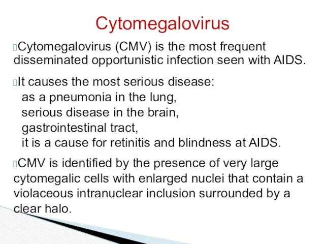
Cytomegalovirus (CMV) is the most frequent disseminated opportunistic infection seen with
Cytomegalovirus (CMV) is the most frequent disseminated opportunistic infection seen with
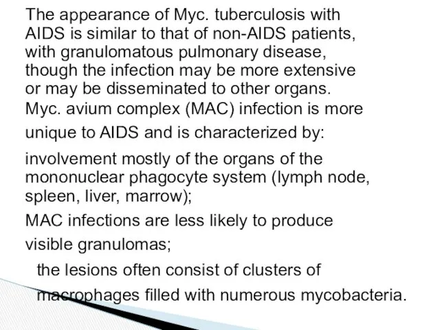
The appearance of Myc. tuberculosis with AIDS is similar to that
The appearance of Myc. tuberculosis with AIDS is similar to that
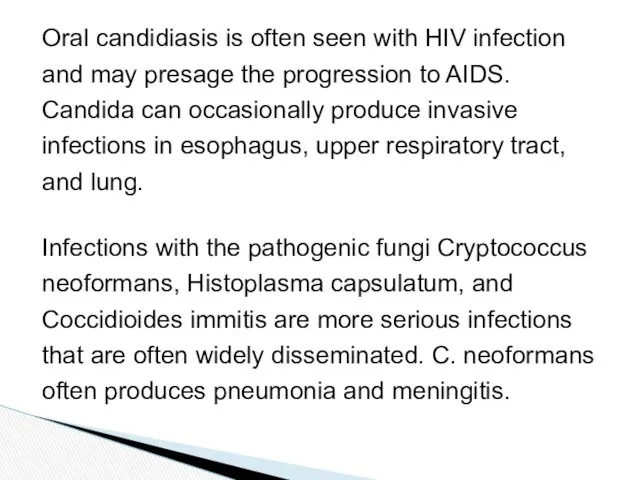
Oral candidiasis is often seen with HIV infection and may presage
Oral candidiasis is often seen with HIV infection and may presage
 Жұқпалы аурулар iндетi пайда болу қаупi төнген жағдайда шектеу шараларын қарастыру
 Закаливание. Польза закаливания
 Лекарственные средства используемые в анестезиологии и интенсивной терапии
 Тіс, тіс қатарлары ақауын емдеу
 Диссеминированный туберкулез легких
 Острая массивная кровопотеря
 Бронхоэктаз ауруы
 Венозный доступ в интенсивной терапии
 Бронхиальная астма и хобл: особенности диагностики и лечения
 Антигены. Антитела
 Диффузный токсический зоб и беременность
 Диспансеризация беременных женщин
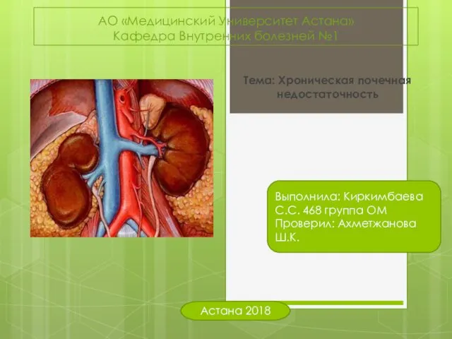 Хроническая почечная недостаточность
 Аппендицит. Диагностика. Техника оперативного лечения
 Біохімічні механізми розвитку та зміни при карієсі. Роль фтору та інших мікроелементів в карієсрезистентності
 Обзор рекомендаций ESC 2017 по лечению заболеваний периферических артерий
 Реабилитация больных, перенесших инсульт
 Инсульт. Первая медицинская помощь при инсульте
 Аллергия. Провоцирующие факторы и факторы риска развития аллергии
 Система гигиенических мероприятий по созданию охранительного режима и благоприятных условий пребывания больных в ЛПУ
 ВІЛ-інфекція. СНІД-асоційовані інфекції та інвазії Історія відкриття. Етіологія. Епідеміологія. Патогенез. Клінічні прояви
 Артериальная гипертония, факторы риска и их коррекция
 Нейропсихологическая характеристика младших школьников (лекция 4)
 Нарушение кровообращения. Лекция
 Правовое обеспечение профессиональной деятельности фармацевтов (1)
 Психология здоровья
 Созылмалы гастриттердің визуальды диагностикасы
 Выявление факторов риска и диагностика железодефицитной анемии у пациентов детского возраста