- Basic dental instrumentation

Содержание
- 2. Lecture plan: 1. Viewing set. The main tools included in the viewing kit. Their purpose. 2.
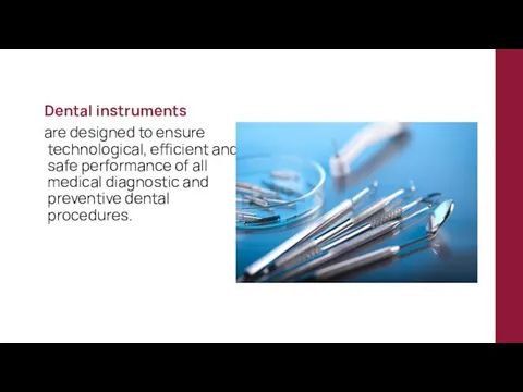
- 3. Dental instruments are designed to ensure technological, efficient and safe performance of all medical diagnostic and
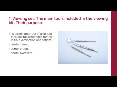
- 4. 1. Viewing set. The main tools included in the viewing kit. Their purpose. The examination set
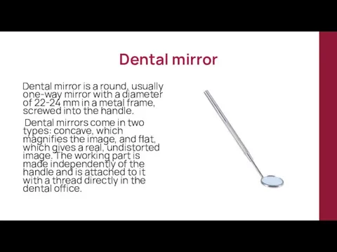
- 5. Dental mirror Dental mirror is a round, usually one-way mirror with a diameter of 22-24 mm
- 6. The dental mirror is intended for the following manipulations: - additional illumination of darkened areas of
- 7. Dental probe The dental probe (pointed probe) is a multifunctional diagnostic tool. The most commonly used
- 8. Appointment of pointed dental probes: - detection of carious cavities, demineralized dentin, areas of soreness of
- 9. Appointment of pointed dental probes: - detection of supragingival dental deposits; - introduction into the cavity
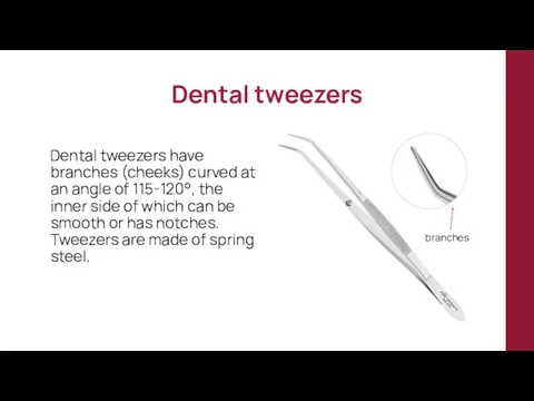
- 10. Dental tweezers Dental tweezers have branches (cheeks) curved at an angle of 115-120°, the inner side
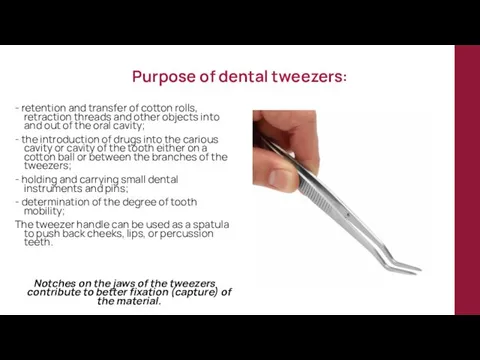
- 11. Purpose of dental tweezers: - retention and transfer of cotton rolls, retraction threads and other objects
- 12. 2. INSTRUMENTS USED IN THERAPEUTIC DENTISTRY
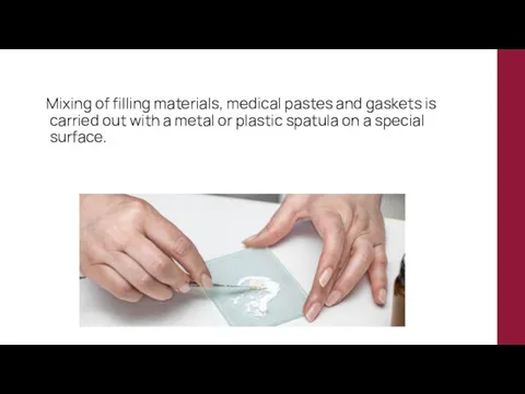
- 13. Mixing of filling materials, medical pastes and gaskets is carried out with a metal or plastic
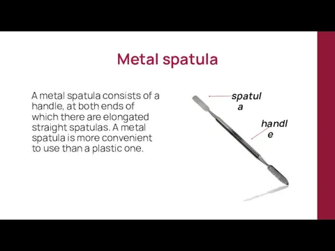
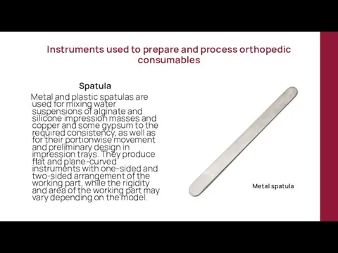
- 14. Metal spatula A metal spatula consists of a handle, at both ends of which there are
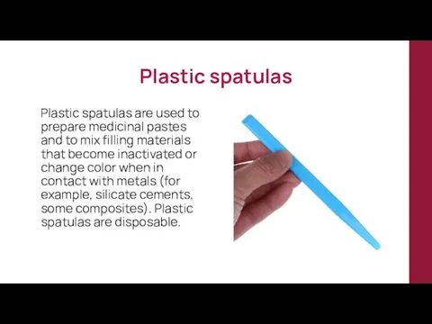
- 15. Plastic spatulas Plastic spatulas are used to prepare medicinal pastes and to mix filling materials that
- 16. The following devices are used as a mixing surface: - glass plates, one side of which
- 17. A number of requirements are imposed on the instruments used for filling cavities: - The instrument
- 18. Dental trowel A trowel is a dental tool with a working part in the form of
- 19. Plugfer Plugfer - a dental instrument with a working part of a spherical, pear-shaped, cylindrical, olive-shaped
- 20. Plugfers are conditionally divided into condensing and modeling Condensing pluggers have the shape of a working
- 21. Condensing pluggers Modeling pluggers
- 22. Pluggers and floaters are available in various sizes, single-sided, double-sided, and also as a combination of
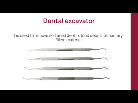
- 23. Dental excavator It is used to remove softened dentin, food debris, temporary filling material.
- 24. There are complete sets of standard instruments for periodontal examination, for example, the set described by
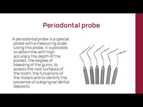
- 25. Periodontal probe A periodontal probe is a special probe with a measuring scale. Using this probe,
- 26. 3. INSTRUMENTS USED IN SURGICAL DENTISTRY
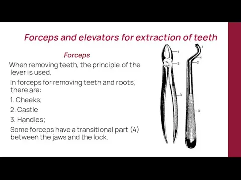
- 27. Forceps and elevators for extraction of teeth Forceps When removing teeth, the principle of the lever
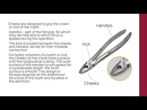
- 28. Cheeks are designed to grip the crown or root of the tooth. Handles - part of
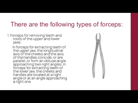
- 29. There are the following types of forceps: 1. Forceps for removing teeth and roots of the
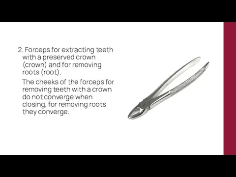
- 30. 2. Forceps for extracting teeth with a preserved crown (crown) and for removing roots (root). The
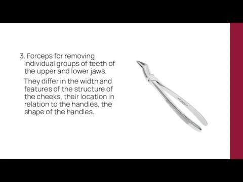
- 31. 3. Forceps for removing individual groups of teeth of the upper and lower jaws. They differ
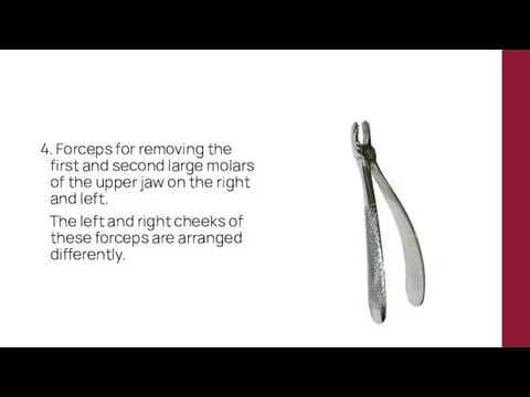
- 32. 4. Forceps for removing the first and second large molars of the upper jaw on the
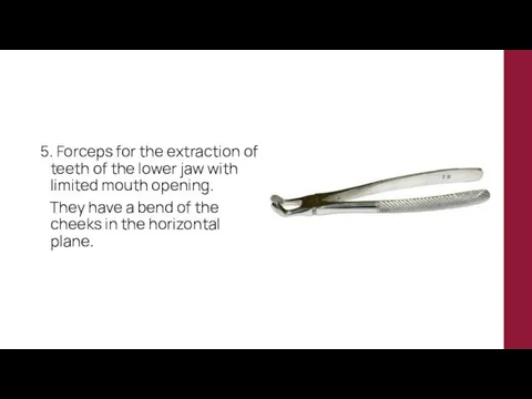
- 33. 5. Forceps for the extraction of teeth of the lower jaw with limited mouth opening. They
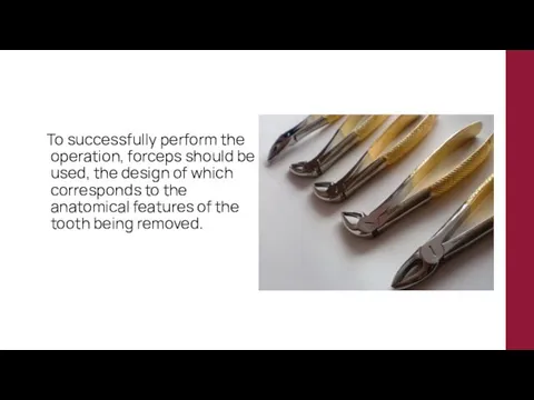
- 34. To successfully perform the operation, forceps should be used, the design of which corresponds to the
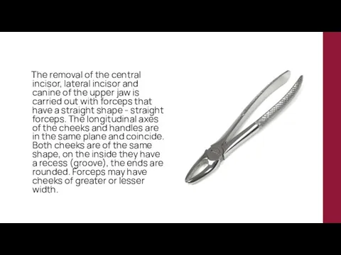
- 35. The removal of the central incisor, lateral incisor and canine of the upper jaw is carried
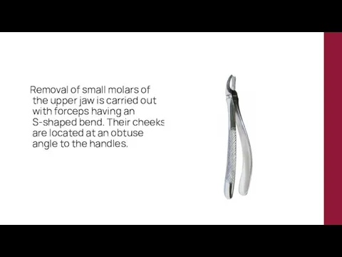
- 36. Removal of small molars of the upper jaw is carried out with forceps having an S-shaped
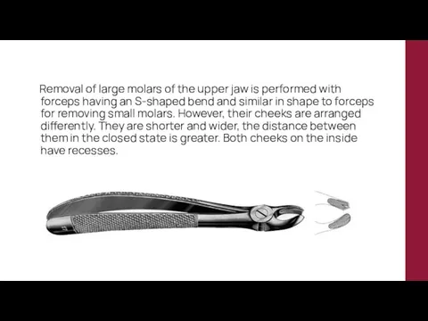
- 37. Removal of large molars of the upper jaw is performed with forceps having an S-shaped bend
- 38. At one cheek, the end is semicircular or flat, at the other it ends with a
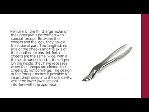
- 39. Removal of the third large molar of the upper jaw is performed with special forceps. Between
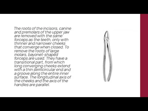
- 40. The roots of the incisors, canine and premolars of the upper jaw are removed with the
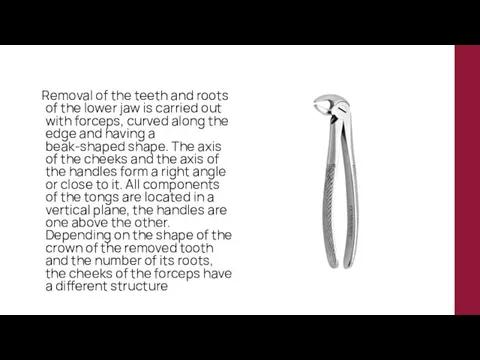
- 41. Removal of the teeth and roots of the lower jaw is carried out with forceps, curved
- 42. To remove the incisors of the lower jaw, the cheeks of the forceps are narrow with
- 43. Forceps for removing large molars have wide cheeks that do not converge when closing. Each of
- 44. The roots of all the teeth of the lower jaw are removed with forceps of the
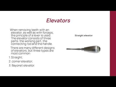
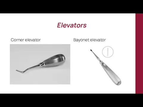
- 45. Elevators When removing teeth with an elevator, as well as with forceps, the principle of a
- 46. Elevators Corner elevator Bayonet elevator
- 47. 4. INSTRUMENTS USED IN ORTHOPEDIC DENTISTRY
- 48. Instruments used to prepare and process orthopedic consumables Spatula Metal and plastic spatulas are used for
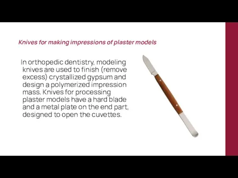
- 49. Knives for making impressions of plaster models In orthopedic dentistry, modeling knives are used to finish
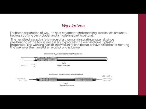
- 50. Wax knives For batch separation of wax, its heat treatment and modeling, wax knives are used,
- 51. Tools used to remove prosthetic structures Forceps To remove fixed structures fixed on the teeth of
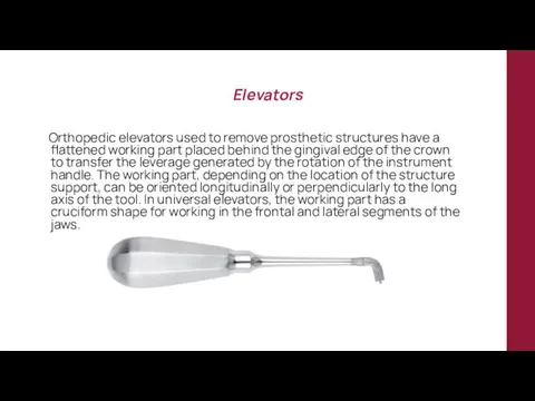
- 52. Elevators Orthopedic elevators used to remove prosthetic structures have a flattened working part placed behind the
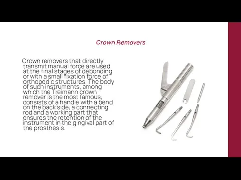
- 53. Crown Removers Crown removers that directly transmit manual force are used at the final stages of
- 56. Скачать презентацию
Lecture plan:
1. Viewing set. The main tools included in the viewing
Lecture plan:
1. Viewing set. The main tools included in the viewing
Dental instruments
are designed to ensure technological, efficient and
Dental instruments
are designed to ensure technological, efficient and
1. Viewing set. The main tools included in the viewing kit.
1. Viewing set. The main tools included in the viewing kit.
Dental mirror
Dental mirror is a round, usually one-way mirror with
Dental mirror
Dental mirror is a round, usually one-way mirror with
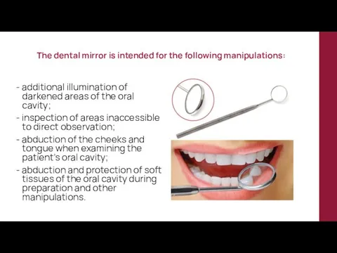
The dental mirror is intended for the following manipulations:
- additional illumination
The dental mirror is intended for the following manipulations:
- additional illumination
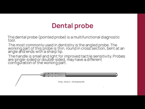
Dental probe
The dental probe (pointed probe) is a multifunctional diagnostic
Dental probe
The dental probe (pointed probe) is a multifunctional diagnostic
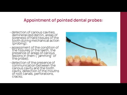
Appointment of pointed dental probes:
- detection of carious cavities, demineralized dentin,
Appointment of pointed dental probes:
- detection of carious cavities, demineralized dentin,
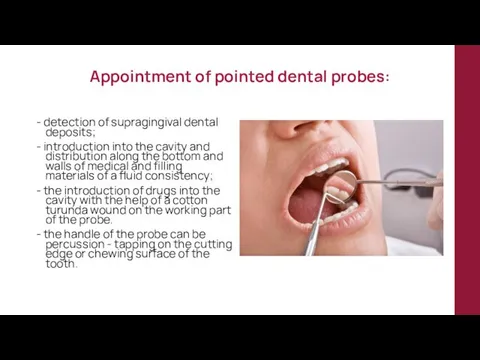
Appointment of pointed dental probes:
- detection of supragingival dental deposits;
-
Appointment of pointed dental probes:
- detection of supragingival dental deposits;
-
Dental tweezers
Dental tweezers have branches (cheeks) curved at an angle
Dental tweezers
Dental tweezers have branches (cheeks) curved at an angle
Purpose of dental tweezers:
- retention and transfer of cotton rolls, retraction
Purpose of dental tweezers:
- retention and transfer of cotton rolls, retraction
2. INSTRUMENTS USED IN THERAPEUTIC DENTISTRY
2. INSTRUMENTS USED IN THERAPEUTIC DENTISTRY
Mixing of filling materials, medical pastes and gaskets is carried
Mixing of filling materials, medical pastes and gaskets is carried
Metal spatula
A metal spatula consists of a handle, at both
Metal spatula
A metal spatula consists of a handle, at both
Plastic spatulas
Plastic spatulas are used to prepare medicinal pastes and
Plastic spatulas
Plastic spatulas are used to prepare medicinal pastes and
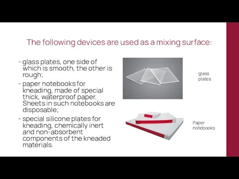
The following devices are used as a mixing surface:
- glass plates,
The following devices are used as a mixing surface:
- glass plates,
A number of requirements are imposed on the instruments used for
A number of requirements are imposed on the instruments used for
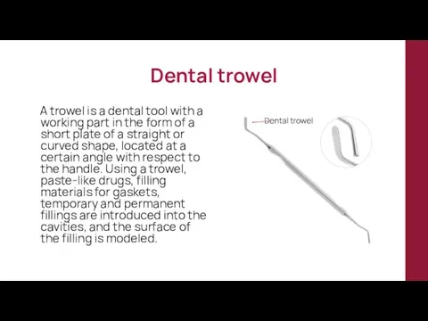
Dental trowel
A trowel is a dental tool with a working
Dental trowel
A trowel is a dental tool with a working
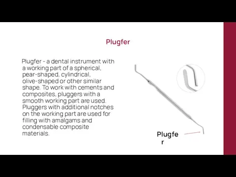
Plugfer
Plugfer - a dental instrument with a working part of
Plugfer
Plugfer - a dental instrument with a working part of
Plugfers are conditionally divided into condensing and modeling
Condensing pluggers have
Plugfers are conditionally divided into condensing and modeling
Condensing pluggers have
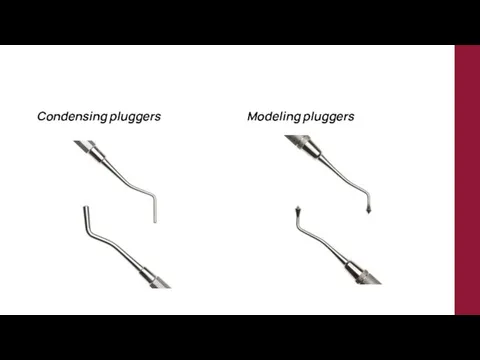
Condensing pluggers
Modeling pluggers
Condensing pluggers
Modeling pluggers
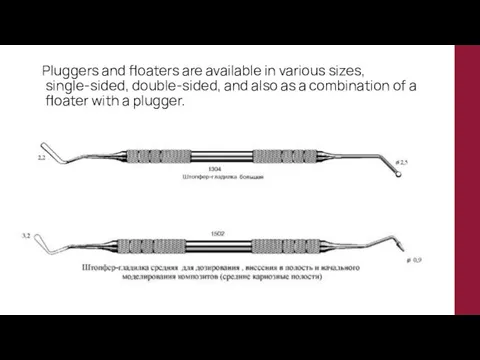
Pluggers and floaters are available in various sizes, single-sided, double-sided,
Pluggers and floaters are available in various sizes, single-sided, double-sided,
Dental excavator
It is used to remove softened dentin, food debris, temporary
Dental excavator
It is used to remove softened dentin, food debris, temporary
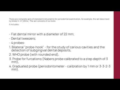
There are complete sets of standard instruments for periodontal examination, for
There are complete sets of standard instruments for periodontal examination, for
Periodontal probe
A periodontal probe is a special probe with a
Periodontal probe
A periodontal probe is a special probe with a
3. INSTRUMENTS USED IN SURGICAL DENTISTRY
3. INSTRUMENTS USED IN SURGICAL DENTISTRY
Forceps and elevators for extraction of teeth
Forceps
When removing teeth,
Forceps and elevators for extraction of teeth
Forceps
When removing teeth,
Cheeks are designed to grip the crown or root of
Cheeks are designed to grip the crown or root of
There are the following types of forceps:
1. Forceps for removing teeth
There are the following types of forceps:
1. Forceps for removing teeth
2. Forceps for extracting teeth with a preserved crown (crown) and
2. Forceps for extracting teeth with a preserved crown (crown) and
3. Forceps for removing individual groups of teeth of the upper
3. Forceps for removing individual groups of teeth of the upper
4. Forceps for removing the first and second large molars of
4. Forceps for removing the first and second large molars of
5. Forceps for the extraction of teeth of the lower jaw
5. Forceps for the extraction of teeth of the lower jaw
To successfully perform the operation, forceps should be used, the
To successfully perform the operation, forceps should be used, the
The removal of the central incisor, lateral incisor and canine
The removal of the central incisor, lateral incisor and canine
Removal of small molars of the upper jaw is carried
Removal of small molars of the upper jaw is carried
Removal of large molars of the upper jaw is performed
Removal of large molars of the upper jaw is performed
At one cheek, the end is semicircular or flat, at
At one cheek, the end is semicircular or flat, at
Removal of the third large molar of the upper jaw
Removal of the third large molar of the upper jaw
The roots of the incisors, canine and premolars of the
The roots of the incisors, canine and premolars of the
Removal of the teeth and roots of the lower jaw
Removal of the teeth and roots of the lower jaw
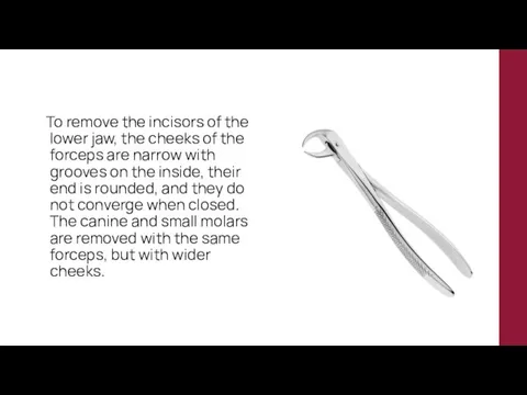
To remove the incisors of the lower jaw, the cheeks
To remove the incisors of the lower jaw, the cheeks
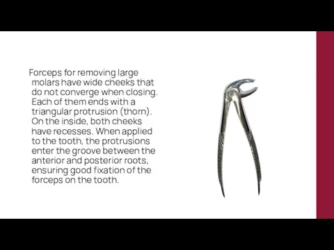
Forceps for removing large molars have wide cheeks that do
Forceps for removing large molars have wide cheeks that do
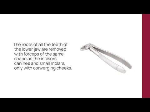
The roots of all the teeth of the lower jaw
The roots of all the teeth of the lower jaw
Elevators
When removing teeth with an elevator, as well as with
Elevators
When removing teeth with an elevator, as well as with
Elevators
Corner elevator
Bayonet elevator
Elevators
Corner elevator
Bayonet elevator
4. INSTRUMENTS USED IN ORTHOPEDIC DENTISTRY
4. INSTRUMENTS USED IN ORTHOPEDIC DENTISTRY
Instruments used to prepare and process orthopedic consumables
Spatula
Metal and plastic
Instruments used to prepare and process orthopedic consumables
Spatula
Metal and plastic
Knives for making impressions of plaster models
In orthopedic dentistry, modeling
Knives for making impressions of plaster models
In orthopedic dentistry, modeling
Wax knives
For batch separation of wax, its heat treatment and
Wax knives
For batch separation of wax, its heat treatment and
Tools used to remove prosthetic structures
Forceps
To remove fixed structures
Tools used to remove prosthetic structures
Forceps
To remove fixed structures
Elevators
Orthopedic elevators used to remove prosthetic structures have a flattened
Elevators
Orthopedic elevators used to remove prosthetic structures have a flattened
Crown Removers
Crown removers that directly transmit manual force are used
Crown Removers
Crown removers that directly transmit manual force are used
 Пузырные дерматозы
 Кардиогенный шок
 Реабилитация инвалидов. Услуги по профессиональной реабилитации инвалидов
 Сенсорные и гностические зрительные расстройства. Зрительные агнозии
 Оказание первой помощи при ушибах, растяжениях, вывихах и переломах
 Diagnosis and mangement of abnormal labour
 Симптомы заболеваний почек и мочевыводящих путей
 Средства, действующие на ЦНС
 Рак легкого: есть ли перспективы снижения смертности
 Амбулатория жағдайында эндокринді синдромдар кезіндегі рационалды дифференциалды диагностика алгоритмі
 Неэпилептические пароксизмальные расстройства сознания
 Правила личной гигиены и здоровья
 Эпилепсия. Этиологиялық факторлары
 Эндопротез тазобедренного сустава
 Влияние алкоголя на развитие организма
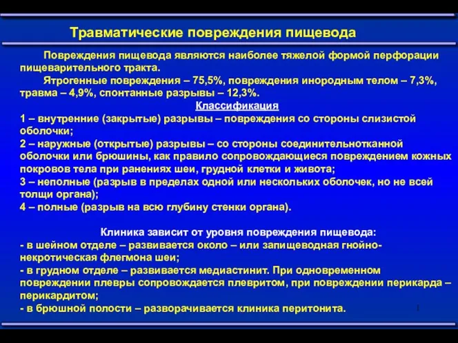 Травматические повреждения пищевода
 Балалардағы қант диабетінің алдын алу
 АКДС-вакцина
 Облачные технологии управления. Модуль скорая помощь
 Самай төменгі жақ буыны ауруларының ортопедиялық емі
 Скрининг дегеніміз не?
 Диафизарные переломы бедренной кости
 Алгоритм информационного поиска
 Forensic or legal medicine
 Психическое здоровье детей. Круглый стол в Государственной думе
 Столбняк. Симптомы
 Биохимия минерализованных тканей
 Клинический случай (амилоидоз сердца)