- Forensic or legal medicine

Содержание
- 2. FORENSIC OR LEGAL MEDICINE Forensic or legal medicine deals with the application of medical knowledge to
- 3. FORENSIC MEDICINE Forensic medicine deals almost entirely with crimes against the person, in which medical examination
- 4. FORENSIC MEDICINE Its particular field of activity is judicial investigation, both civil and criminal. In all
- 5. The medical expert should be very careful when he is examining living people. He should not
- 6. Three things are needed for success: the power of observation, 2) a wide range of exact
- 7. Forensic medicine is not an exact science. Unexpected results are produced due to biological variations. In
- 8. The doctor should be ready to defend every finding and conclusion on the report on clinical
- 9. For the purpose of illustrating and clarifying his testimony, the medical expert may employ photographs, maps,
- 10. Forensic-medical examination is performed only when there is a written resolution or direction from the investigative
- 11. Objects of forensic-medical examination: 3. Material evidences. 4. Materials of crime cases. FORENSIC-MEDICINE EXAMINATION
- 12. Examination or research of these objects is produced in Bureau of forensic-medical examination or Institute of
- 13. INVESTIGATION OF THE SCENE OF DEATH The first research of dead body an expert conducts on
- 14. INVESTIGATION OF THE SCENE OF DEATH Medico-legal Masquerades: Many cases of homicide go undetected because of
- 15. INVESTIGATION OF THE SCENE OF DEATH Accidental deaths and suicides can occur under circumstances which suggest
- 16. INVESTIGATION OF THE SCENE OF DEATH The doctor must look for any possible inconsistencies between the
- 17. INVESTIGATION OF THE SCENE OF DEATH In such cases, the real cause of death can be
- 18. INVESTIGATION OF THE SCENE OF DEATH The answers to the following questions have to be found:
- 19. INVESTIGATION OF THE SCENE OF DEATH The answers to the following questions have to be found:
- 20. CONDUCT AND DUTIES OF THE DOCTOR AT THE SCENE OF CRIME Complete and accurate recording of
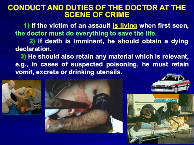
- 21. CONDUCT AND DUTIES OF THE DOCTOR AT THE SCENE OF CRIME 1) If the victim of
- 22. CONDUCT AND DUTIES OF THE DOCTOR AT THE SCENE OF CRIME 4) He must make sure
- 23. CONDUCT AND DUTIES OF THE DOCTOR AT THE SCENE OF CRIME 9) He should ask the
- 24. CONDUCT AND DUTIES OF THE DOCTOR AT THE SCENE OF CRIME c) A list of all
- 25. CONDUCT AND DUTIES OF THE DOCTOR AT THE SCENE OF CRIME f) Make a sketch noting
- 26. CONDUCT AND DUTIES OF THE DOCTOR AT THE SCENE OF CRIME Note the amount of bleeding
- 27. CONDUCT AND DUTIES OF THE DOCTOR AT THE SCENE OF CRIME h) Free hair, fibers or
- 28. CONDUCT AND DUTIES OF THE DOCTOR AT THE SCENE OF CRIME k) The objects on premises,
- 29. CONDUCT AND DUTIES OF THE DOCTOR AT THE SCENE OF CRIME m) If a weapon is
- 30. MEDICO-LEGAL AUTOPSY Objects: To find out the cause of death, whether unviolent or violent. 2) To
- 31. MEDICO-LEGAL AUTOPSY Rules for Medico-legal Autopsies: The autopsy should be conducted in a mortuary and never
- 32. MEDICO-LEGAL AUTOPSY Reasons of Forensic Autopsy Violent death or marks of suspicion of it. Sudden death
- 33. MEDICO-LEGAL AUTOPSY The approach of the forensic pathologist to the investigation of death is different from
- 34. MEDICO-LEGAL AUTOPSY Forensic pathologist has to determine time of death and age of injuries. He has
- 35. DOCUMENTS OF FORENSIC-MEDICAL EXAMINATION In all cases of forensic-medical examination is made: Conclusion of forensic-medical examination
- 36. Thanatology Thanatos is Greek god of death Thanatology deals with death in all its aspects. Three
- 37. Thanatology POSTMORTEM CHANGES Signs of death appear in the following order: 1.Immediate (somatic death): a. Insensibility
- 38. Thanatology POSTMORTEM CHANGES 2.Early (cellular death): a. Pallor and loss of elasticity of skin b. Changes
- 39. Thanatology POSTMORTEM CHANGES 3. Late (decomposition and decay): a. Putrefaction b. Adipocere formation c. Mummification
- 40. Thanatology POSTMORTEM CHANGES LIVOR MORTIS (Postmortem Staining) Synonyms of postmortem staining are cadaveric or postmortem lividity,
- 41. Thanatology POSTMORTEM CHANGES LIVOR MORTIS (Postmortem Staining) - It is usually well developed within 4 hours
- 42. Thanatology POSTMORTEM CHANGES LIVOR MORTIS (Postmortem Staining) Location of Postmortem hypostasis In case of Hanging: Hypostasis
- 43. Thanatology POSTMORTEM CHANGES LIVOR MORTIS (Postmortem Staining) Location of Postmortem hypostasis Distribution of Livores mortis depends
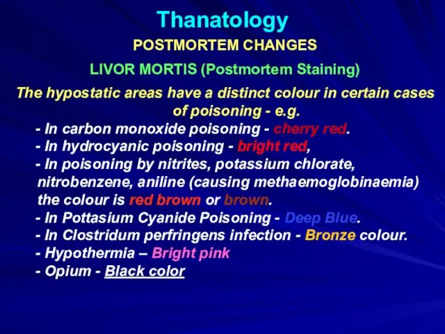
- 44. Thanatology POSTMORTEM CHANGES LIVOR MORTIS (Postmortem Staining) The hypostatic areas have a distinct colour in certain
- 45. Thanatology POSTMORTEM CHANGES: LIVOR MORTIS
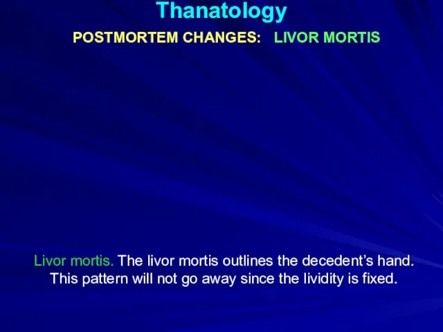
- 46. Thanatology POSTMORTEM CHANGES: LIVOR MORTIS Livor mortis. The livor mortis outlines the decedent’s hand. This pattern
- 47. Thanatology POSTMORTEM CHANGES: LIVOR MORTIS Occasionally, livor mortis may appear as an unusual pattern or look
- 48. Thanatology POSTMORTEM CHANGES: Rigor Mortis Definition This is stage of stiffening of muscles with shortening of
- 49. Thanatology POSTMORTEM CHANGES: Rigor Mortis The Order of Appearance of Rigor Mortis - All muscles of
- 50. Thanatology POSTMORTEM CHANGES: Rigor Mortis Time of Onset In India, it begins 1 to 2 hours
- 51. Thanatology POSTMORTEM CHANGES: Rigor Mortis This man was found in this position the day after he
- 52. Thanatology POSTMORTEM CHANGES: Rigor Mortis The man’s knee remains bent after he is moved because the
- 53. Thanatology POSTMORTEM CHANGES: Putrefaction Putrefaction is the final stage following death, produced mainly by the action
- 54. Thanatology POSTMORTEM CHANGES: Putrefaction The first external sign of putrefaction in a body lying in air
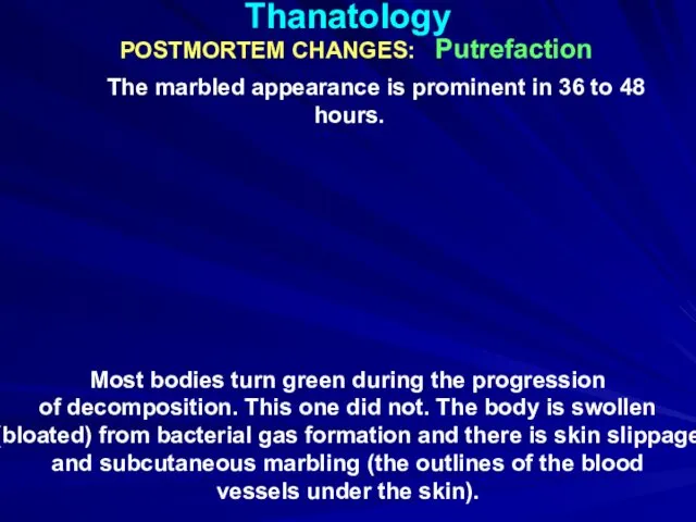
- 55. Thanatology POSTMORTEM CHANGES: Putrefaction The marbled appearance is prominent in 36 to 48 hours. Most bodies
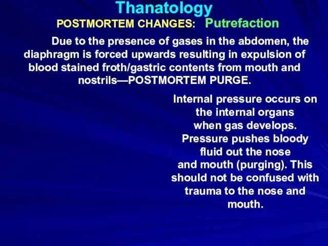
- 56. Thanatology POSTMORTEM CHANGES: Putrefaction Due to the presence of gases in the abdomen, the diaphragm is
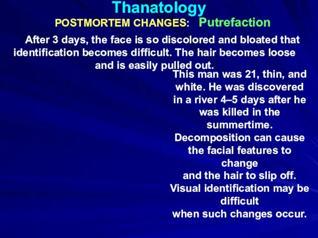
- 57. Thanatology POSTMORTEM CHANGES: Putrefaction After 3 days, the face is so discolored and bloated that identification
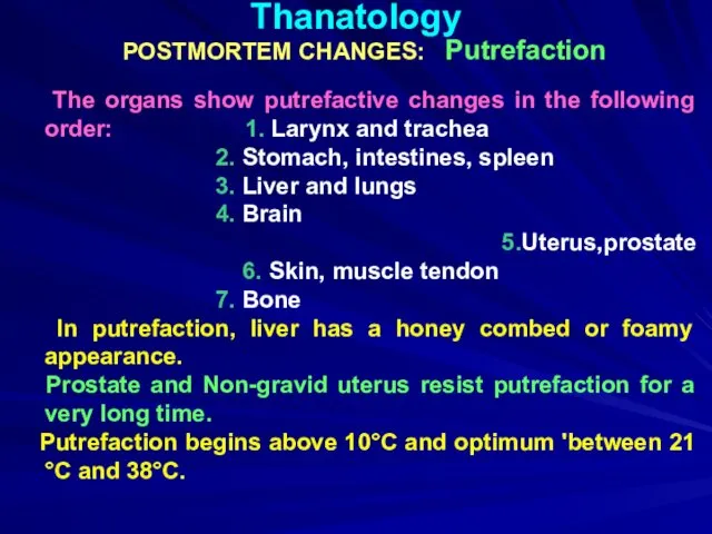
- 58. Thanatology POSTMORTEM CHANGES: Putrefaction The organs show putrefactive changes in the following order: 1. Larynx and
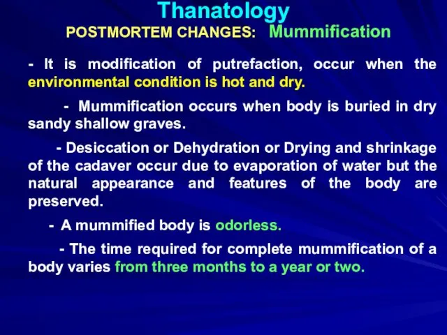
- 59. Thanatology POSTMORTEM CHANGES: Mummification - It is modification of putrefaction, occur when the environmental condition is
- 60. Thanatology POSTMORTEM CHANGES: Mummification The skin dries out and turns leathery. This man’s head mummified within
- 62. Скачать презентацию
FORENSIC OR LEGAL MEDICINE
Forensic or legal medicine deals with the application
FORENSIC OR LEGAL MEDICINE
Forensic or legal medicine deals with the application

FORENSIC MEDICINE
Forensic medicine deals almost entirely with crimes against the person,
FORENSIC MEDICINE
Forensic medicine deals almost entirely with crimes against the person,
FORENSIC MEDICINE
Its particular field of activity is judicial investigation, both civil
FORENSIC MEDICINE
Its particular field of activity is judicial investigation, both civil
The medical expert should be very careful when he is examining
The medical expert should be very careful when he is examining
Three things are needed for success:
the power of observation,
2)
Three things are needed for success:
the power of observation,
2)
Forensic medicine is not an exact science. Unexpected results are produced
Forensic medicine is not an exact science. Unexpected results are produced

The doctor should be ready to defend every finding and conclusion
The doctor should be ready to defend every finding and conclusion
For the purpose of illustrating and clarifying his testimony, the medical
For the purpose of illustrating and clarifying his testimony, the medical
Forensic-medical examination is performed only when there is a written resolution
Forensic-medical examination is performed only when there is a written resolution
Objects of forensic-medical examination:
3. Material evidences.
4. Materials of crime cases.
FORENSIC-MEDICINE
Objects of forensic-medical examination:
3. Material evidences.
4. Materials of crime cases.
FORENSIC-MEDICINE
Examination or research of these objects is produced in Bureau of
Examination or research of these objects is produced in Bureau of
INVESTIGATION OF THE SCENE OF DEATH
The first research of dead
INVESTIGATION OF THE SCENE OF DEATH
The first research of dead
INVESTIGATION OF THE SCENE OF DEATH
Medico-legal Masquerades:
Many cases of
INVESTIGATION OF THE SCENE OF DEATH
Medico-legal Masquerades:
Many cases of
INVESTIGATION OF THE SCENE OF DEATH
Accidental deaths and suicides can
INVESTIGATION OF THE SCENE OF DEATH
Accidental deaths and suicides can
INVESTIGATION OF THE SCENE OF DEATH
The doctor must look for
INVESTIGATION OF THE SCENE OF DEATH
The doctor must look for
INVESTIGATION OF THE SCENE OF DEATH
In such cases, the real
INVESTIGATION OF THE SCENE OF DEATH
In such cases, the real
INVESTIGATION OF THE SCENE OF DEATH
The answers to the following
INVESTIGATION OF THE SCENE OF DEATH
The answers to the following
INVESTIGATION OF THE SCENE OF DEATH
The answers to the following
INVESTIGATION OF THE SCENE OF DEATH
The answers to the following

CONDUCT AND DUTIES OF THE DOCTOR AT THE SCENE OF CRIME
Complete
CONDUCT AND DUTIES OF THE DOCTOR AT THE SCENE OF CRIME
Complete
CONDUCT AND DUTIES OF THE DOCTOR AT THE SCENE OF CRIME
CONDUCT AND DUTIES OF THE DOCTOR AT THE SCENE OF CRIME
CONDUCT AND DUTIES OF THE DOCTOR AT THE SCENE OF CRIME
4)
CONDUCT AND DUTIES OF THE DOCTOR AT THE SCENE OF CRIME
4)
CONDUCT AND DUTIES OF THE DOCTOR AT THE SCENE OF CRIME
9)
CONDUCT AND DUTIES OF THE DOCTOR AT THE SCENE OF CRIME
9)
CONDUCT AND DUTIES OF THE DOCTOR AT THE SCENE OF CRIME
CONDUCT AND DUTIES OF THE DOCTOR AT THE SCENE OF CRIME
CONDUCT AND DUTIES OF THE DOCTOR AT THE SCENE OF CRIME
CONDUCT AND DUTIES OF THE DOCTOR AT THE SCENE OF CRIME
CONDUCT AND DUTIES OF THE DOCTOR AT THE SCENE OF CRIME
Note
CONDUCT AND DUTIES OF THE DOCTOR AT THE SCENE OF CRIME
Note
CONDUCT AND DUTIES OF THE DOCTOR AT THE SCENE OF CRIME
CONDUCT AND DUTIES OF THE DOCTOR AT THE SCENE OF CRIME
CONDUCT AND DUTIES OF THE DOCTOR AT THE SCENE OF CRIME
CONDUCT AND DUTIES OF THE DOCTOR AT THE SCENE OF CRIME
CONDUCT AND DUTIES OF THE DOCTOR AT THE SCENE OF CRIME
CONDUCT AND DUTIES OF THE DOCTOR AT THE SCENE OF CRIME
MEDICO-LEGAL AUTOPSY
Objects:
To find out the cause of death,
MEDICO-LEGAL AUTOPSY
Objects:
To find out the cause of death,

MEDICO-LEGAL AUTOPSY
Rules for Medico-legal Autopsies:
The autopsy should be
MEDICO-LEGAL AUTOPSY
Rules for Medico-legal Autopsies:
The autopsy should be
MEDICO-LEGAL AUTOPSY
Reasons of Forensic Autopsy
Violent death or marks of
MEDICO-LEGAL AUTOPSY
Reasons of Forensic Autopsy
Violent death or marks of

MEDICO-LEGAL AUTOPSY
The approach of the forensic pathologist to the investigation
MEDICO-LEGAL AUTOPSY
The approach of the forensic pathologist to the investigation
MEDICO-LEGAL AUTOPSY
Forensic pathologist has to determine time of death and
MEDICO-LEGAL AUTOPSY
Forensic pathologist has to determine time of death and
DOCUMENTS OF FORENSIC-MEDICAL EXAMINATION
In all cases of forensic-medical examination is
DOCUMENTS OF FORENSIC-MEDICAL EXAMINATION
In all cases of forensic-medical examination is
Thanatology
Thanatos is Greek god of death
Thanatology deals with death in
Thanatology
Thanatos is Greek god of death
Thanatology deals with death in
Thanatology
POSTMORTEM CHANGES
Signs of death appear in the following order:
1.Immediate
Thanatology
POSTMORTEM CHANGES
Signs of death appear in the following order:
1.Immediate
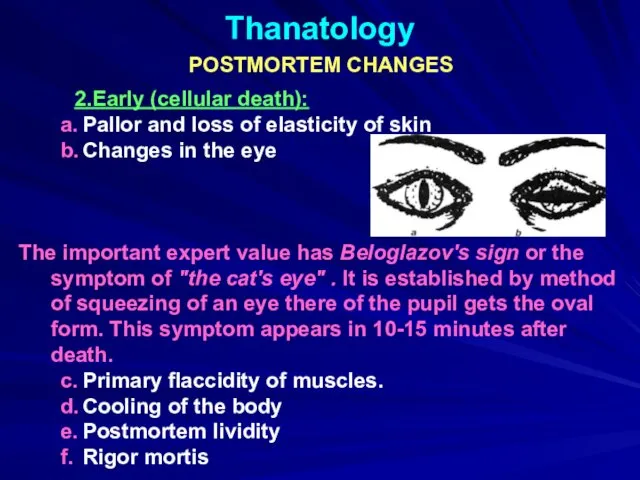
Thanatology
POSTMORTEM CHANGES
2.Early (cellular death):
a. Pallor and loss of elasticity
Thanatology
POSTMORTEM CHANGES
2.Early (cellular death):
a. Pallor and loss of elasticity
Thanatology
POSTMORTEM CHANGES
3. Late (decomposition and decay):
a. Putrefaction
b. Adipocere formation
Thanatology
POSTMORTEM CHANGES
3. Late (decomposition and decay):
a. Putrefaction
b. Adipocere formation
Thanatology
POSTMORTEM CHANGES
LIVOR MORTIS (Postmortem Staining)
Synonyms of postmortem staining are
Thanatology
POSTMORTEM CHANGES
LIVOR MORTIS (Postmortem Staining)
Synonyms of postmortem staining are
Thanatology
POSTMORTEM CHANGES
LIVOR MORTIS (Postmortem Staining)
- It is usually well
Thanatology
POSTMORTEM CHANGES
LIVOR MORTIS (Postmortem Staining)
- It is usually well
Thanatology
POSTMORTEM CHANGES
LIVOR MORTIS (Postmortem Staining)
Location of Postmortem hypostasis
In
Thanatology
POSTMORTEM CHANGES
LIVOR MORTIS (Postmortem Staining)
Location of Postmortem hypostasis
In
Thanatology
POSTMORTEM CHANGES
LIVOR MORTIS (Postmortem Staining)
Location of Postmortem hypostasis
Distribution of
Thanatology
POSTMORTEM CHANGES
LIVOR MORTIS (Postmortem Staining)
Location of Postmortem hypostasis
Distribution of
Thanatology
POSTMORTEM CHANGES
LIVOR MORTIS (Postmortem Staining)
The hypostatic areas have a
Thanatology
POSTMORTEM CHANGES
LIVOR MORTIS (Postmortem Staining)
The hypostatic areas have a
Thanatology
POSTMORTEM CHANGES: LIVOR MORTIS
Thanatology
POSTMORTEM CHANGES: LIVOR MORTIS
Thanatology
POSTMORTEM CHANGES: LIVOR MORTIS
Livor mortis. The livor mortis outlines the
Thanatology
POSTMORTEM CHANGES: LIVOR MORTIS
Livor mortis. The livor mortis outlines the
Thanatology
POSTMORTEM CHANGES: LIVOR MORTIS
Occasionally, livor mortis may appear as
Thanatology
POSTMORTEM CHANGES: LIVOR MORTIS
Occasionally, livor mortis may appear as
Thanatology
POSTMORTEM CHANGES: Rigor Mortis
Definition
This is stage of stiffening of
Thanatology
POSTMORTEM CHANGES: Rigor Mortis
Definition
This is stage of stiffening of
Thanatology
POSTMORTEM CHANGES: Rigor Mortis
The Order of Appearance of Rigor
Thanatology
POSTMORTEM CHANGES: Rigor Mortis
The Order of Appearance of Rigor
Thanatology
POSTMORTEM CHANGES: Rigor Mortis
Time of Onset
In India, it begins
Thanatology
POSTMORTEM CHANGES: Rigor Mortis
Time of Onset
In India, it begins
Thanatology
POSTMORTEM CHANGES: Rigor Mortis
This man was found in
Thanatology
POSTMORTEM CHANGES: Rigor Mortis
This man was found in
Thanatology
POSTMORTEM CHANGES: Rigor Mortis
The man’s knee remains bent
Thanatology
POSTMORTEM CHANGES: Rigor Mortis
The man’s knee remains bent
Thanatology
POSTMORTEM CHANGES: Putrefaction
Putrefaction is the final stage following death,
Thanatology
POSTMORTEM CHANGES: Putrefaction
Putrefaction is the final stage following death,
Thanatology
POSTMORTEM CHANGES: Putrefaction
The first external sign of putrefaction
in a
Thanatology
POSTMORTEM CHANGES: Putrefaction
The first external sign of putrefaction
in a
Thanatology
POSTMORTEM CHANGES: Putrefaction
The marbled appearance is prominent in 36
Thanatology
POSTMORTEM CHANGES: Putrefaction
The marbled appearance is prominent in 36
Thanatology
POSTMORTEM CHANGES: Putrefaction
Due to the presence of gases in
Thanatology
POSTMORTEM CHANGES: Putrefaction
Due to the presence of gases in
Thanatology
POSTMORTEM CHANGES: Putrefaction
After 3 days, the face is so discolored
Thanatology
POSTMORTEM CHANGES: Putrefaction
After 3 days, the face is so discolored
Thanatology
POSTMORTEM CHANGES: Putrefaction
The organs show putrefactive changes in the
Thanatology
POSTMORTEM CHANGES: Putrefaction
The organs show putrefactive changes in the
Thanatology
POSTMORTEM CHANGES: Mummification
- It is modification of putrefaction, occur when
Thanatology
POSTMORTEM CHANGES: Mummification
- It is modification of putrefaction, occur when

Thanatology
POSTMORTEM CHANGES: Mummification
The skin dries out and turns
leathery. This man’s
Thanatology
POSTMORTEM CHANGES: Mummification
The skin dries out and turns
leathery. This man’s
 Основы рационального питания детей дошкольного возраста
 Қазақстан Республикасының Президентінің жыл сайынғы халқына жолдауындағы денсаулық тақырыбы
 Невідкладна медична допомога постраждалим
 Особенности оказания помощи при экстремальных воздействиях. Медицина катастроф. Тема 3
 Гнойная инфекция кисти (панариций, флегмона)
 Особенности косметических средств и процедуры салонного ухода при мелкоморщинистом типе старения
 Синдром Картагенера
 Вирус папилломы человека
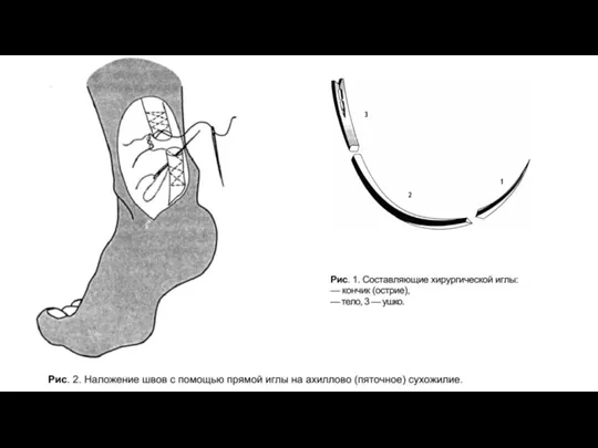 Иглы и шовные материалы
 Врожденные аномалии ЖКТ
 Общественное здоровье-высшая ценность человечества
 Предраковые заболевания челюстно-лицевой области
 Мүгедектік
 Головные боли. Мигрень
 Травмы глаза
 Балаларда Helicobacter pylori – инфекциясымен шақырылған асқорыту жолының жоғары бөлігін емдеу схемасы
 Depression and the consumption of alcohol and tobacco among youth, cross-sectional analysis
 Фізична реабілітація для дітей з особливими потребами
 Педиатрия, как наука о здоровом и больном ребенке
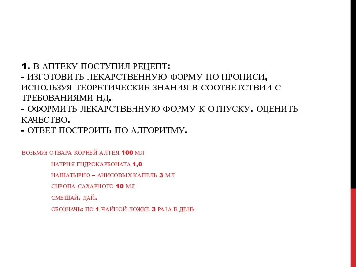 Изготовление лекарственной формы по прописи, используя теоретические знания в соответствии с требованиями нд
 Средства, влияющие на систему крови
 Neuro-oncology
 Belotero. Презентация-тренинг
 СПИД-ассоцированные инфекции. Хронические неинфекционные заболевания при ВИЧ-инфекции
 Бет-жақ ауытқулары бар балаларды диспансерлік бақылау
 Анатомо-физиологические особенности формирования потребностей человека. (Лекция 1)
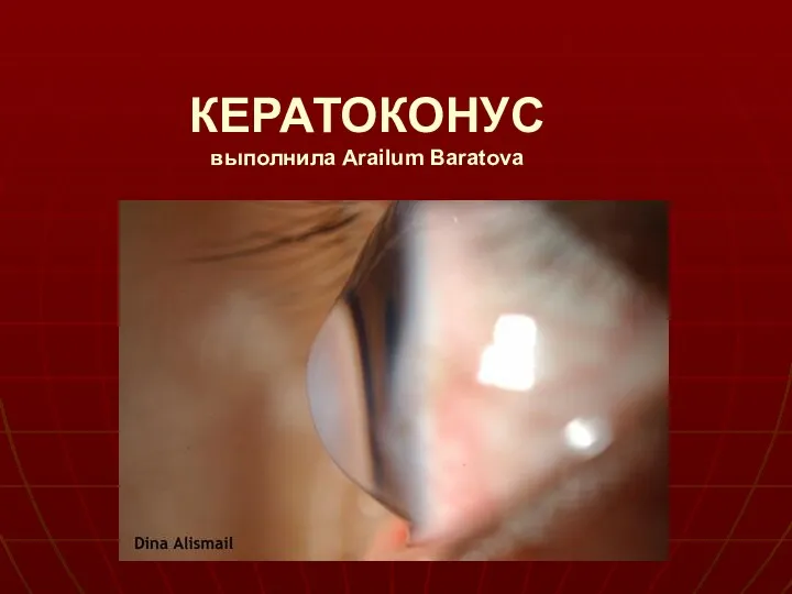 Кератоконус. Классификация, симптомы
 Нәрестелердің физиологиялық сары аурулары