Слайд 2Malignant Tumors
In the United States, the vast majority of tumors involving the liver
are metastatic.
Hepatocellular carcinoma is the most common primary liver cancer;
cholangiocarcinomas are much less common.
Angiosarcomas of the liver resemble those occurring elsewhere. Interestingly, liver angiosarcomas can be associated with exposure to vinyl chloride, arsenic
Слайд 3Hepatoblastoma
the most common liver tumor of early childhood.
hepatoblastomas are also associated
with familial polyposis syndrome
• The epithelial type vaguely recapitulates liver development.
• The mixed epithelial and mesenchymal type contains foci of mesenchymal differentiation including osteoid, cartilage, or striated muscle.
Hepatoblastomas are usually fatal if untreated, but resection and chemotherapy yield 80% 5-year survival rates.
Слайд 4Hepatocellular Carcinoma
(HCC) occurs most commonly in developing countries with high rates of HBV
infection;
it is the third most common cause of cancer deaths worldwide.
The male to female ratio is 2.4:1.
Слайд 5Pathogenesis
HCC usually arises in the background of chronic liver disease.
The major
etiologic factors are
chronic viral infection (HBV or HCV),
chronic alcoholism,
food contaminants (e.g., aflatoxins);
lesser causes include hemochromatosis, tyrosinemia, and a1-antitrypsin deficiency.
Слайд 6the high co-incidence of HCC with HBV and HCV infections suggests that viral
factors can also contribute:
Morphology
HCC can present as a solitary mass, as multifocal nodules, or
as a diffusely infiltrative cancer with massive liver enlargement,
frequently in a background of cirrhosis; intrahepatic spread and vascular invasion are common.
Слайд 7Clinical Features
include hepatomegaly,
right upper quadrant pain,
weight loss, and
elevated serum a-fetoprotein.
Prognosis depends on the resectability of the tumor;
mortality is secondary to
cachexia,
gastrointestinal or esophageal variceal bleeding, liver failure with hepatic coma, or
tumor rupture and fatal hemorrhage.
Слайд 8Cholangiocarcinoma
(CCA) arises from elements of the intra- and extrahepatic biliary tree; 50% to
60% are perihilar (called Klatskin
tumors), 20% to 30% are distal, and 10% are intrahepatic. CCA accounts for 3% of cancer deaths in the United States and 7.6% of cancer deaths worldwide. Clinical outlook is dismal because CCA is rarely resectable at diagnosis.
Morphology CCA can manifest as a single large mass or as multifocal
nodules, or it can be diffusely infiltrative.
In contrast to HCC, CAA is typically pale since biliary epithelium does not secrete bilirubin pigment.
Слайд 9Metastatic Tumors
Any cancer in the body—including those of the blood-forming elements—can spread to
the liver;
colon, breast, lung, and pancreas primaries are most common.
Typically, multiple implants are present, with massive hepatic enlargement.
Large implants tend to have defective vascular supplies and become centrally necrotic.
Massive involvement of the liver is usually present before hepatic failure develops.
Слайд 11SUMMARY
Viral Hepatitis
• In the alphabet of hepatotropic viruses, some easy mnemonic devices may
be useful:
The vowels (hepatitis A and E) never cause chronic hepatitis, only acute hepatitis . ) Fecal-oral(
Only the consonants (hepatitis B, C, D) have the potential to cause chronic disease (C for consonant and for chronic). ) Parenteral(
Hepatitis C is the single virus that is more often chronic than not (almost never detected acutely; 85% or more of patients develop chronic hepatitis, 20% of whom will develop cirrhosis).
Hepatitis D, the delta agent, is a defective virus, requiring hepatitis B coinfection for its own capacity to infect and replicate.
Hepatitis E is endemic in equatorial regions and frequently epidemic.
• The inflammatory cells in both acute and chronic viral hepatitis are mainly T cells; it is the pattern of injury that is different, not the nature of the infiltrate.
• Biopsy assessment in chronic viral hepatitis is most important for grading and staging of disease, which are used to decide whether a patient undergoes often arduous antiviral treatments.
• Patients with long-standing HBV or HCV infections are at increased risk for the development of hepatocellular carcinomas, even in the absence of established cirrhosis.
Слайд 12Incubation period
A 2–6 weeks
B 4–26 weeks
C 2–26 weeks
D Same
as for HBV
E 2–8 weeks
Слайд 13Carcinoma of the Gallbladder
is slightly more common in women,
typically occurring in women older
than 70 years.
Gallstones coexist in 95% of U.S. patients;
chronic gallbladder inflammation (with or without stones) is a critical risk factor.
Gallstones are less common in Asian populations, where pyogenic and parasitic disease
dominate as causes.
Morphology
Tumors may be infiltrating, with diffuse gallbladder thickening and
induration, or they may be
exophytic—growing into the lumen as an irregular, cauliflower-like mass.
Most gallbladder carcinomas are adenocarcinomas;
Слайд 14Tumors spread by
local invasion of the liver,
extension to cystic duct and portohepatic
lymph nodes, and
metastatic seeding of peritoneum, viscera, and lungs.
Clinical Features
Symptoms are insidious and indistinguishable from those caused by cholelithiasis.
Tumors are usually unresectable when discovered.
Слайд 15Neoplasms of the pancreas
are broadly grouped as cystic or solid.
Cystic Neoplasms
Constitute less
than 5% of pancreatic neoplasms;
they typically occur as painless, slow-growing masses.
• Serous cystadenoma:
These are almost always benign and resection is curative.
• Mucinous cystic neoplasm: One third of these lesions harbor an invasive adenocarcinoma.
Слайд 16Pancreatic Carcinoma
It is an infiltrating ductal adenocarcinoma;
it is the fourth leading cause
of cancer deaths in the USA
Precursors to Pancreatic Cancer
There is a progression from non-neoplastic epithelium to small ductal non-invasive lesions, to invasive carcinoma.
The precursor lesions are called pancreatic intraepithelial neoplasms
Слайд 17Pathogenesis
About 80% of cases occur in individuals between the ages of 60 and
80 years;
smoking increases the risk roughly two-fold.
Chronic pancreatitis, consumption of a diet rich in fats, a
family history of pancreatic cancer, and diabetes mellitus impose a modestly increased risk.
Слайд 18Morphology
60% of pancreatic cancers arise in the head of the gland,
15% occur in
the body, 5% occur in the tail, and 20% diffusely involve the organ.
These are typically highly invasive and elicit an intense host scarring response (desmoplasia).
Most carcinomas in the head of the pancreas obstruct the distal common bile duct, leading to jaundice;
conversely, cancers of the body and tail can remain clinically silent for long periods of time and are often large or widely metastatic when initially discovered.
Extensive perineural and vascular invasion are common.
Слайд 19Clinical Features
Weight loss and pain are typical presenting symptoms;
obstructive jaundice develops with
tumors in the head of the gland.
Metastases are common, and
more than 80% of adenocarcinomas are unresectable at presentation; massive liver metastasis frequently develops.
The outlook is dismal: first-year mortality rate exceeds
80% and the 5-year survival rate is less than 5%. Migratory thrombophlebitis can occur with pancreatic neoplasms

 Бронхиальді тал құрылысы
 ЛФК для беременных
 Сепсис
 Ультраструктура десны, десневой борозды, десневой жидкости
 Геморрагический инсульт
 Муталлапова 301М Орг. мед. пом. жен. и детям
 Первичный туберкулезный комплекс
 Принципы диагностики гельминтозов
 Телескопические съемные протезы
 Половое воспитание. Инфекции, передаваемые половым путём. 9 класс
 Три функциональных блока мозга
 Біль у бігунів, шляхи діагностики, відновлення та профілактика
 Питание различных групп взрослого населения
 Заболевания кишечника
 Миодистрофия Дюшенна-Беккера
 ОРЗ, грипп: клиника, диагностика, временная нетрудоспособность
 Физиологические основы психических функций человека
 Эссенциальді артериалды гипертензиясы бар науқастарда каптоприл мен нифедипиннің емдік және эффективтілік әсерін салыстыру
 Еріту. Сұйықтықтарды араластыру. Қатты заттарды және сұйықтықтарды бөліп алу
 Тағамдық аллергия мен тағаммен улану кезіндегі емдік тамақтану
 Геннің құрылысы. ДНҚ-ның кодтаушы және реттеуші жиіліктері
 Микобактерии – возбудители микобактериозов, туберкулеза и лепры
 Дезинфекция медицинских перчаток
 Colegiul de Medicină Ungheni Studiu individual la igiena generală
 Режимы лечебно-профилактических учреждений (ЛПУ)
 Рентгенодиагностика пневмоний
 Ожирение – как фактор риска эндокринных и сердечно - сосудистых заболеваний
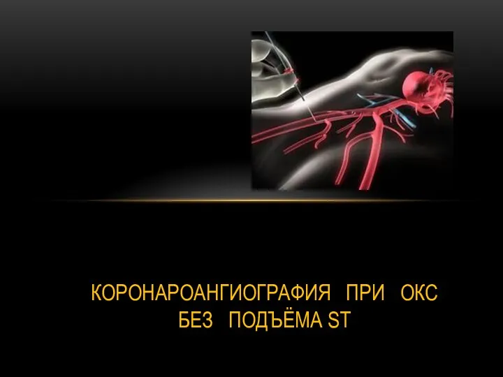 Коронароангиография при ОКС без подъёма ST