- Cardiovascular system

Содержание
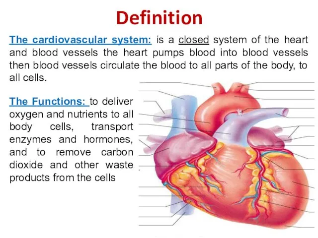
- 2. Definition The cardiovascular system: is a closed system of the heart and blood vessels the heart
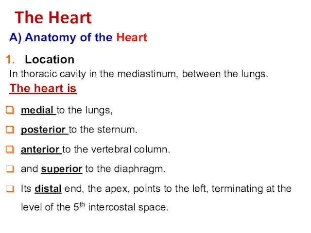
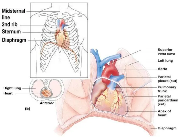
- 3. A) Anatomy of the Heart Location In thoracic cavity in the mediastinum, between the lungs. The
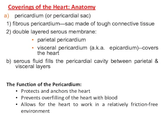
- 5. Coverings of the Heart: Anatomy The Function of the Pericardium: Protects and anchors the heart Prevents
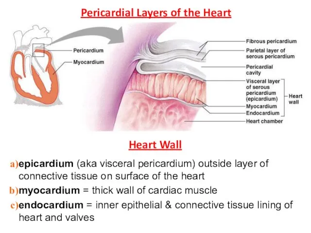
- 6. Pericardial Layers of the Heart Heart Wall epicardium (aka visceral pericardium) outside layer of connective tissue
- 7. Chambers of the heart (4) atrium (R & L)—receive blood each atria extends into a smaller,
- 8. External Heart: Major Vessels of the Heart Vessels returning blood to the heart include: Superior and
- 9. External Heart: Anterior View
- 10. External Heart: Posterior View
- 11. Atria of the Heart Atria are the receiving chambers of the heart Pectinate muscles mark atrial
- 12. Gross Anatomy of Heart: Frontal Section
- 13. Thickness of myocardium varies according to the function of the chamber Atria are thin walled, deliver
- 14. Pathway of Blood Through the Heart and Lungs Right atrium ? ? tricuspid valve ? ?
- 15. Pulmonary circulation The flow of blood between the heart and lungs. Systemic circulation The flow of
- 16. Coronary Circulation Coronary circulation is the functional blood supply to the heart muscle itself Collateral routes
- 17. Coronary Circulation Veins
- 18. Heart Valves Heart valves ensure uni-directional blood flow through the heart Atrioventricular (AV) valves lie between
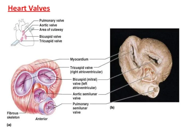
- 19. Heart Valves
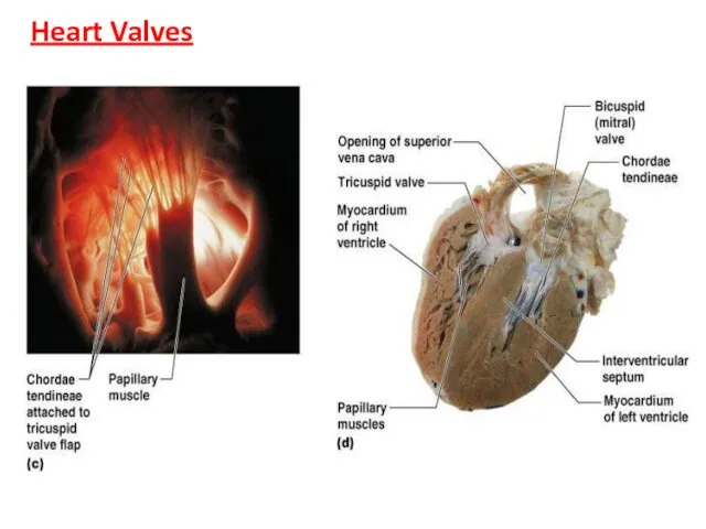
- 20. Heart Valves
- 21. Atrio-ventricular Valve Function (b)
- 22. Semilunar Valve Function
- 24. Скачать презентацию
Definition
The cardiovascular system: is a closed system of the heart and
Definition
The cardiovascular system: is a closed system of the heart and
A) Anatomy of the Heart
Location
In thoracic cavity in the mediastinum, between
A) Anatomy of the Heart
Location
In thoracic cavity in the mediastinum, between
Coverings of the Heart: Anatomy
The Function of the Pericardium:
Protects and anchors
Coverings of the Heart: Anatomy
The Function of the Pericardium:
Protects and anchors
Pericardial Layers of the Heart
Heart Wall
epicardium (aka visceral pericardium) outside layer
Pericardial Layers of the Heart
Heart Wall
epicardium (aka visceral pericardium) outside layer
Chambers of the heart (4)
atrium (R & L)—receive blood
each atria extends
Chambers of the heart (4)
atrium (R & L)—receive blood
each atria extends

External Heart: Major Vessels of the Heart
Vessels returning blood to the
External Heart: Major Vessels of the Heart
Vessels returning blood to the
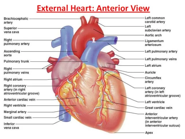
External Heart: Anterior View
External Heart: Anterior View
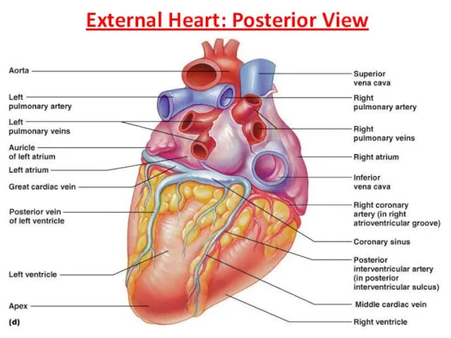
External Heart: Posterior View
External Heart: Posterior View
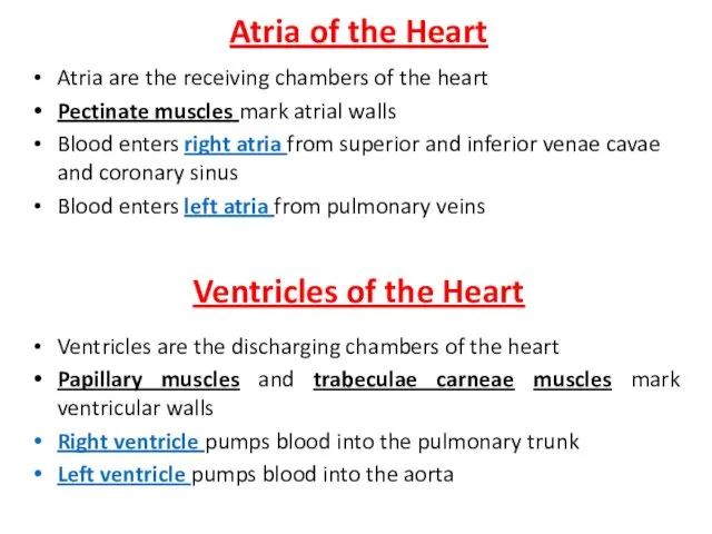
Atria of the Heart
Atria are the receiving chambers of the heart
Pectinate
Atria of the Heart
Atria are the receiving chambers of the heart
Pectinate
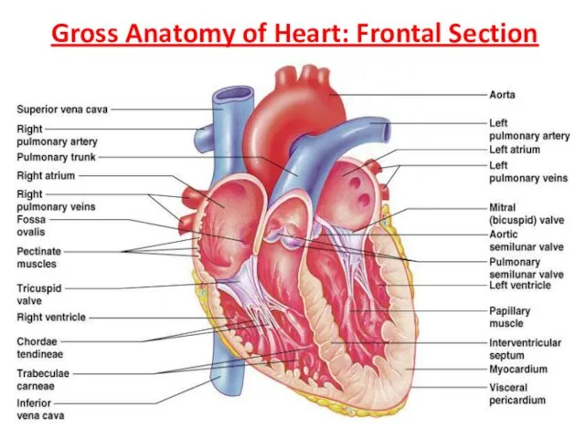
Gross Anatomy of Heart: Frontal Section
Gross Anatomy of Heart: Frontal Section
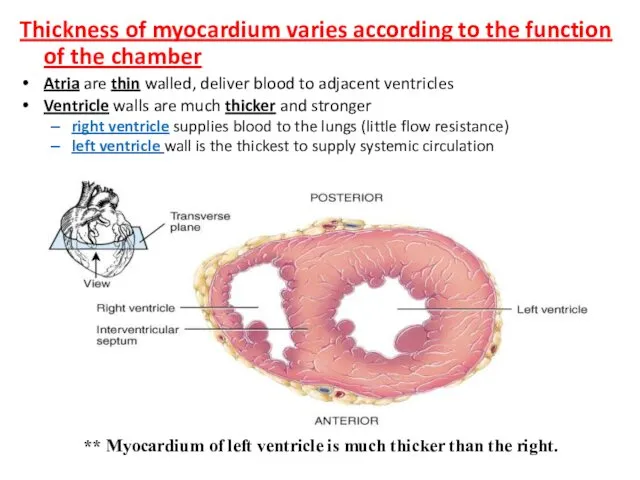
Thickness of myocardium varies according to the function of the chamber
Atria
Thickness of myocardium varies according to the function of the chamber
Atria
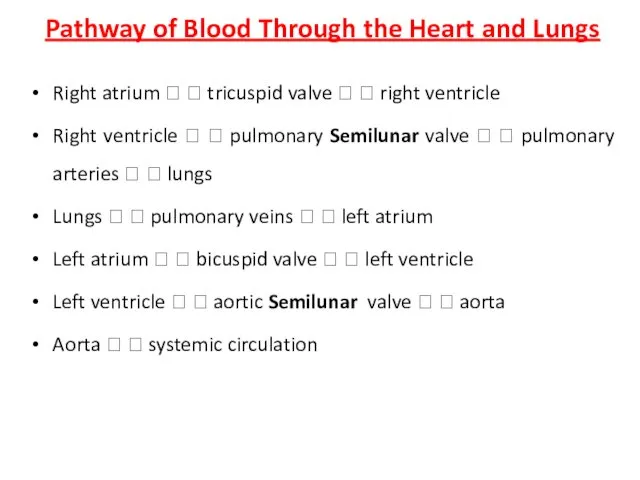
Pathway of Blood Through the Heart and Lungs
Right atrium ? ?
Pathway of Blood Through the Heart and Lungs
Right atrium ? ?
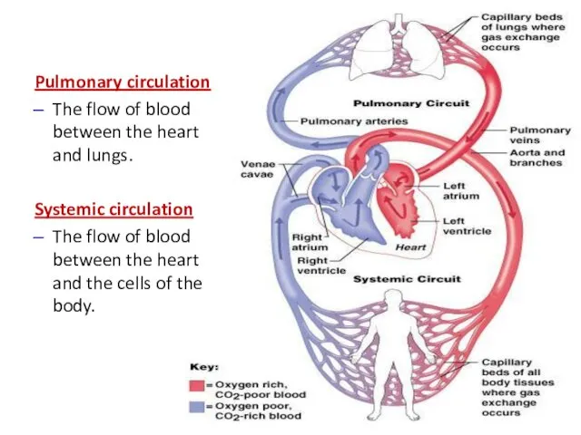
Pulmonary circulation
The flow of blood between the heart and lungs.
Systemic
Pulmonary circulation
The flow of blood between the heart and lungs.
Systemic
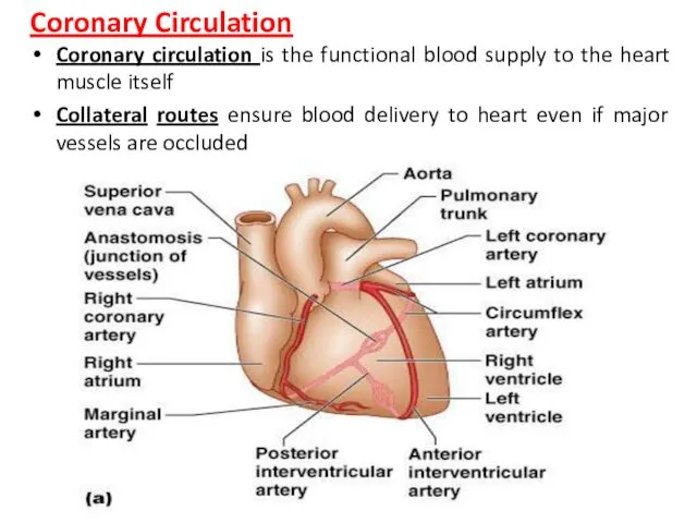
Coronary Circulation
Coronary circulation is the functional blood supply to the heart
Coronary Circulation
Coronary circulation is the functional blood supply to the heart
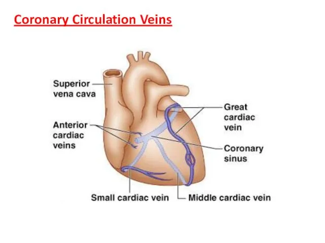
Coronary Circulation Veins
Coronary Circulation Veins
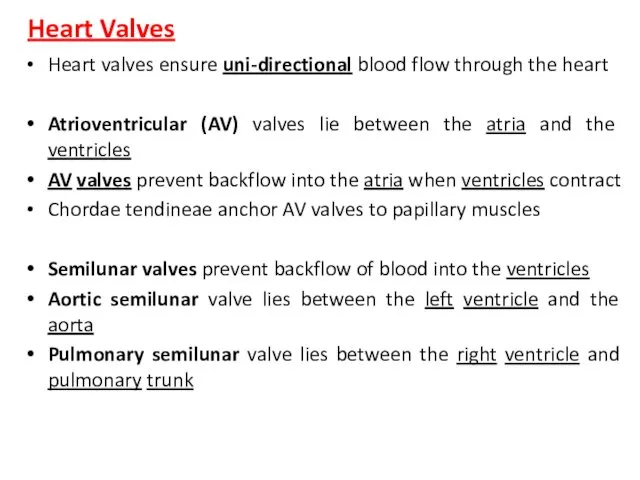
Heart Valves
Heart valves ensure uni-directional blood flow through the heart
Atrioventricular (AV)
Heart Valves
Heart valves ensure uni-directional blood flow through the heart
Atrioventricular (AV)
Heart Valves
Heart Valves
Heart Valves
Heart Valves
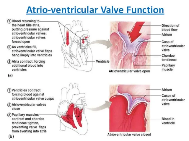
Atrio-ventricular Valve Function
(b)
Atrio-ventricular Valve Function
(b)
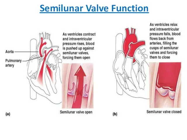
Semilunar Valve Function
Semilunar Valve Function
 Наркоз (общее обезболивание)
 Sistem nervos - generalitati
 Законы движения крови по сосудам. Основные гемодинамические показатели
 Medical insurance for employees and their family members
 Лихорадка Эбола
 Заболеваемость населения как медико-социальная проблема. Эпидемиологические методы изучения заболеваемости
 Государственное автономное учреждение здравоохранения Московской области Центральная городская клиническая больница г. Реутов
 Патология и физиология климактерия. Индивидуализация ЗГТ
 Миоксигенирующий массаж лица
 Фізіологічний післяпологовий період
 Радиобиологические основы лучевой терапии. Реакции и осложнения при лучевой терапии. Лучевая терапия неопухолевых заболеваний
 Кишечная непроходимость. Динамическая кишечная непроходимость
 Медициналық – генетикалық кеңес беру
 Фотометрические методы биохимического анализа. Турбидиметрия и нефелометрия
 Распорядок дня для здорового образа жизни: основы правильного режима дня
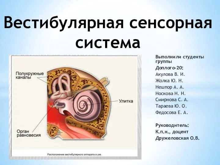 Вестибулярная сенсорная система
 Сенситивные периоды детей раннего возраста
 Қант диабеті
 Миокардиты. Этиология
 Серологические реакции
 Чувствительность: общие понятия
 Профилактика некариозных поражений:флоороза, гипоплазия эмали. Факторы риска возникновения флюороза, местной и системной
 Бруцеллез (сарып) ауруы
 Неязвенные желудочные диспепсии
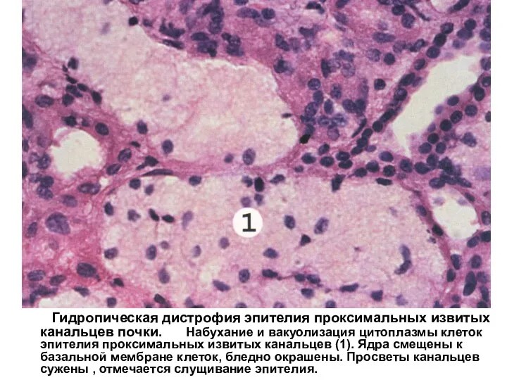 Гидропическая дистрофия эпителия проксимальных извитых канальцев почки
 Классификация энцефалитов. Аутоиммунные энцефалиты
 Выхаживание детей с экстремально низкой массой тела
 Мектептегі оқу жұмысының кейбір түрлеріне қойылатын гигиеналық талаптар