- Defects of eye

Содержание
- 2. At rest, the light rays focus behind instead of on the retina. This type of eye
- 3. Red-green color blindness is the inability to distinguish red and green colors in dim light (and
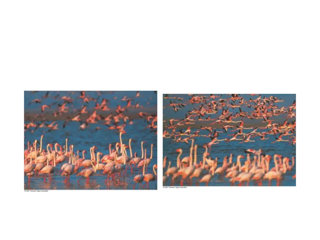
- 4. Nearsightedness (myopia) results when the image is focused in front of the retina. Farsightedness (hyperopia) is
- 5. Fig. 14.21 (top), p. 264 (focal point) distant object (focal point) close object
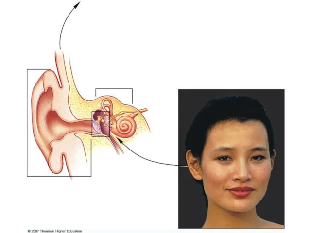
- 7. The human ear has 2 sensory functions. One of them is hearing. Other is maintaning balance
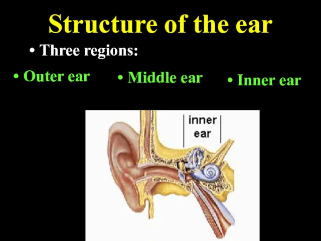
- 8. Structure of ears Ears contains 3 main parts; Outer ear, The middle ear Inner ear
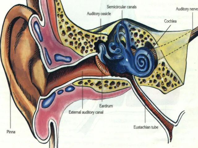
- 10. OUTER EAR Outer ear is composed of 3 parts. These are pinna, auditory canal and eardrum.
- 11. Auditory canal is a canal which is found between pinna and eardrum. It has hairs and
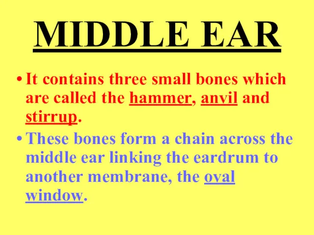
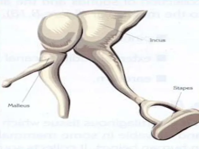
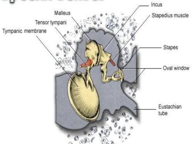
- 12. MIDDLE EAR It contains three small bones which are called the hammer, anvil and stirrup. These
- 14. The hammer attached to the eardrum, the anvil connects the hammer to the stirrup. Stirrup is
- 16. EUSTACHIAN TUBE It is located between pharynx and the middle ear. It equalizes in the middle
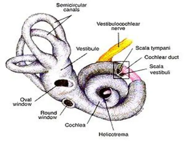
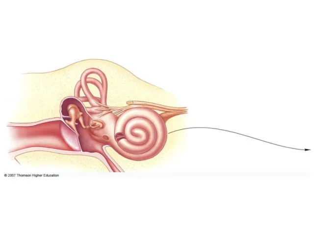
- 18. THE INNER EAR It consists of the cochlea and semicircular canals. Cochlea is organ of hearing
- 19. They are separated from another by membranes. Lining of the membranes are specialized hair cells that
- 20. Semicircular canals enable the body to maintain balance. These canals contain fluid and hairlike projenctions that
- 21. Sound waves collected by outer ear pass down the auditory canal to the eardrum. They cause
- 22. Vibration of stirrup cause vibrations in the oval window which in turn cause the fluid within
- 26. Structure of the ear Three regions: Outer ear Middle ear Inner ear
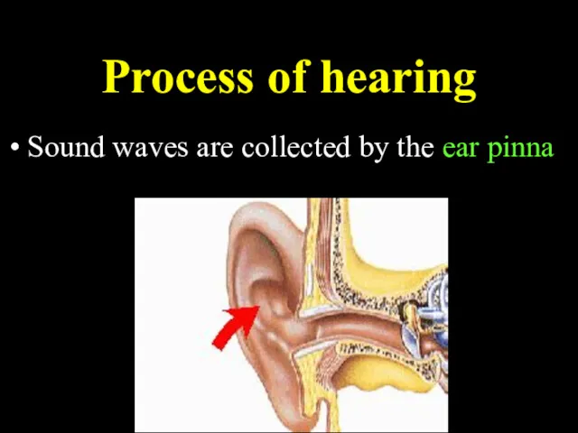
- 27. Process of hearing Sound waves are collected by the ear pinna
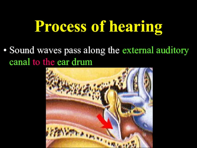
- 28. Process of hearing Sound waves pass along the external auditory canal to the ear drum
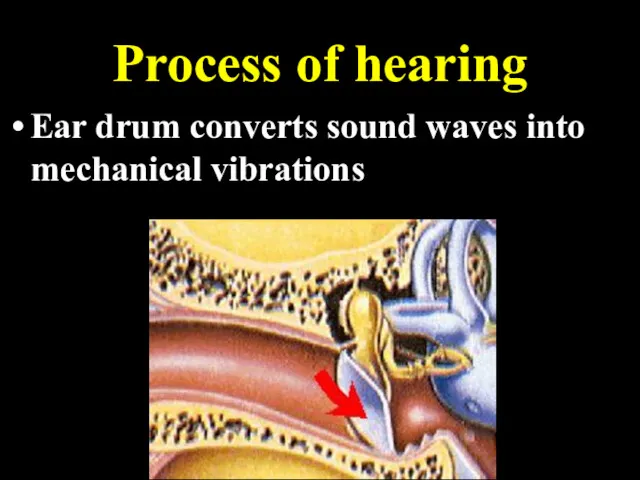
- 29. Process of hearing Ear drum converts sound waves into mechanical vibrations
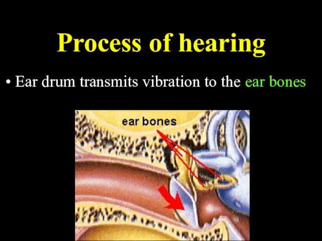
- 30. Process of hearing Ear drum transmits vibration to the ear bones
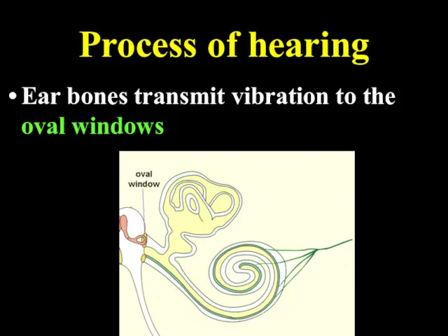
- 31. Process of hearing Ear bones transmit vibration to the oval windows
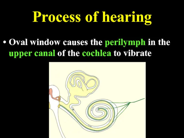
- 32. Process of hearing Oval window causes the perilymph in the upper canal of the cochlea to
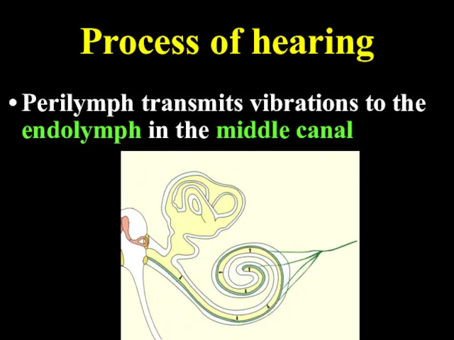
- 33. Process of hearing Perilymph transmits vibrations to the endolymph in the middle canal
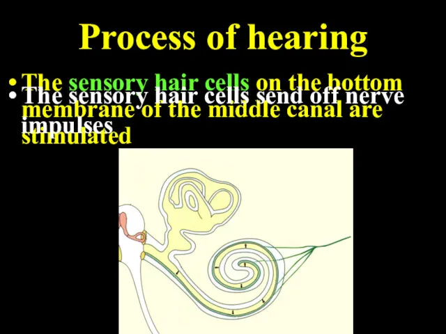
- 34. Process of hearing The sensory hair cells on the bottom membrane of the middle canal are
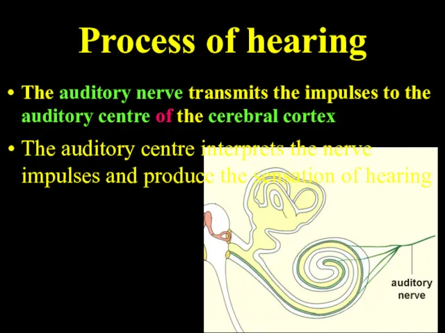
- 35. Process of hearing The auditory nerve transmits the impulses to the auditory centre of the cerebral
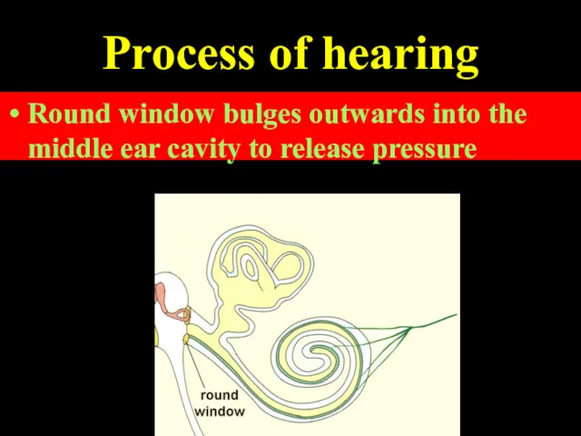
- 36. Process of hearing The vibrations of perilymph are transmitted to the round window Round window bulges
- 37. All multicellular organisms have a skin composed of one or more layers. THE SKIN
- 38. Functions of Skin It protects the inner layers of the body from physical and chemical effects.
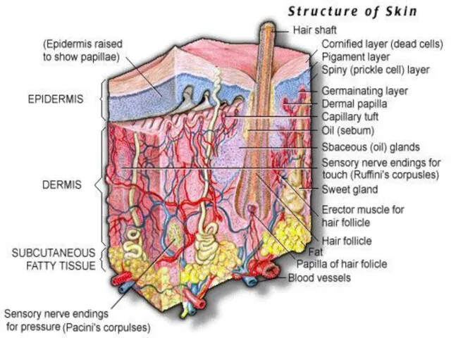
- 39. Structure of the skin Epidermis Dermis Accesory structure of the skin Skin gland Hair follicles Nails
- 40. EPIDERMIS is outermost layer of skin. This layer composed of keratinised epithelial cells. Epidermis contains no
- 41. DERMIS is rich in blood vessels and nerve ending. The receptors located in the skin are
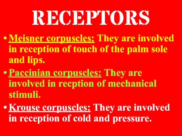
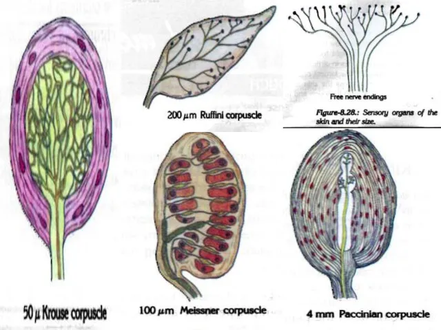
- 43. RECEPTORS Meisner corpuscles: They are involved in reception of touch of the palm sole and lips.
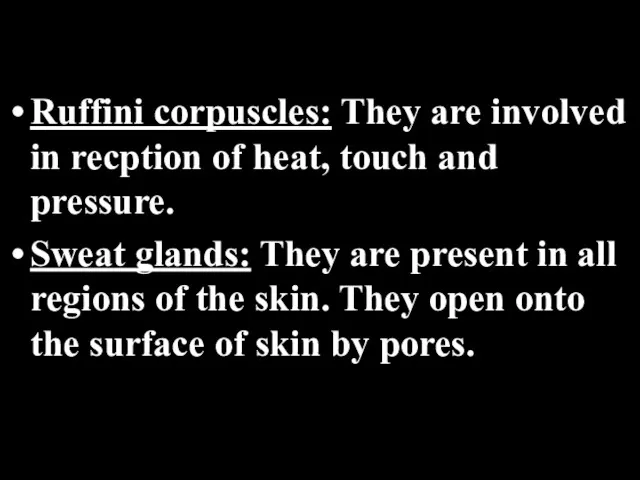
- 44. Ruffini corpuscles: They are involved in recption of heat, touch and pressure. Sweat glands: They are
- 46. They are involved in removal of water, minerals, urea and other substances. The main function of
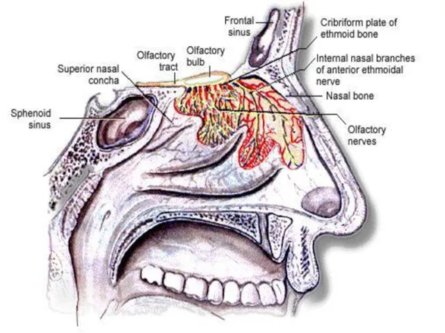
- 47. Nose is the organ of the body involved in both respiration and smell. The reception of
- 49. Smelling is fundemantal in the detection of food, maintenance of relationship, reproduction and communication of some
- 51. The surface of the tongue is covered with small projections called papillae. There are the taste
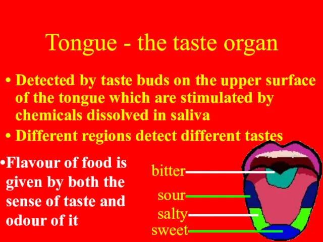
- 52. The taste buds are sensitive to only four basic tastes; SWEET, SOUR, SALT AND BITTER Each
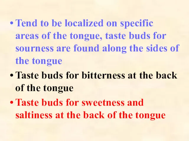
- 53. Tend to be localized on specific areas of the tongue, taste buds for sourness are found
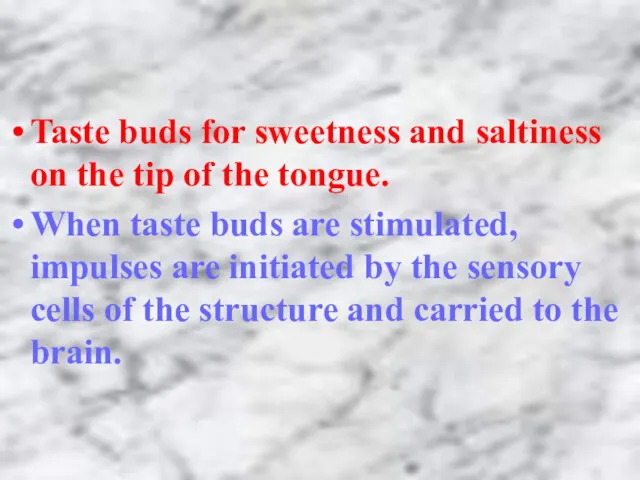
- 54. Taste buds for sweetness and saltiness on the tip of the tongue. When taste buds are
- 55. Tongue - the taste organ Detected by taste buds on the upper surface of the tongue
- 57. Скачать презентацию
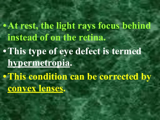
At rest, the light rays focus behind instead of on the
At rest, the light rays focus behind instead of on the

Red-green color blindness is the inability to distinguish red and green
Red-green color blindness is the inability to distinguish red and green
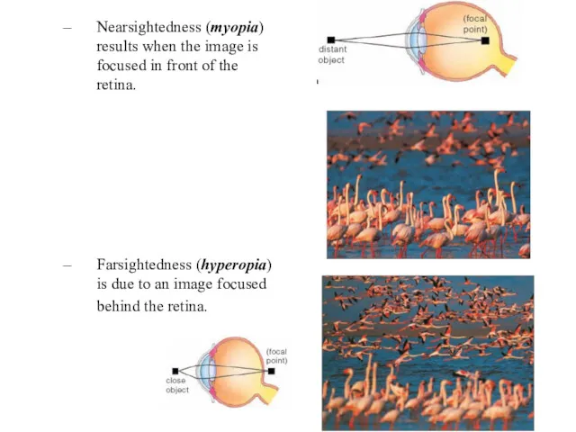
Nearsightedness (myopia) results when the image is focused in front of
Nearsightedness (myopia) results when the image is focused in front of
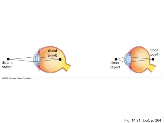
Fig. 14.21 (top), p. 264
(focal
point)
distant
object
(focal
point)
close
object
Fig. 14.21 (top), p. 264
(focal
point)
distant
object
(focal
point)
close
object
The human ear has 2 sensory functions.
One of them is
The human ear has 2 sensory functions.
One of them is
Structure of ears
Ears contains 3 main parts;
Outer ear,
The middle ear
Inner
Structure of ears
Ears contains 3 main parts;
Outer ear,
The middle ear
Inner
OUTER EAR
Outer ear is composed of 3 parts.
These are pinna, auditory
OUTER EAR
Outer ear is composed of 3 parts.
These are pinna, auditory
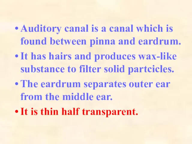
Auditory canal is a canal which is found between pinna and
Auditory canal is a canal which is found between pinna and
MIDDLE EAR
It contains three small bones which are called the hammer,
MIDDLE EAR
It contains three small bones which are called the hammer,
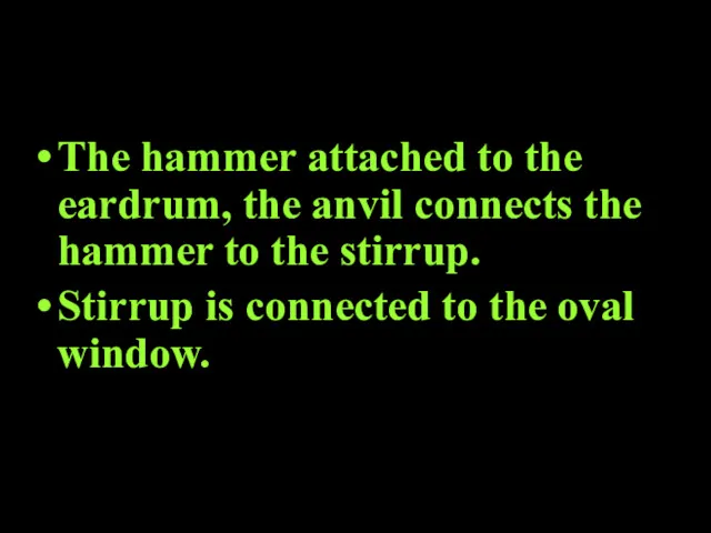
The hammer attached to the eardrum, the anvil connects the hammer
The hammer attached to the eardrum, the anvil connects the hammer
EUSTACHIAN TUBE
It is located between pharynx and the middle ear.
It equalizes
EUSTACHIAN TUBE
It is located between pharynx and the middle ear.
It equalizes
THE INNER EAR
It consists of the cochlea and semicircular canals.
Cochlea is
THE INNER EAR
It consists of the cochlea and semicircular canals.
Cochlea is
They are separated from another by membranes.
Lining of the membranes are
They are separated from another by membranes.
Lining of the membranes are
Semicircular canals enable the body to maintain balance.
These canals contain fluid
Semicircular canals enable the body to maintain balance.
These canals contain fluid
Sound waves collected by outer ear pass down the auditory canal
Sound waves collected by outer ear pass down the auditory canal
Vibration of stirrup cause vibrations in the oval window which in
Vibration of stirrup cause vibrations in the oval window which in

Structure of the ear
Three regions:
Outer ear
Middle ear
Inner ear
Structure of the ear
Three regions:
Outer ear
Middle ear
Inner ear
Process of hearing
Sound waves are collected by the ear pinna
Process of hearing
Sound waves are collected by the ear pinna
Process of hearing
Sound waves pass along the external auditory canal to
Process of hearing
Sound waves pass along the external auditory canal to
Process of hearing
Ear drum converts sound waves into mechanical vibrations
Process of hearing
Ear drum converts sound waves into mechanical vibrations
Process of hearing
Ear drum transmits vibration to the ear bones
Process of hearing
Ear drum transmits vibration to the ear bones
Process of hearing
Ear bones transmit vibration to the oval windows
Process of hearing
Ear bones transmit vibration to the oval windows
Process of hearing
Oval window causes the perilymph in the upper canal
Process of hearing
Oval window causes the perilymph in the upper canal
Process of hearing
Perilymph transmits vibrations to the endolymph in the middle
Process of hearing
Perilymph transmits vibrations to the endolymph in the middle
Process of hearing
The sensory hair cells on the bottom membrane of
Process of hearing
The sensory hair cells on the bottom membrane of
Process of hearing
The auditory nerve transmits the impulses to the auditory
Process of hearing
The auditory nerve transmits the impulses to the auditory
Process of hearing
The vibrations of perilymph are transmitted to the round
Process of hearing
The vibrations of perilymph are transmitted to the round
All multicellular organisms have a skin composed of one or more
All multicellular organisms have a skin composed of one or more
Functions of Skin
It protects the inner layers of the body from
Functions of Skin
It protects the inner layers of the body from
Structure of the skin
Epidermis
Dermis
Accesory structure of the skin
Skin gland
Hair
Structure of the skin
Epidermis
Dermis
Accesory structure of the skin
Skin gland
Hair
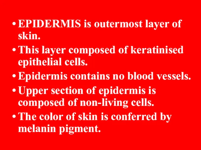
EPIDERMIS is outermost layer of skin.
This layer composed of keratinised epithelial
EPIDERMIS is outermost layer of skin.
This layer composed of keratinised epithelial
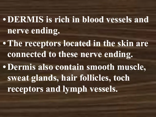
DERMIS is rich in blood vessels and nerve ending.
The receptors located
DERMIS is rich in blood vessels and nerve ending.
The receptors located
RECEPTORS
Meisner corpuscles: They are involved in reception of touch of the
RECEPTORS
Meisner corpuscles: They are involved in reception of touch of the
Ruffini corpuscles: They are involved in recption of heat, touch and
Ruffini corpuscles: They are involved in recption of heat, touch and
They are involved in removal of water, minerals, urea and other
They are involved in removal of water, minerals, urea and other
Nose is the organ of the body involved in both respiration
Nose is the organ of the body involved in both respiration
Smelling is fundemantal in the detection of food, maintenance of relationship,
Smelling is fundemantal in the detection of food, maintenance of relationship,

The surface of the tongue is covered with small projections called
The surface of the tongue is covered with small projections called
The taste buds are sensitive to only four basic tastes;
SWEET, SOUR,
The taste buds are sensitive to only four basic tastes;
SWEET, SOUR,
Tend to be localized on specific areas of the tongue, taste
Tend to be localized on specific areas of the tongue, taste
Taste buds for sweetness and saltiness on the tip of the
Taste buds for sweetness and saltiness on the tip of the
Tongue - the taste organ
Detected by taste buds on the upper
Tongue - the taste organ
Detected by taste buds on the upper
 Эндоскопические исследования
 Флеботромбоз. Варикозное расширение вен нижних конечностей
 Распорядок дня для здорового образа жизни: основы правильного режима дня
 Лечебно-профилактическое питание
 Розвиток урології як клінічної дисципліни. Неспецифічні запальні захворювання органів сечової системи
 Трансплантация легких
 Воспаление. Классификация воспаления
 Каннабиноиды и их синтетические аналоги
 Балалардағы асқазан мен он екі елі ішектің жара аурулары. Синдромдық негізде диагностика, ем және профилактика жүргізу
 Остеопороз
 Ребёнок с ранним детским аутизмом (РДА) и РАС
 Смерть. Некроз. Апоптоз
 Основы организации обеспечения медицинским имуществом и техникой
 Невідкладна допомога на догоспітальному етапі та в стаціонарі. Судомний, гіпертермічний, токсичний та коматозний синдроми
 История становления травматологии и ортопедии в мире
 Диареегенные эшерихии
 Ведение физиологических родов и методы наблюдения во время родов за новорожденным
 Организация питания пациентов терапевтического и хирургического профиля
 Қызыл иектің ультрақұрылысы, қызыл иек сайы, қызыл иек сұйықтығы
 Стационарды алмастыратын көмек
 Пульпа. Поддерживающий аппарат зуба
 Алгоритм ранней диагностики злокачественной меланомы на уровне ПМСП
 Профессия врача. Одна из самых нужных и важных профессий
 Тромбоэмболия легочной артерии
 Инфекционный процесс. Внутрибольничная инфекция (ВБИ)
 Биологиялық сағат және физикалық сағат
 Трофобластическая болезнь
 Организация работы процедурного кабинета