- Dental anatomy

Содержание
- 2. For the general description and information on human teeth, see Teeth (human). For other uses, see
- 3. Tooth development is the complex process by which teeth form from embryonic cells, grow, and erupt
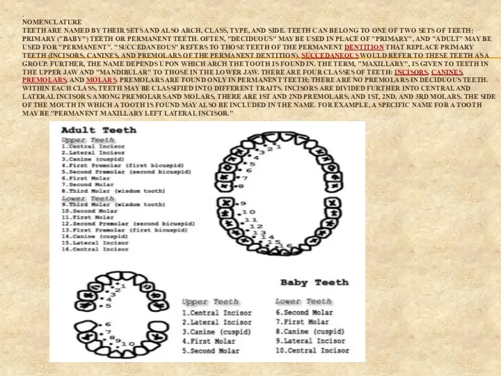
- 4. . NOMENCLATURE TEETH ARE NAMED BY THEIR SETS AND ALSO ARCH, CLASS, TYPE, AND SIDE. TEETH
- 5. THE TOOTH IS ATTACHED TO THE SURROUNDING GINGIVAL TISSUE AND ALVEOLAR BONE (C) BY FIBROUS ATTACHMENTS.
- 6. Surfaces Surfaces that are nearest the cheeks or lips are referred to as facial, and those
- 8. Скачать презентацию
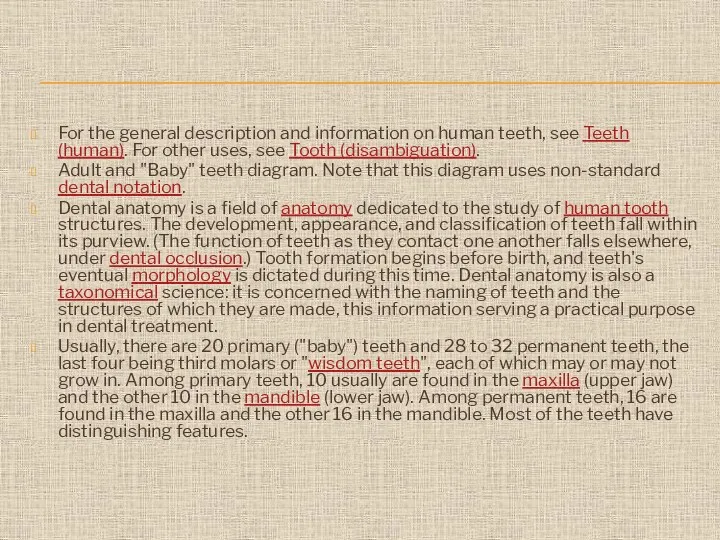
For the general description and information on human teeth, see Teeth
For the general description and information on human teeth, see Teeth
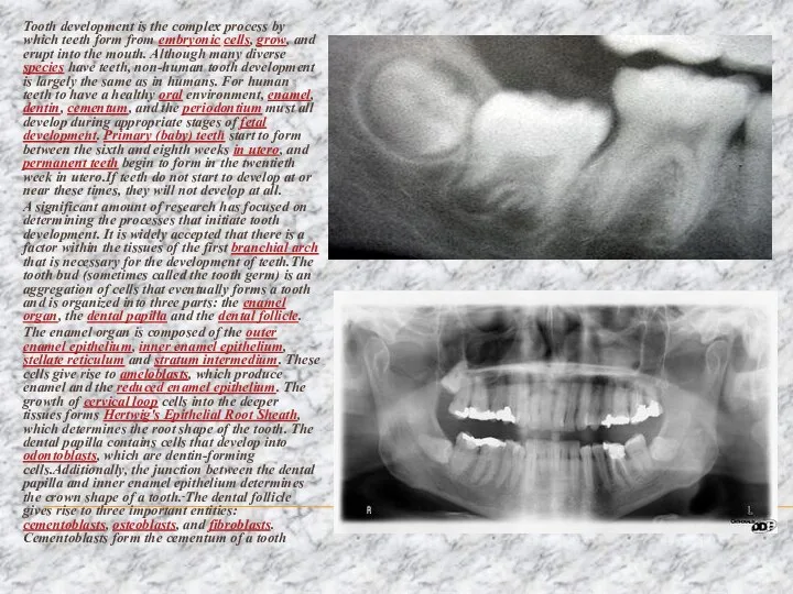
Tooth development is the complex process by which teeth form from
Tooth development is the complex process by which teeth form from
.
NOMENCLATURE
TEETH ARE NAMED BY THEIR SETS AND ALSO ARCH, CLASS, TYPE,
. NOMENCLATURE TEETH ARE NAMED BY THEIR SETS AND ALSO ARCH, CLASS, TYPE,
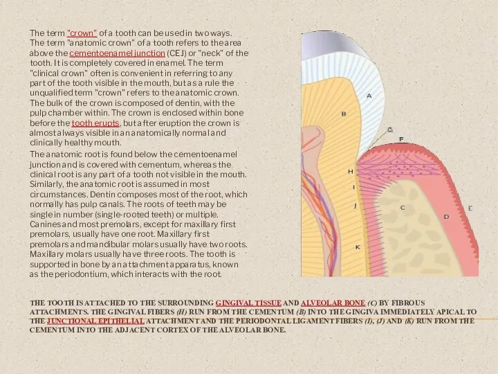
THE TOOTH IS ATTACHED TO THE SURROUNDING GINGIVAL TISSUE AND ALVEOLAR
THE TOOTH IS ATTACHED TO THE SURROUNDING GINGIVAL TISSUE AND ALVEOLAR
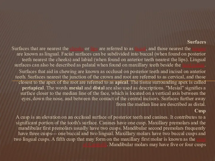
Surfaces
Surfaces that are nearest the cheeks or lips are referred to
Surfaces
Surfaces that are nearest the cheeks or lips are referred to
 Правила хранения лекарственных огнеопасных и наркотических средств. Лекция 8
 Дифференциальная диагностика внутрикостных остеолитических образований
 Практикум по ЭКГ
 Отруйність спиртів та їх згубна дія на організм людини
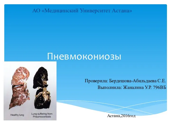 Пневмокониозы. Этиология и патогенез
 Гігієнічна оцінка умов та характеру праці в парфумерно-косметичній галузі
 ВИЧ. Основные факты
 Омегалицин. Профилактика атеросклероза,
 Хронический пылевой бронхит
 Характеристика крови, как части внутренней среды организма
 Понятие инфекционного процесса и инфекционной болезни. Эпидемический процесс
 Антигендер. Антиденелер
 Средства изоляции от слюны в стоматологии
 Профилактика онкогинекологических заболеваний
 Лечебные диеты. Лечебно-профилактическое питание
 ЗВУР. Маловесный к сроку гестации. Гравидограмма
 Антенатальды кезеңдегі инвазивті тексеру
 Невропатология как наука
 Промежуточный мозг. Эпифиз
 Внеаудиторная самостоятельная работа по теме: Техника промывания желудка
 Профилактика инфицирования медицинского персонала гриппом и другими ОРВИ
 Этика и деонтология медицинских работников
 Заболевания щитовидной железы
 Аккредитация специалистов в системе непрерывного медицинского образования
 Оказание помощи при септическом шоке
 Вакцинация от клещевого энцефалита
 Клинический случай. Менингококковая инфекция
 Ампутации и экзартикуляции конечностей