- Dermatomycosis. Pathogenesis

Содержание
- 2. Pathogenesis Despite the abundance of fungi in the surroundings of man, only a few of them
- 3. Classification of fungal diseases There differentiate 4 basic groups: Keratomycosis: pityriasis versicolor; conditional: erythrasma, nodosal trisporum;
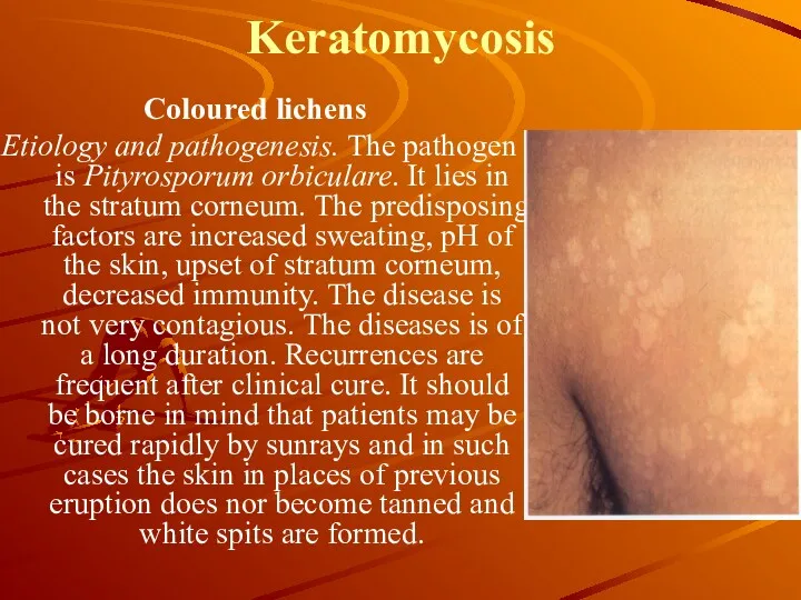
- 4. Keratomycosis Coloured lichens Etiology and pathogenesis. The pathogen is Pityrosporum orbiculare. It lies in the stratum
- 5. Keratomycosis Histopathology. In the absence of inflammatory phenomena, there is looseness of the horny layer, in
- 6. Erythrasma Etiology and pathogenesis. The pathogen is cornebacteria, which infects only stratum corneum, usually in big
- 7. Erythrasma Treatment. The same agents as in pityriasis versicolor are applied in the treatment but in
- 8. Dermatomycosis This is a large group of fungus diseases, in which not only the skin but
- 9. Epidermophytosis (Epidermophytia) Epidermophytosis is a contagious disease of the superficial layers of the smooth skin and
- 10. Epidermophytosis of the Large Skin Folds Etiology. The causative agent is the fungus Epidermophyton inguinale Sabouraud
- 11. Epidermophytosis of feet is a widespread disease. Etiology. The pathogen is Trichophyton mentagrophytes. The disease is
- 12. The intertriginous form Мay occur independently but more frequently it develops when there is a mildly
- 13. The squamous form Moderate scaling on a slightly hyperemic skin is revealed on the arches of
- 14. The dyshidrotic form Is characterized by the formation of a group of vesicles on the arch
- 15. Rubromycosis or rubrophytes. The pathogen is tinea rubrum. It occupies the central position between Epidermophyton and
- 16. Trichophytosis Trichophytosis corporis and chronic trichophytosis, purulent infiltrative trichophytosis. Such fungi include large spored and small
- 17. Microsporia Etiology. Pathogen is anthropophilic fungi and zoo-antropophilic. Epidemiology is the same as in trichophytosis. Affection
- 18. Favus Etiology. Pathogen is Trichophyton schoenleinii of endothrix species. Epidemiology. Favus is less contagious. Chronic in
- 19. Treatment of trichophyton, microsporum and favus. During the infection of the skin iodine solutions are used.
- 20. Candidiasis Is an infection of the skin, mucous membrane, nail plates and internal organs, caused by
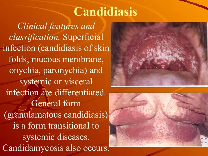
- 21. Candidiasis Clinical features and classification. Superficial infection (candidiasis of skin folds, mucous membrane, onychia, paronychia) and
- 22. Candidiasis Treatment. First of all, remove the factors causing the diseases. Locally spirit and water solutions.
- 24. Скачать презентацию
Pathogenesis
Despite the abundance of fungi in the surroundings of man, only
Pathogenesis
Despite the abundance of fungi in the surroundings of man, only
Classification of fungal diseases
There differentiate 4 basic groups:
Keratomycosis: pityriasis versicolor;
Classification of fungal diseases
There differentiate 4 basic groups:
Keratomycosis: pityriasis versicolor;
Keratomycosis
Coloured lichens
Etiology and pathogenesis. The pathogen is Pityrosporum orbiculare. It
Keratomycosis
Coloured lichens
Etiology and pathogenesis. The pathogen is Pityrosporum orbiculare. It
Keratomycosis
Histopathology. In the absence of inflammatory phenomena, there is looseness
Keratomycosis
Histopathology. In the absence of inflammatory phenomena, there is looseness
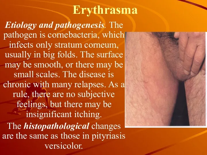
Erythrasma
Etiology and pathogenesis. The pathogen is cornebacteria, which infects only
Erythrasma
Etiology and pathogenesis. The pathogen is cornebacteria, which infects only
Erythrasma
Treatment. The same agents as in pityriasis versicolor are applied in
Erythrasma
Treatment. The same agents as in pityriasis versicolor are applied in
Dermatomycosis
This is a large group of fungus diseases, in which not
Dermatomycosis
This is a large group of fungus diseases, in which not
Epidermophytosis (Epidermophytia)
Epidermophytosis is a contagious disease of the superficial layers of
Epidermophytosis (Epidermophytia)
Epidermophytosis is a contagious disease of the superficial layers of
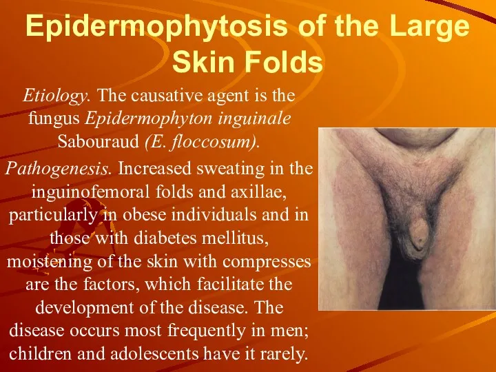
Epidermophytosis of the Large Skin Folds
Etiology. The causative agent is
Epidermophytosis of the Large Skin Folds
Etiology. The causative agent is
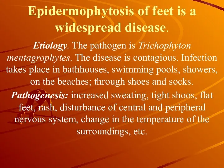
Epidermophytosis of feet is a widespread disease.
Etiology. The pathogen is
Epidermophytosis of feet is a widespread disease.
Etiology. The pathogen is
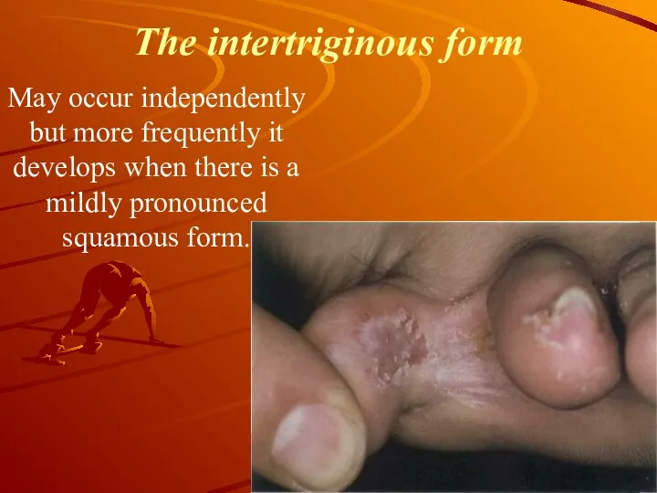
The intertriginous form
Мay occur independently but more frequently it develops
The intertriginous form
Мay occur independently but more frequently it develops
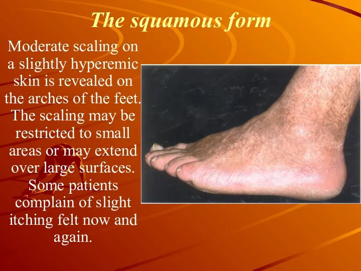
The squamous form
Moderate scaling on a slightly hyperemic skin is
The squamous form
Moderate scaling on a slightly hyperemic skin is
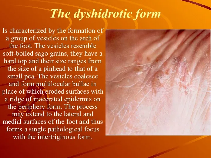
The dyshidrotic form
Is characterized by the formation of a group
The dyshidrotic form
Is characterized by the formation of a group
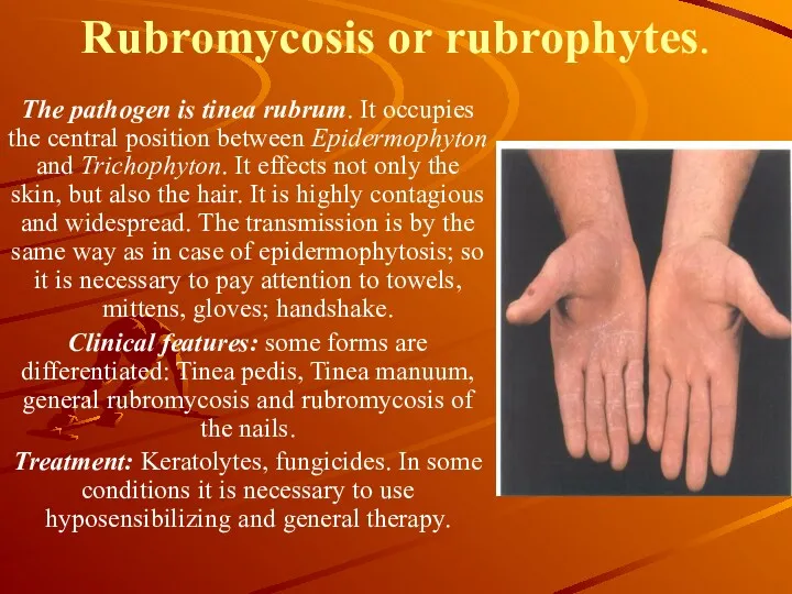
Rubromycosis or rubrophytes.
The pathogen is tinea rubrum. It occupies the
Rubromycosis or rubrophytes.
The pathogen is tinea rubrum. It occupies the
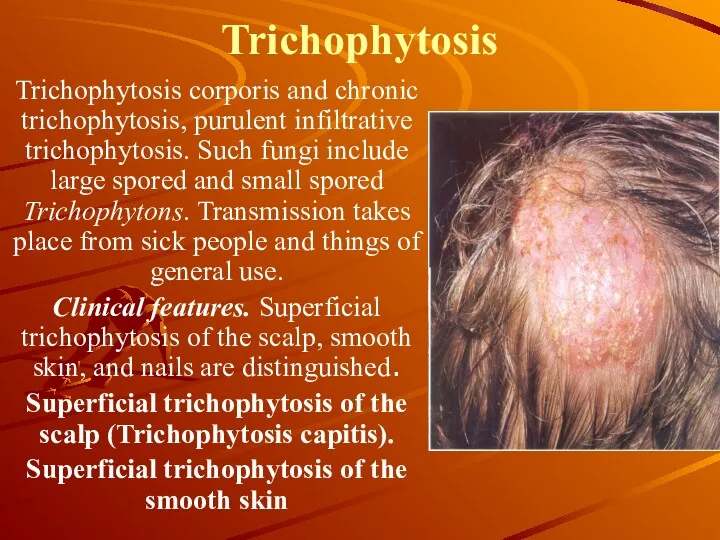
Trichophytosis
Trichophytosis corporis and chronic trichophytosis, purulent infiltrative trichophytosis. Such fungi
Trichophytosis
Trichophytosis corporis and chronic trichophytosis, purulent infiltrative trichophytosis. Such fungi
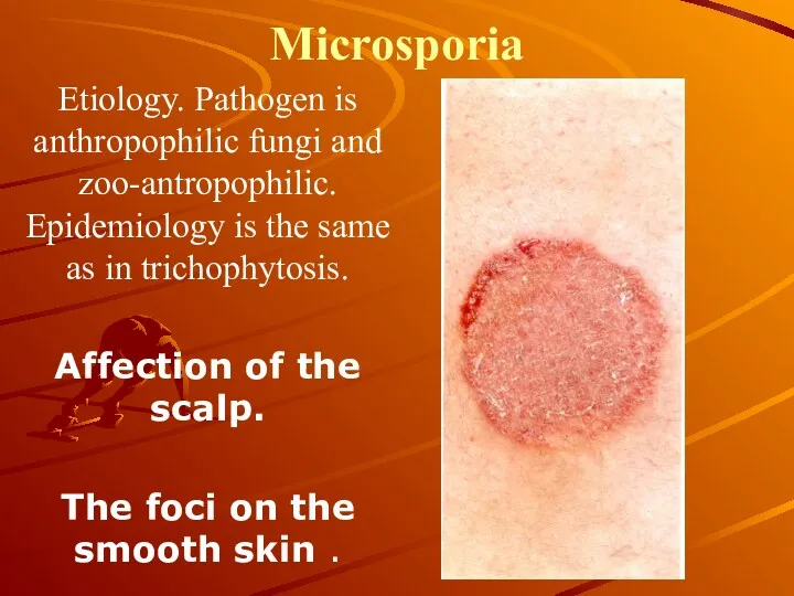
Microsporia
Etiology. Pathogen is anthropophilic fungi and zoo-antropophilic. Epidemiology is the same
Microsporia
Etiology. Pathogen is anthropophilic fungi and zoo-antropophilic. Epidemiology is the same
Favus
Etiology. Pathogen is Trichophyton schoenleinii of endothrix species.
Epidemiology. Favus is less
Favus
Etiology. Pathogen is Trichophyton schoenleinii of endothrix species.
Epidemiology. Favus is less
Treatment of trichophyton, microsporum and favus.
During the infection of the
Treatment of trichophyton, microsporum and favus.
During the infection of the
Candidiasis
Is an infection of the skin, mucous membrane, nail plates
Candidiasis
Is an infection of the skin, mucous membrane, nail plates
Candidiasis
Clinical features and classification. Superficial infection (candidiasis of skin folds, mucous
Candidiasis
Clinical features and classification. Superficial infection (candidiasis of skin folds, mucous
Candidiasis
Treatment. First of all, remove the factors causing the diseases. Locally
Candidiasis
Treatment. First of all, remove the factors causing the diseases. Locally
 Ауадаға бактерия санын есептеу. Судағы микроағзалардың санын есептеу
 Оценки клинических руководств,с позиций доказательной медицины. Уровни доказательной медицины
 Огнестрельные ранения
 Воспаление. Этиология, патогенез и виды хронического воспаления
 Неотложная помощь детям с анафилактическим шоком. Роль медицинской сестры
 Медицинская информационная система ЕМИАС
 Травмы. Травматический шок
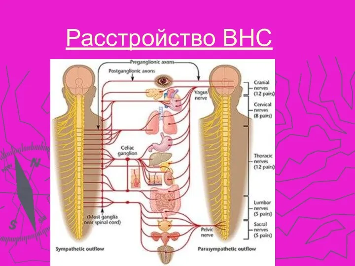 Расстройство ВНС
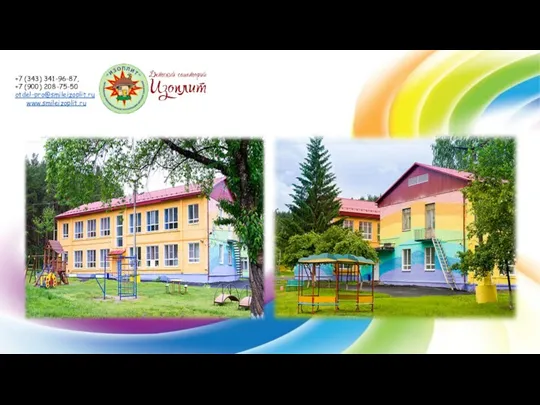 Детский санаторий Изоплит, г. Екатеринбуг. На оздоровление приглашаются дети от 4 до 14 лет
 Образование МГУ. Его роль в развитии медицины
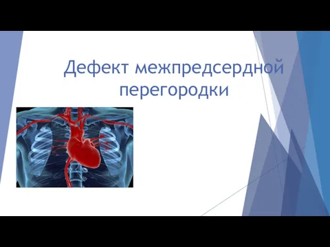 Дефект межпредсердной перегородки
 Ойлау және интеллект бұзылыстарының клиникалық мініздемесі
 Операции на нервных стволах
 Диагностика и оказание скорой помощи при угрожающих жизни поражениях центральной нервной системы
 Тривалість життя людини
 Особенности анестезии в торакальной и в абдоминальной хирургии у детей
 Суставной синдром
 Науқас пен оның туыстарымен денсаулық сақтау саласында жұмыс істейтін қызметкерлер мен тиімді қарымқатынасқа
 Иммундық жүйенің ісікке қарсы қорғанысының себептері
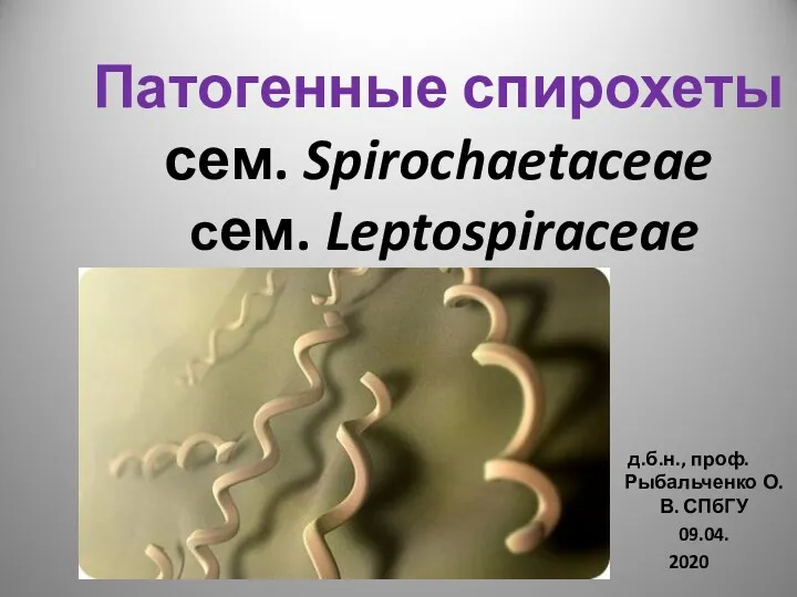 Патогенные спирохеты сем. Spirochaetaceae сем. Leptospiraceae
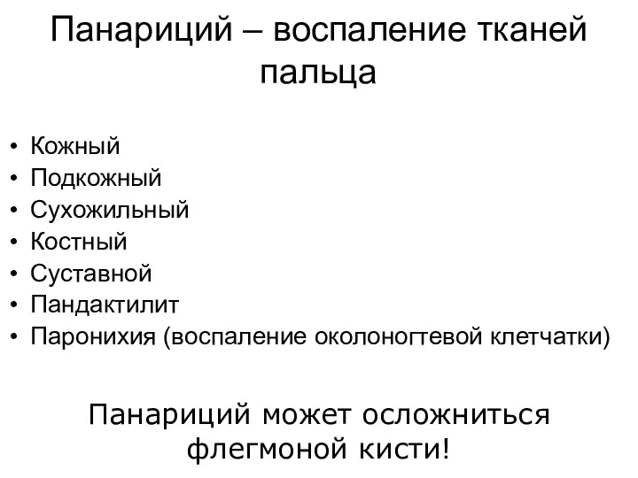 Панариций – воспаление тканей пальца
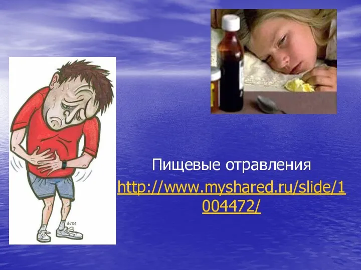 Пищевые отравления
 Заикание у детей и взрослых
 Биоэквивалентность лекарственных средств
 Антигены, основные свойства. Антигены гистосовместимости. (Лекция 11)
 Вопросы общей онкологии. Структура и организация онкологической службы в России
 Система гемостаза и ее нарушения. Лекция 9
 Мутация