- Doppler ultrasound

Содержание
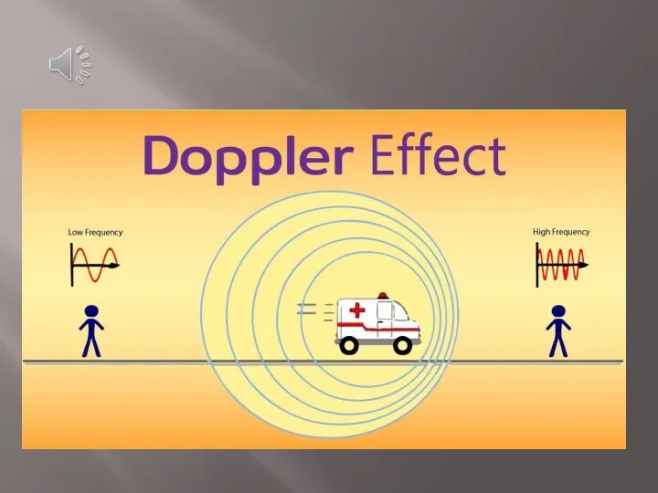
- 2. Doppler effect We are all aware that the pitch of an ambulance siren changes as we
- 4. Physics Ultrasound images are formed by reflected echoes. These waves have an amplitude (strength) and a
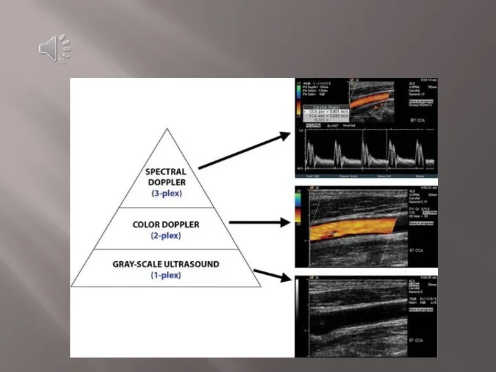
- 5. Application of Doppler Three basic levels of US can be performed, with each level adding information
- 7. The Doppler effect in diagnostic imaging can be used to study blood flow, for example, and
- 8. Doppler shift Blood flow moving towards the transducer produces positive Doppler shifted signals and conversely blood
- 9. Angle of insonation (Doppler angle) The angle of insonation is very important. The angle of insonation
- 10. Magnified view (right) of the color Doppler interrogation region shows the components used to acquire the
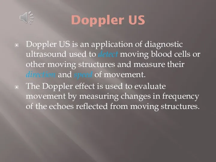
- 11. Doppler US Doppler US is an application of diagnostic ultrasound used to detect moving blood cells
- 12. In many instances, Doppler ultrasound has replaced x-ray methods such as angiography, as a method to
- 13. Types of Doppler ultrasound include: Color Doppler Power Doppler Spectral Doppler
- 14. Color Doppler Color Doppler uses a computer to convert the Doppler measurements into an array of
- 15. Color Doppler
- 16. Power Doppler Power Doppler is a technique that uses the amplitude of Doppler signal to detect
- 17. Power Doppler Power Doppler: is independent of velocity and direction of flow. is independent of angle,
- 18. Spectral Doppler Instead of displaying Doppler measurements visually as in the color and power Doppler methods,
- 19. Spectral Doppler US findings are displayed with the spectral waveform at the bottom of the screen
- 21. Introduction to Doppler ultrasound https://www.youtube.com/watch?v=tQn8jKtwk6o
- 23. Скачать презентацию
Doppler effect
We are all aware that the pitch of an
Doppler effect
We are all aware that the pitch of an
Physics
Ultrasound images are formed by reflected echoes. These waves have an
Physics
Ultrasound images are formed by reflected echoes. These waves have an
Application of Doppler
Three basic levels of US can be performed,
Application of Doppler
Three basic levels of US can be performed,
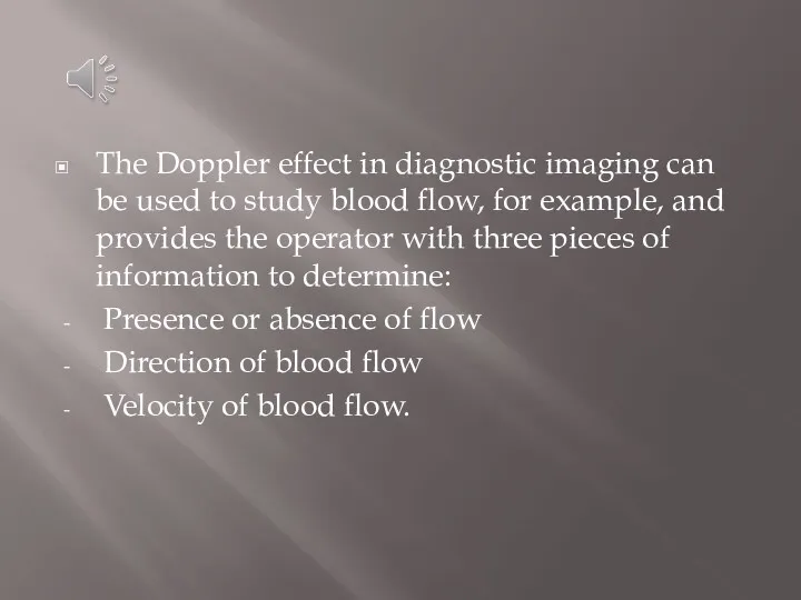
The Doppler effect in diagnostic imaging can be used to study
The Doppler effect in diagnostic imaging can be used to study
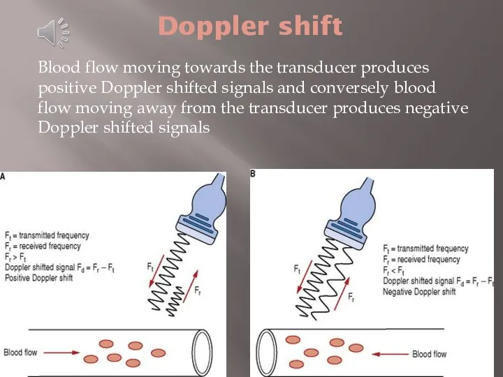
Doppler shift
Blood flow moving towards the transducer produces positive Doppler shifted
Doppler shift
Blood flow moving towards the transducer produces positive Doppler shifted
Angle of insonation (Doppler angle)
The angle of insonation is very
Angle of insonation (Doppler angle)
The angle of insonation is very
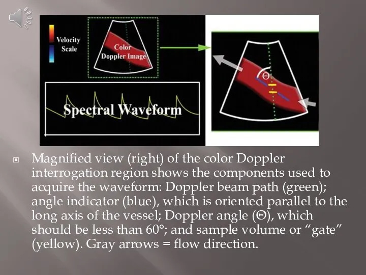
Magnified view (right) of the color Doppler interrogation region shows the
Magnified view (right) of the color Doppler interrogation region shows the
Doppler US
Doppler US is an application of diagnostic ultrasound used to detect moving
Doppler US
Doppler US is an application of diagnostic ultrasound used to detect moving
In many instances, Doppler ultrasound has replaced x-ray methods such as
In many instances, Doppler ultrasound has replaced x-ray methods such as
Types of Doppler ultrasound include:
Color Doppler
Power Doppler
Spectral Doppler
Types of Doppler ultrasound include:
Color Doppler
Power Doppler
Spectral Doppler
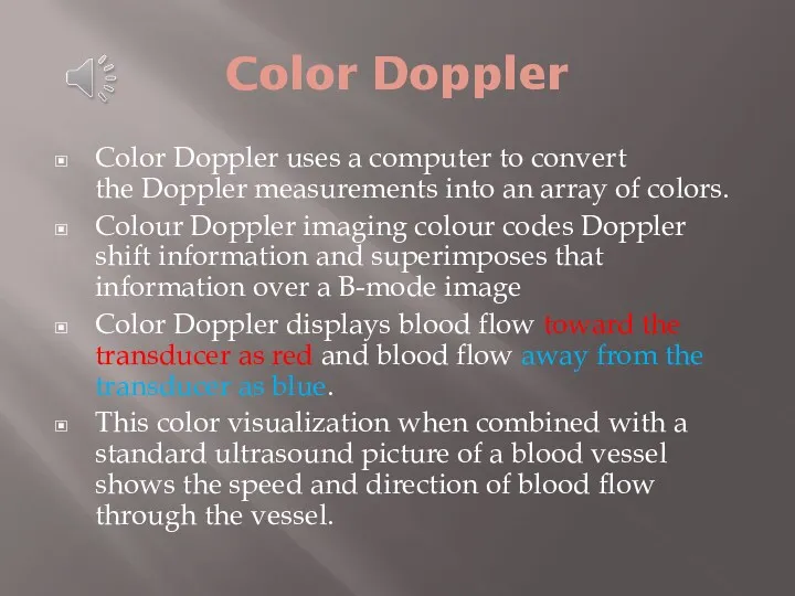
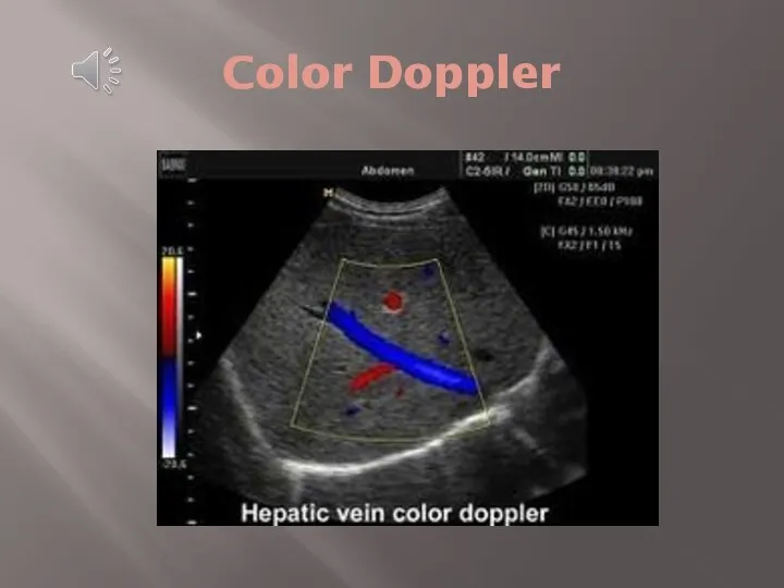
Color Doppler
Color Doppler uses a computer to convert the Doppler measurements into an
Color Doppler
Color Doppler uses a computer to convert the Doppler measurements into an
Color Doppler
Color Doppler
Power Doppler
Power Doppler is a technique that uses the amplitude of Doppler
Power Doppler
Power Doppler is a technique that uses the amplitude of Doppler
Power Doppler
Power Doppler:
is independent of velocity and direction of flow.
is independent
Power Doppler
Power Doppler:
is independent of velocity and direction of flow.
is independent
Spectral Doppler
Instead of displaying Doppler measurements visually as in the color and power Doppler methods, spectral Doppler
Spectral Doppler
Instead of displaying Doppler measurements visually as in the color and power Doppler methods, spectral Doppler
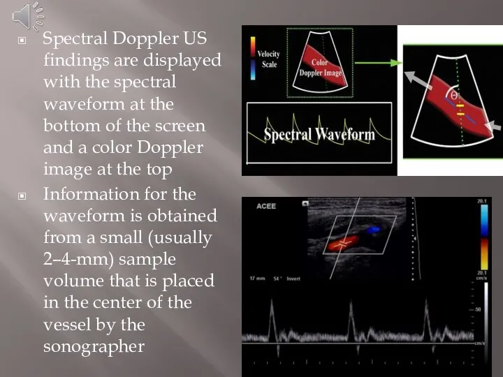
Spectral Doppler US findings are displayed with the spectral waveform at
Spectral Doppler US findings are displayed with the spectral waveform at
Introduction to Doppler ultrasound
https://www.youtube.com/watch?v=tQn8jKtwk6o
Introduction to Doppler ultrasound
https://www.youtube.com/watch?v=tQn8jKtwk6o
 Туа біткен жүрек ақаулары. Жүйелі қызыл жегі, ревматоидты артрит, дерматомиозит, жүйелі склеродермия
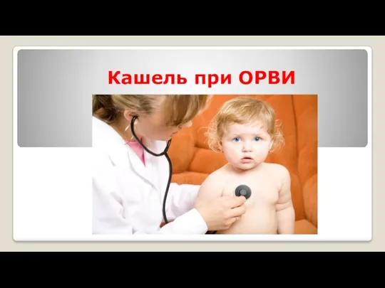 Кашель при ОРВИ
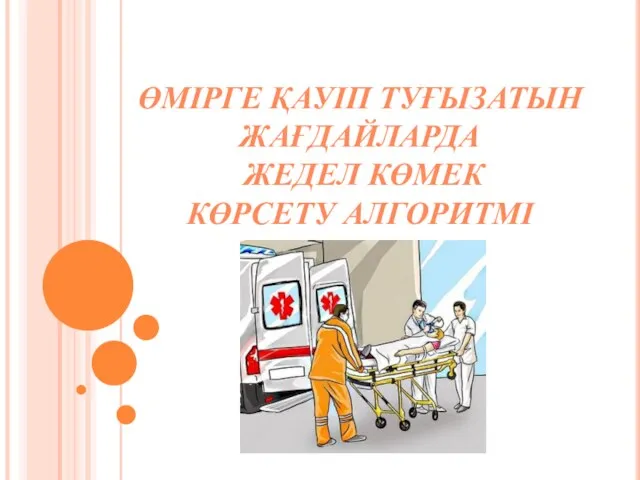 Өмірге қауіп туғызатын жағдайларда жедел көмек көрсету алгоритмі
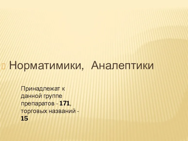 Норматимики, Аналептики
 Факторы патогенности микроорганизмов
 Лучевые поражения
 Мұрынның алдыңғы тампонадасын өткізу
 Диссеминированный туберкулез легких
 Надання допомоги пораненим під час бойових дій
 Абсцесс головного мозга
 Первая помощь при растяжении связок, вывихах суставов, переломах костей
 Влияние алкоголя на развитие организма
 Тема: Психотропные средства
 Пациентам с ХБП о трансплантации почки
 Репродуктивті денсаулық және мінез құлық. Сырқаттылықты жеке және жалпы тіркеу. Медициналық сақтандыру
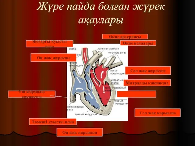 Жүре пайда болған жүрек ақаулары
 Течение родов. Гипоксия плода и асфиксия новорожденного
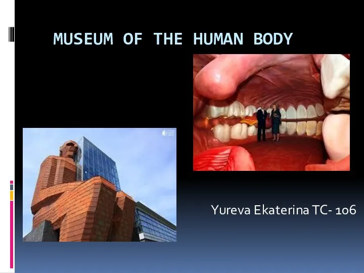 Museum of the human body
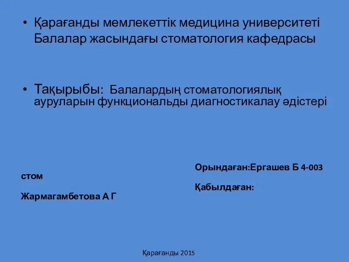 Балалардың стоматологиялық ауруларын функциональды диагностикалау әдістері
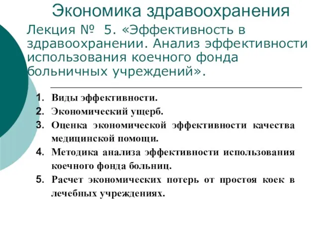 Эффективность в здравоохранении. Анализ эффективности использования коечного фонда больничных учреждений
 Лечение больных с сахарным диабетом
 Оттискные материалы
 Учение об инфекции. Понятие об инфекционном процессе
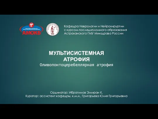 Мультисистемная атрофия. Оливопонтоцеребеллярная атрофия
 Дефицит магния. Причины и клинические проявления
 Ароматные воды. Технология получения ароматных вод
 Основы здорового образа жизни студентов
 К вопросу о преодолении резистентности к терапии АГ