- ECG complication of treatment

Содержание
- 2. Case 1
- 3. A 65-year-old woman presents to the Emergency Department (ED) with generalized fatigue and palpitations. She was
- 6. Life-Threatening Condition (I): Hyperkalemia. The tracing shows a regular rhythm at 75 beats/min. A P wave
- 7. Case 2
- 8. An 83-year-old man with known ischemic cardiomyopathy has an out-of-hospital cardiac arrest. He is rushed to
- 11. Life-Threatening Condition (II): Long QT Interval and T-Wave Alternans. The tracing shows a sinus rhythm at
- 12. Case 3
- 13. A 74-year-old man with mild dementia presents to the Emergency Department with worsening confusion. His medications
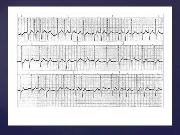
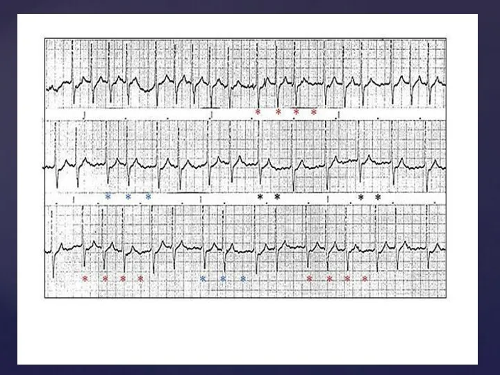
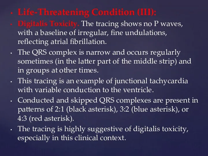
- 16. Life-Threatening Condition (III): Digitalis Toxicity. The tracing shows no P waves, with a baseline of irregular,
- 17. Case 4
- 18. A 25-year-old man arrives at the Emergency Department with a heavy cough after getting caught outside
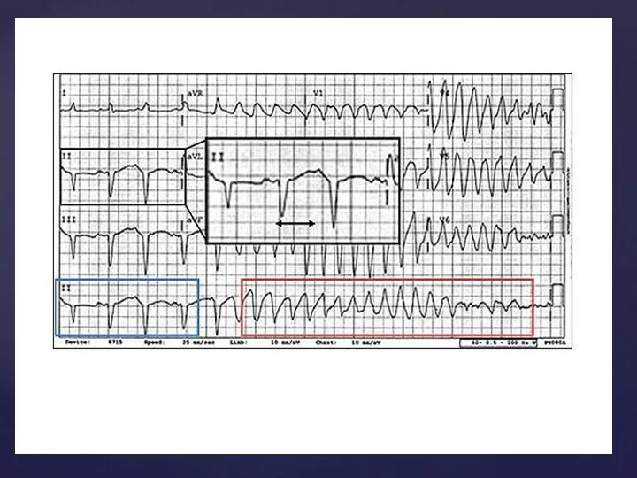
- 21. Life-Threatening Condition (IV): Artifact Simulating a Run of Ventricular Tachycardia. At first glance, this ECG suggests
- 22. Case 5
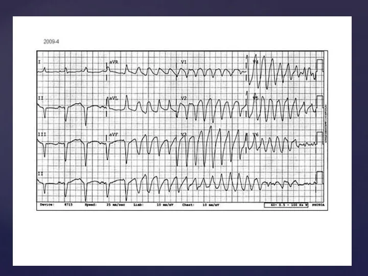
- 23. A 32-year-old woman comes to the Emergency Department complaining of light-headedness and sweating. She is 5'6"
- 27. Скачать презентацию
Case 1
Case 1
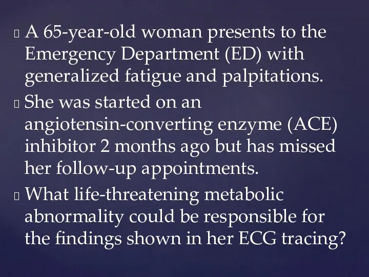
A 65-year-old woman presents to the Emergency Department (ED) with generalized
A 65-year-old woman presents to the Emergency Department (ED) with generalized
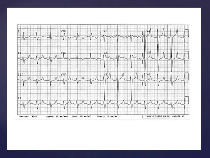
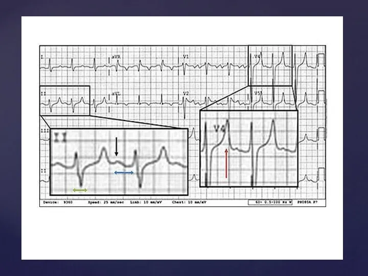
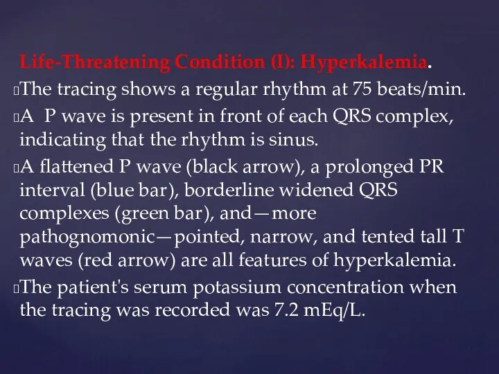
Life-Threatening Condition (I): Hyperkalemia.
The tracing shows a regular rhythm at
Life-Threatening Condition (I): Hyperkalemia.
The tracing shows a regular rhythm at

Case 2
Case 2

An 83-year-old man with known ischemic cardiomyopathy has an out-of-hospital cardiac
An 83-year-old man with known ischemic cardiomyopathy has an out-of-hospital cardiac
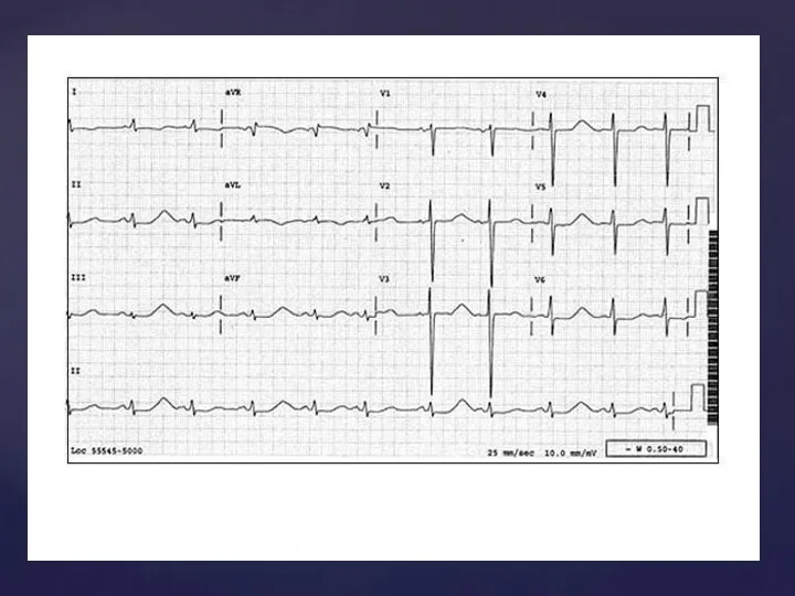
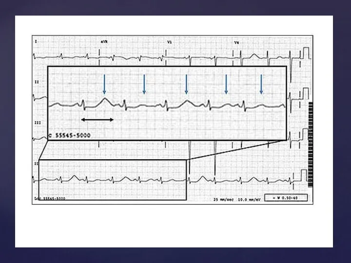
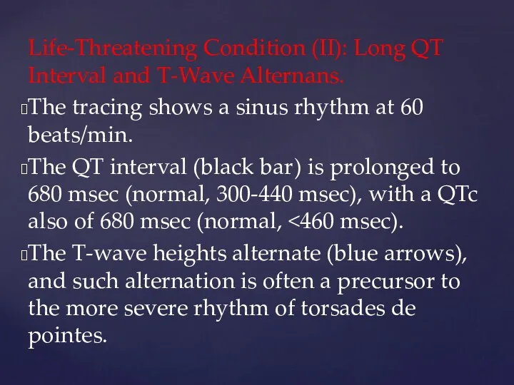
Life-Threatening Condition (II): Long QT Interval and T-Wave Alternans.
The tracing
Life-Threatening Condition (II): Long QT Interval and T-Wave Alternans.
The tracing

Case 3
Case 3
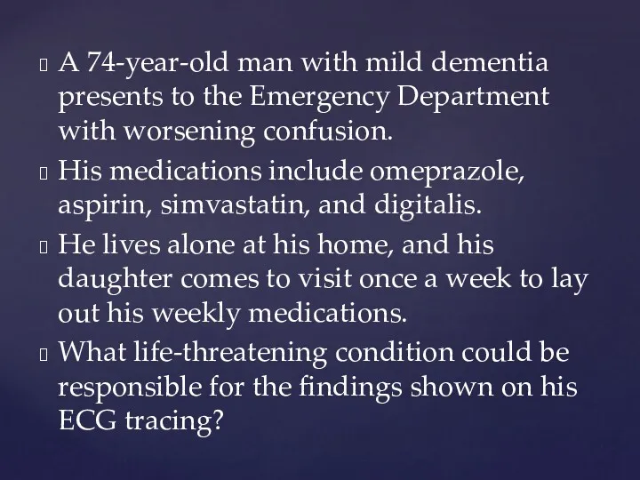
A 74-year-old man with mild dementia presents to the Emergency Department
A 74-year-old man with mild dementia presents to the Emergency Department
Life-Threatening Condition (III):
Digitalis Toxicity. The tracing shows no P waves,
Life-Threatening Condition (III):
Digitalis Toxicity. The tracing shows no P waves,
Case 4
Case 4
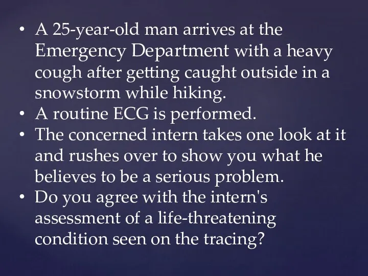
A 25-year-old man arrives at the Emergency Department with a heavy
A 25-year-old man arrives at the Emergency Department with a heavy
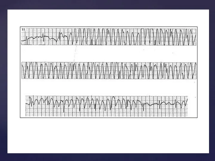
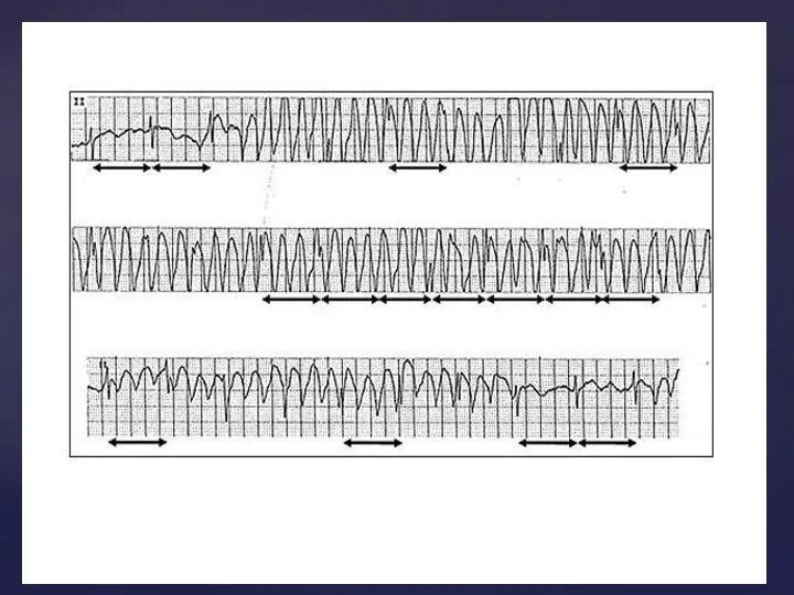
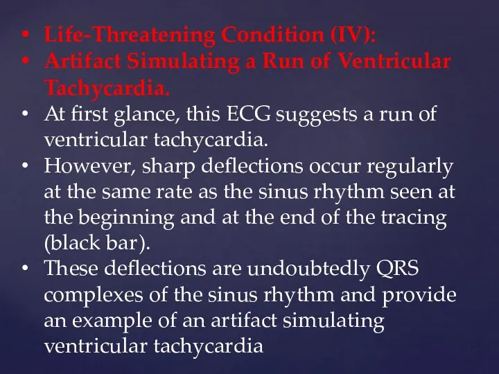
Life-Threatening Condition (IV):
Artifact Simulating a Run of Ventricular Tachycardia.
At
Life-Threatening Condition (IV):
Artifact Simulating a Run of Ventricular Tachycardia.
At
Case 5
Case 5
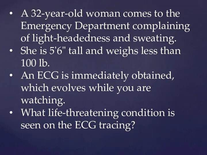
A 32-year-old woman comes to the Emergency Department complaining of light-headedness
A 32-year-old woman comes to the Emergency Department complaining of light-headedness
 Біомедична спілка - біоетика як галузь медицини
 Лечение псориаза
 Физиотерапевтические методы лечения заболеваний пародонта у детей
 Ангиогенин – естественное восстановление красоты и здоровья
 Німісил
 Визуальная диагностика при гипоталамо-гипофизарном ожирении
 Засоби, що діють в ділянці закінчень аферентних нервів
 Лапароцентез және диагностикалық лапароскопия
 Неодимовый лазер. Удаление тату. Удаление татуажа. Карбоновый пилинг
 Қан тобын анықтау
 Эпигенетика. Эпигенетическая патология в процессах канцерогенеза. Часть 3
 Менінгококова інфекція
 Фармакологія. Загальна рецептура
 Классификация опухолей ЦНС. ВОЗ 2016
 Недоношенный ребенок – особенности ухода. Ранняя реабилитация недоношенного новорожденного
 Никотиновая зависимость и как от нее избавится
 Осложнения инфаркта миокарда
 Болезни, передаваемые половым путём
 ХТІ, сальмонельоз, ботулізм
 Условия и факторы нормального и аномального развития личности
 Диагностика острого трахеита, острых и хронических бронхитов, эмфиземы легких
 Повреждения мочеполовых органов
 Использование логопедического массажа в коррекционной работе с детьми с тяжёлыми нарушениями речи
 Климактерический синдром
 Зардап шеккендер мен науқастарды жедел көмек көлігімен тасымалдау
 Неврологические осложнения гипотиреоза
 Денсаулық сақтаудағы менеджмент
 Физиологические основы питания. (Лекция 11)