- Fibroadenoma fibrocytic and mastitis

Содержание
- 2. Mastitis 1) Acute Mastitis : Occurs during the first month of breastfeeding. Caused by a local
- 3. Infection may spread to entire breast. Staphylococcal abscesses- single or multiple, Streptococci- spread infection in the
- 4. 2) Duct Ectasia Presents as a palpable periareolar mass with thick, white nipple secretions and occasionally
- 5. Morphology Ectatic dilated ducts are filled with inspissated secretions and numerous lipid-laden macrophages. When ruptured?marked periductal
- 6. deposits Granulomas may form around and cholesterol secretions. Subsequent mass with retraction. fibrosis? skin and irregular
- 7. 3) Granulomatous Mastitis: Can be a manifestation of systemic granulomatous diseases (e.g. polyangiitis, sarcoidosis, TB) or
- 8. Granulomatous lobular mastitis: Uncommon disease, occurs in parous women. Granulomas are closely associated with lobules, suggesting
- 9. Localized infections are most common in immunocompromised patients or adjacent to foreign objects such as breast
- 10. FIBROCYSTIC CHANGES Changes in female breast that range from innocuous to patterns associated with increased risk
- 11. Alterations subdivided into nonproliferative and proliferative patterns. Nonproliferative lesions- cysts and/or fibrosis and adenosis focally. Proliferative
- 12. Nonproliferative Change Most common type of alteration. Involved areas show ill-defined, diffusely increased density and discrete
- 13. Cysts: Brown to blue cysts filled with serous, turbid fluid. Secretory products may calcify, appear as
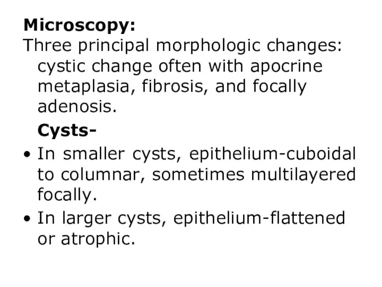
- 14. Microscopy: Three principal morphologic changes: cystic change often with apocrine metaplasia, fibrosis, and focally adenosis. Cysts-
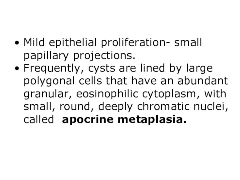
- 15. Mild epithelial proliferation- small papillary projections. Frequently, cysts are lined by large polygonal cells that have
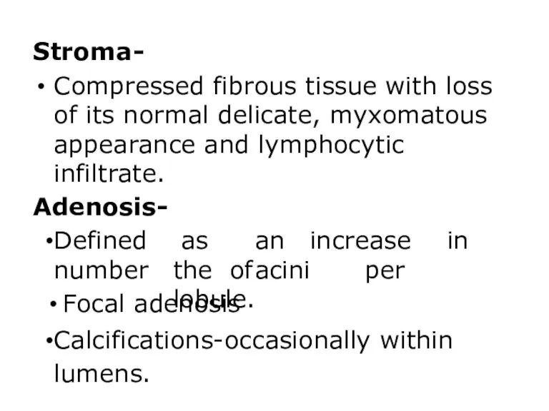
- 16. Stroma- Compressed fibrous tissue with loss of its normal delicate, myxomatous appearance and lymphocytic infiltrate. Adenosis-
- 17. Apocrine cysts. Cells with round nuclei and abundant granular eosinophilic cytoplasm, resembling cells of normal apocrine
- 18. Proliferative Change Disease Without Atypia Lesions characterized by proliferation of epithelial cells without atypia. Small increase
- 19. Microscopy- Wide spectrum Ducts, ductules, or lobules may be filled with orderly cuboidal cells, within which
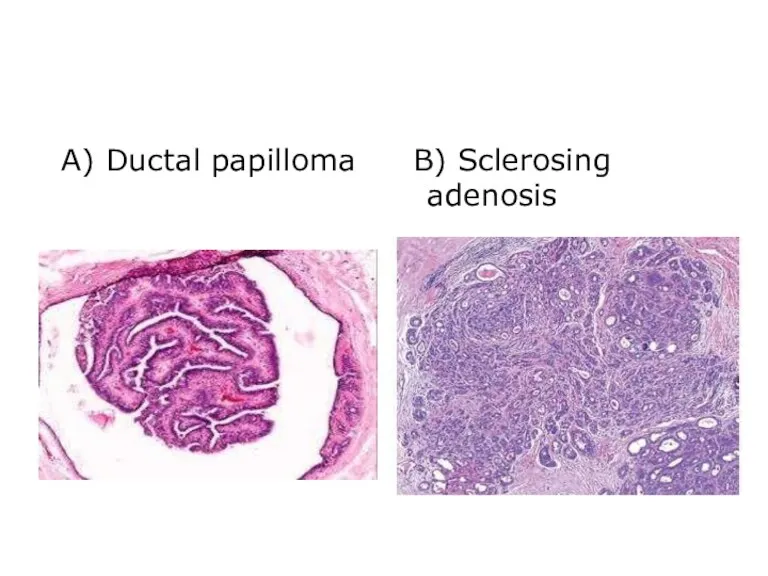
- 20. Papilloma within a dilated duct, composed of multiple branching fibrovascular cores into ductal lumen. Sclerosing Adenosis-Increased
- 21. A) Normal duct or acinus B, Epithelial hyperplasia. With irregular slitlike - fenestrations
- 22. A) Ductal papilloma B) Sclerosing adenosis
- 23. Proliferative Breast Disease with Atypia Hyperplasia with atypia is present in ducts or lobules. Moderately increased
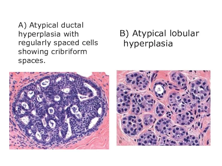
- 24. A) Atypical ductal hyperplasia with regularly spaced cells showing cribriform spaces. B) Atypical lobular hyperplasia
- 25. Fibroadenoma Most common benign fibroepithelial tumor of female breast. Increase in estrogen activity contributes to its
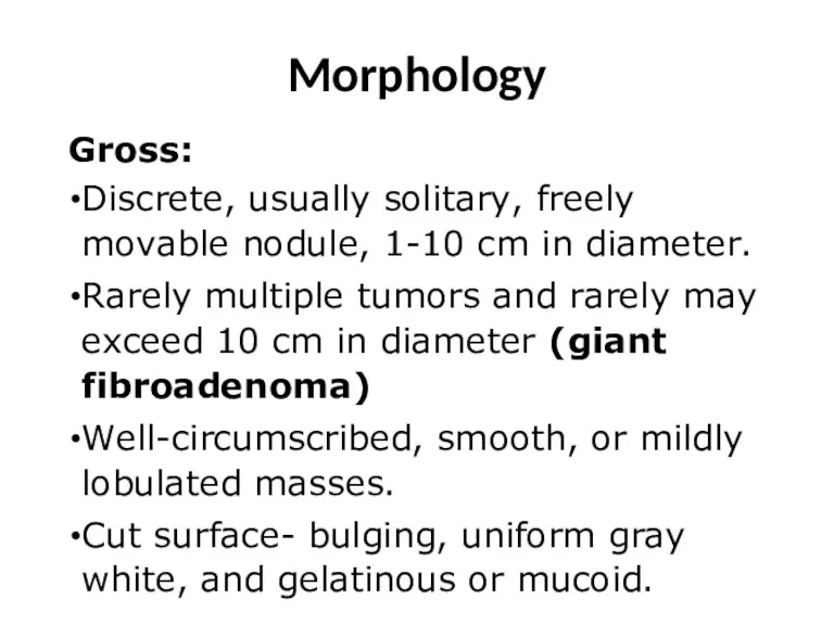
- 26. Morphology Gross: Discrete, usually solitary, freely movable nodule, 1-10 cm in diameter. Rarely multiple tumors and
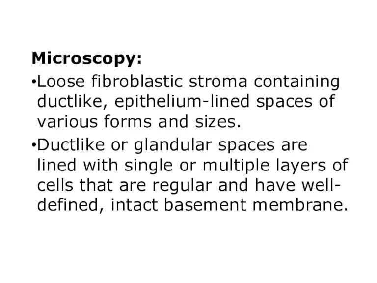
- 27. Microscopy: Loose fibroblastic stroma containing ductlike, epithelium-lined spaces of various forms and sizes. Ductlike or glandular
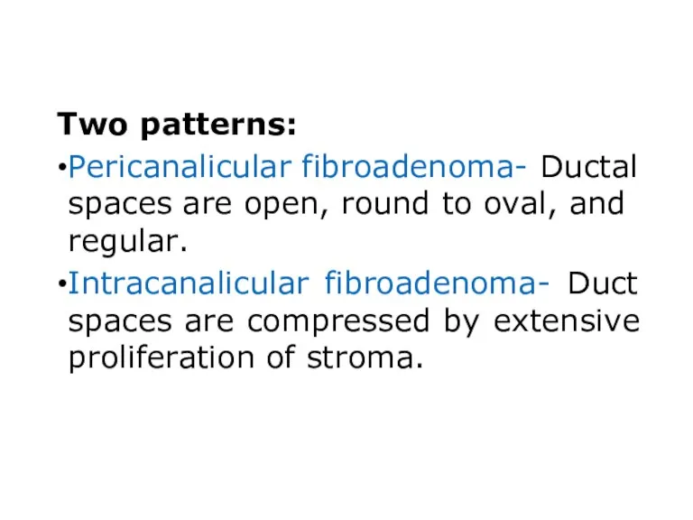
- 28. Two patterns: Pericanalicular fibroadenoma- Ductal spaces are open, round to oval, and regular. Intracanalicular fibroadenoma- Duct
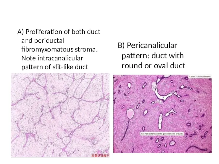
- 29. A) Proliferation of both duct and periductal fibromyxomatous stroma. Note intracanalicular pattern of slit-like duct B)
- 30. Clinical features Present as solitary, discrete, movable painless masses. May enlarge late in menstrual cycle and
- 32. Скачать презентацию
Mastitis
1) Acute Mastitis :
Occurs during the first month of breastfeeding.
Caused by
Mastitis
1) Acute Mastitis :
Occurs during the first month of breastfeeding.
Caused by
Infection may spread to entire breast.
Staphylococcal abscesses- single or multiple,
Streptococci- spread
Infection may spread to entire breast.
Staphylococcal abscesses- single or multiple,
Streptococci- spread
2) Duct Ectasia
Presents as a palpable periareolar mass with thick, white
2) Duct Ectasia
Presents as a palpable periareolar mass with thick, white
Morphology
Ectatic dilated ducts are filled with inspissated secretions and numerous lipid-laden macrophages.
When ruptured?marked periductal and interstitial chronic
inflammatory reaction
consisting of
lymphocytes, macrophages,
and
plasma cells.
Morphology
Ectatic dilated ducts are filled with inspissated secretions and numerous lipid-laden macrophages.
When ruptured?marked periductal and interstitial chronic
inflammatory reaction
consisting of
lymphocytes, macrophages,
and
plasma cells.
deposits
Granulomas may form around
and
cholesterol secretions.
Subsequent mass with retraction.
fibrosis? skin and
irregular nipple
deposits
Granulomas may form around
and
cholesterol secretions.
Subsequent mass with retraction.
fibrosis? skin and
irregular nipple
3) Granulomatous Mastitis:
Can be a manifestation of systemic granulomatous diseases (e.g.
3) Granulomatous Mastitis:
Can be a manifestation of systemic granulomatous diseases (e.g.
Granulomatous lobular mastitis:
Uncommon disease, occurs in parous women.
Granulomas are closely associated
Granulomatous lobular mastitis:
Uncommon disease, occurs in parous women.
Granulomas are closely associated
Localized infections are most common in immunocompromised patients or adjacent to foreign
Localized infections are most common in immunocompromised patients or adjacent to foreign
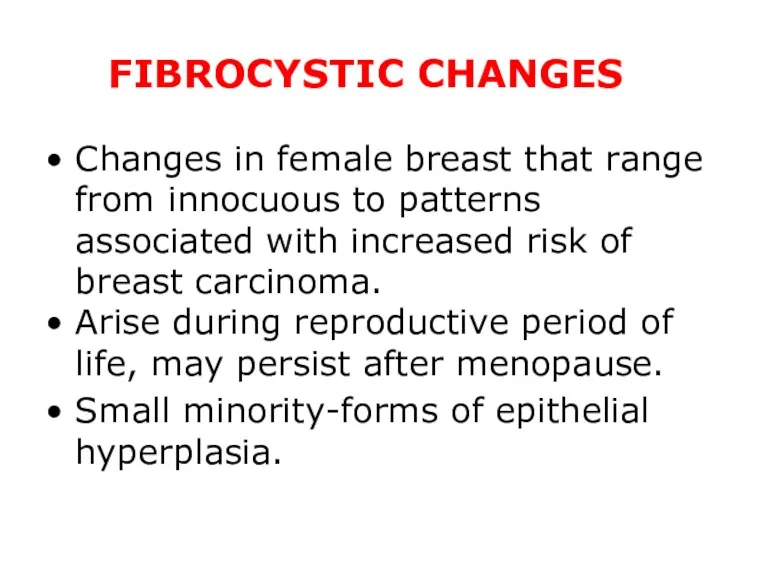
FIBROCYSTIC CHANGES
Changes in female breast that range from innocuous to patterns
FIBROCYSTIC CHANGES
Changes in female breast that range from innocuous to patterns
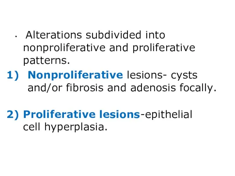
Alterations subdivided into nonproliferative and proliferative patterns.
Nonproliferative lesions- cysts and/or fibrosis
Alterations subdivided into nonproliferative and proliferative patterns.
Nonproliferative lesions- cysts and/or fibrosis
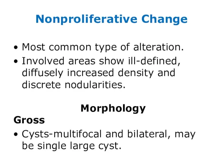
Nonproliferative Change
Most common type of alteration.
Involved areas show ill-defined, diffusely increased
Nonproliferative Change
Most common type of alteration.
Involved areas show ill-defined, diffusely increased
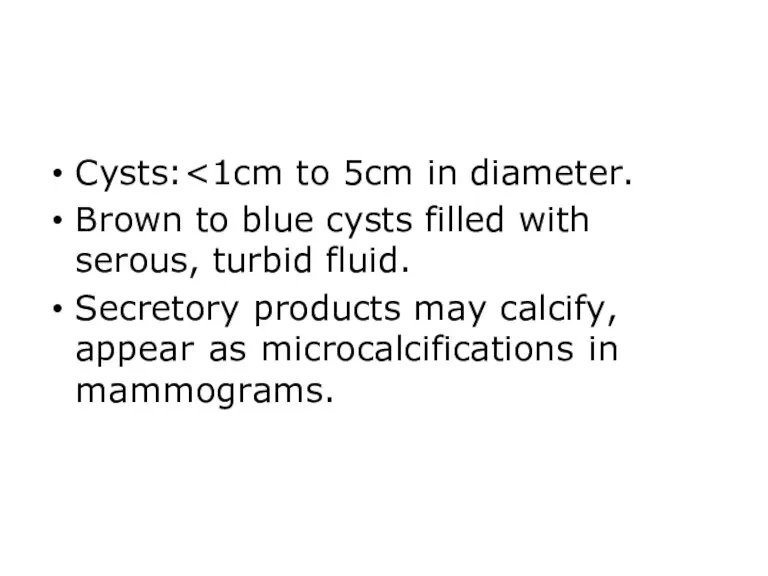
Cysts:<1cm to 5cm in diameter.
Brown to blue cysts filled with serous,
Cysts:<1cm to 5cm in diameter.
Brown to blue cysts filled with serous,
Microscopy:
Three principal morphologic changes: cystic change often with apocrine metaplasia, fibrosis,
Microscopy:
Three principal morphologic changes: cystic change often with apocrine metaplasia, fibrosis,
Mild epithelial proliferation- small papillary projections.
Frequently, cysts are lined by large
Mild epithelial proliferation- small papillary projections.
Frequently, cysts are lined by large
Stroma-
Compressed fibrous tissue with loss of its normal delicate, myxomatous appearance
Stroma-
Compressed fibrous tissue with loss of its normal delicate, myxomatous appearance
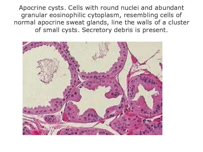
Apocrine cysts. Cells with round nuclei and abundant granular eosinophilic cytoplasm,
Apocrine cysts. Cells with round nuclei and abundant granular eosinophilic cytoplasm,
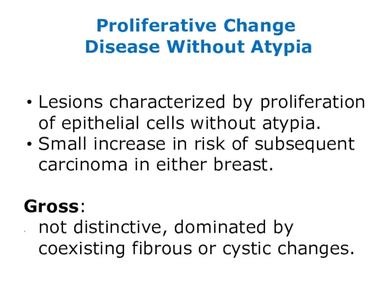
Proliferative Change Disease Without Atypia
Lesions characterized by proliferation of epithelial cells
Proliferative Change Disease Without Atypia
Lesions characterized by proliferation of epithelial cells
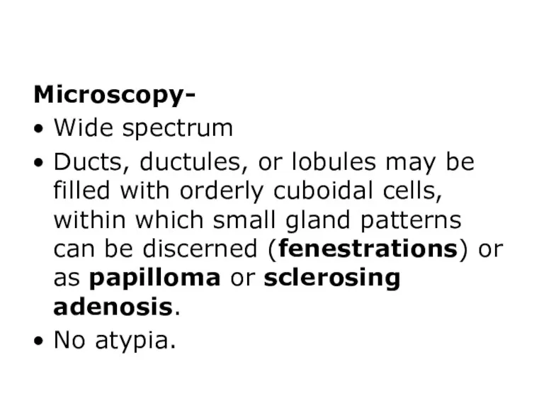
Microscopy-
Wide spectrum
Ducts, ductules, or lobules may be filled with orderly cuboidal
Microscopy-
Wide spectrum
Ducts, ductules, or lobules may be filled with orderly cuboidal
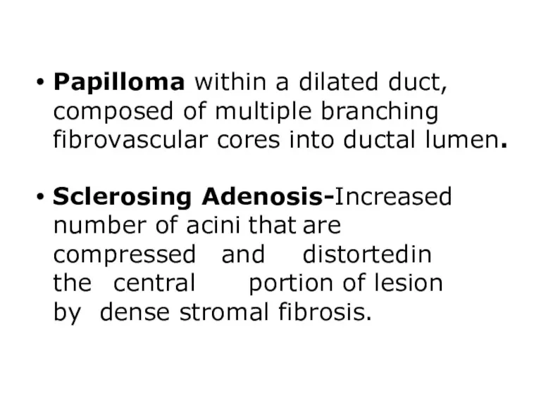
Papilloma within a dilated duct, composed of multiple branching fibrovascular cores into
Papilloma within a dilated duct, composed of multiple branching fibrovascular cores into
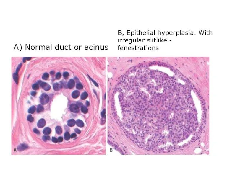
A) Normal duct or acinus
B, Epithelial hyperplasia. With irregular slitlike -
A) Normal duct or acinus
B, Epithelial hyperplasia. With irregular slitlike -
A) Ductal papilloma
B) Sclerosing adenosis
A) Ductal papilloma
B) Sclerosing adenosis
Proliferative Breast Disease with Atypia
Hyperplasia with atypia is present in ducts
Proliferative Breast Disease with Atypia
Hyperplasia with atypia is present in ducts
A) Atypical ductal hyperplasia with regularly spaced cells showing cribriform spaces.
B)
A) Atypical ductal hyperplasia with regularly spaced cells showing cribriform spaces.
B)
Fibroadenoma
Most common benign fibroepithelial tumor of female breast.
Increase in estrogen activity
Fibroadenoma
Most common benign fibroepithelial tumor of female breast.
Increase in estrogen activity
Morphology
Gross:
Discrete, usually solitary, freely movable nodule, 1-10 cm in diameter.
Rarely multiple
Morphology
Gross:
Discrete, usually solitary, freely movable nodule, 1-10 cm in diameter.
Rarely multiple
Microscopy:
Loose fibroblastic stroma containing ductlike, epithelium-lined spaces of various forms and
Microscopy:
Loose fibroblastic stroma containing ductlike, epithelium-lined spaces of various forms and
Two patterns:
Pericanalicular fibroadenoma- Ductal spaces are open, round to oval, and
Two patterns:
Pericanalicular fibroadenoma- Ductal spaces are open, round to oval, and
A) Proliferation of both duct and periductal fibromyxomatous stroma. Note intracanalicular
A) Proliferation of both duct and periductal fibromyxomatous stroma. Note intracanalicular
Clinical features
Present as solitary, discrete, movable painless masses.
May enlarge late in menstrual
Clinical features
Present as solitary, discrete, movable painless masses.
May enlarge late in menstrual
 Пневмония. Этиология. Принципы лечения
 Острые нарушения мозгового кровообращения
 Анатомо-фізіологічні особливості жувального апарату при повній втраті зубів
 Микроорганизмы в окружающей среде
 Дейл Карнеги бойынша қарым-қатынас орнату
 Презентация тема № 2 (из 17) Трепанация черепа, инструменты, ПХО 6 семестр Готово 02.02.23
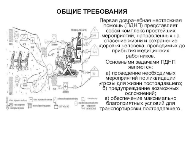 Первая доврачебная неотложная помощь
 Протеи. Причины возникновения инфекций, вызванных протеем
 Жатыр мен қынаптың толықтай емес түсуі бар әйелдерде кольпорафиялық емнің және пессарий қоюдың нәтижелігін анықтау
 Диагностика туберкулеза
 Стационардағы медбикенің қол жуу маңыздылығы
 Нормализация моторики органов артикуляции при дизартрии
 Неврологиялық ауруларды алдын-алу шаралары
 Electrical Processes of the Heart
 Хирургическое лечение эпилепсии и паркинсонизма
 Орнитоз (пситтакоз)
 Комы. Патогенез развития коматозных состояний
 Заболевание глотки
 Тромботические микроангиопатии. Большая проблема или интересная задача
 Бактериальные инфекции
 Гангренозный стоматит. Ангина Симановского-Плаута-Венсана
 Тіндердің шығу тегі мен дамуының заңдылықтары. Тіндердің жіктелуі
 Хто вони: Рослини чи Тварини?
 Жатыр мойны қатерлі ісігі
 Pflege von Menschen mit Erkrankungen der Atemwege und Lunge
 Рентгенанатомия и рентгенпатология желудочно-кишечного тракта
 Аневризмы сосудов ГМ
 A comparison of pill dispensers