Hprogram of subject (syllabus). Recent developments of biotechnology in veterinary medicine and animal husbandry презентация
- Hprogram of subject (syllabus). Recent developments of biotechnology in veterinary medicine and animal husbandry

Содержание
- 2. Distribution of training period
- 3. Course objectives is: to familiarize Masters with new developments and achievements of modern Biotechnology in the
- 4. As a result of studying this subject, masters must know: the latest achievements of world science
- 5. COURSE CONTENT
- 6. List of Practical Classes
- 7. SCHEDULE OF ACCEPTANCE MIW’s themes on discipline "Recent developments of Biotechnology in Veterinary Medicine and Animal
- 8. REFERENCE Basic Literature: The Basic literature of the discipline are articles and reviews published in scientific
- 9. HYBRIDOMA TECHNIQUE TEACHING OBJECTIVES: 1.INTRODUCTION 2. PRINCIPLE INVOLVED IN MONOCLONAL ANTIBODIES PRODUCTION 3. PRODUCTION OF MONOCLONAL
- 10. INTRODUCTION
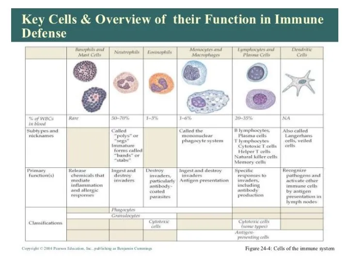
- 13. Two neutrophils in blood film Polymorphonuclear cells are recruited to the site of infection where they
- 18. Bone Marrow Bone marrow (medulla ossea) is the site of B cell maturation in mice and
- 21. The thymus is a two-lobed organ overlying the upper part of the heart. It is large
- 25. Antibodies are produced by a specialized group of cells called B-Lymphocytes. When an foreign antigen enters
- 26. WHAT’S THE ROLE OF ANTIBODY IN IMMUNE SYSTEM? Each Antigen has specific antigen determinants (epitopes) located
- 27. Поликлональность антител при традиционной технологии
- 28. Общие эпитопы гетерогенных антигенов
- 31. WHAT’S THE NEED TO DEVELOP MONOCLONAL ANTIBODIES? In general naturally produced antibodies are non-specific and heterogenous
- 32. WHAT ARE MONOCLONAL ANTIBODIES? MAb is a single type of antibody that is directed against a
- 33. СРАВНЕНИЕ ПОЛИКЛОНАЛЬНЫХ И МОНОКЛОНАЛЬНЫХ АНТИТЕЛ
- 34. History of Mab development 1890 Von Behring and kitasato discovered the serum of vaccinated persons contained
- 35. Structure of MAb
- 36. Antigen- antibody binding
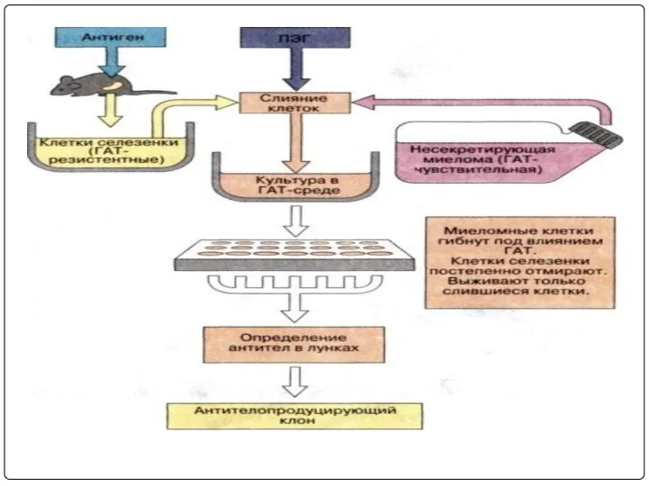
- 37. Hybridoma technology: In this B-Lymphocytes and myeloma cells are mixed together and exposed to PEG for
- 38. Плазмоцитомы
- 39. Слияние клеток человека и мыши
- 41. Immunise Spleen Cell Myeloma Cell Line FUSE HAT sensitive Hybridoma HAT resistant Stable hybrid myeloma producing
- 42. Immunization Cell fusion Selection of hybridomas Screening the products Cloning and propagation Characterization and storage PRODUCTION
- 43. Мыши линии Balb/c
- 44. Immunize an animal usually mouse by injecting with an appropriate antigen along with Freund’s complete or
- 45. Иммунизация мыши линии Balb/c
- 46. Lymphocytes are mixed with HGPRT deficient myeloma cells and is exposed to PEG for a short
- 47. Dihydrofolate Synthesis of nucleotides Tetrahydrofolate Precursors Nucleotides---->DNA Hypoxanthine Thymidine De novo synthesis Salvage pathway Aminopterin HGPRT
- 48. The above mixture is cultured in HAT medium for 7-10 days. Due to lack of HGPRT
- 49. Среда RPMI-1640
- 50. Сыворотка плода коровы
- 51. Слияние иммунных лимфоцитов с миеломой
- 52. 96-луночные планшеты для культуральных работ
- 53. Образование гибридной клетки
- 54. Слияние лифоцитов с миеломой
- 57. Распределение клеток по лункам планшеты
- 58. Культивирование гибридом в СО2 -инкубаторе
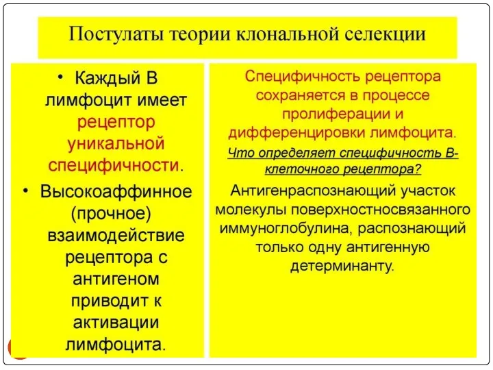
- 60. КЛОНАЛЬНО-СЕЛЕКЦИОННАЯ ТЕОРИЯ БЕРНЕТА
- 62. Виды клеток, образуемые в процессе слияния Неслившиеся клетки лимфоидного органа; Неслившиеся клетки миеломы; Гибриды лимфоцит+лимфоцит и
- 63. The above mixture is cultured in HAT medium for 7-10 days. Due to lack of HGPRT
- 64. Изоляция гибридов лимфоцит+миелома - от неслившихся лимфоцитов и гибридов лимфоцит+лимфоцит избавляться не нужно: через несколько дней
- 65. Screening is done for antibody specificity. For this we need to test the culture medium from
- 67. Схема получения МКА
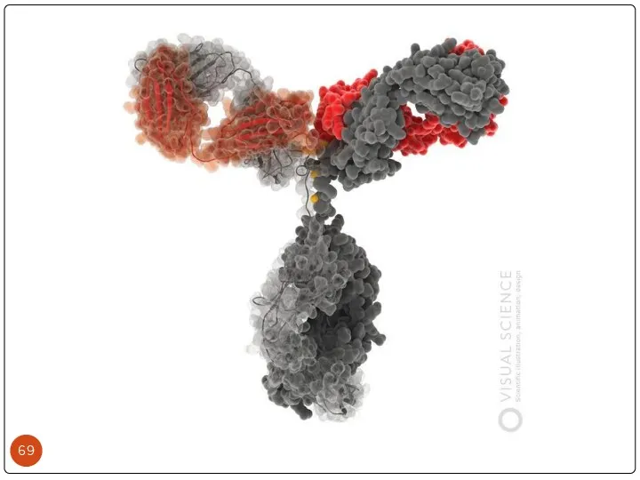
- 68. Electrophoretic separation of serum proteins DEFINITION Immunoglobulin (Ig) Immunoglobulins are glycoprotein molecules that are produced by
- 70. Heavy and Light Chains All immunoglobulins have a four chain structure as their basic unit. They
- 71. The basic structure of immunoglobulins Hinge Region This is the region at which the arms of
- 75. 96-луночный планшет для ИФА
- 76. ПРИНЦИП НЕПРЯМОГО ИФА
- 80. Спектрофотометр для ИФА
- 81. The single hybrid cell producing the desired antibody are isolated and cloned. Usually two techniques are
- 82. Выделение макрофагов для «питающего слоя»
- 83. 96-луночные планшеты для культуральных работ
- 84. «Питающий слой» из макрофагов
- 85. Biochemical and biophysical characterization are made for desired specificity. It is important to note the monoclonal
- 86. Хранение клеток в жидком азоте
- 87. Разморозка гибридомных клеток
- 88. Накопление МКА в матрасах
- 89. Наработка МКА в асцитной жидкости
- 92. Очистка МКА с помощью гель-фильтрации
- 95. Encapsulating the hybridoma cells in alginate gels and using a coating solution containing poly-lysine is employed.
- 96. АФФИННАЯ ХРОМАТОГРАФИЯ
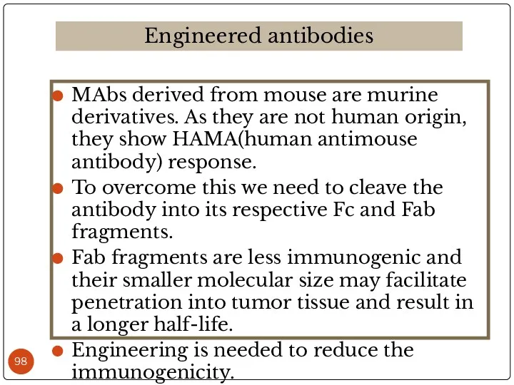
- 98. MAbs derived from mouse are murine derivatives. As they are not human origin, they show HAMA(human
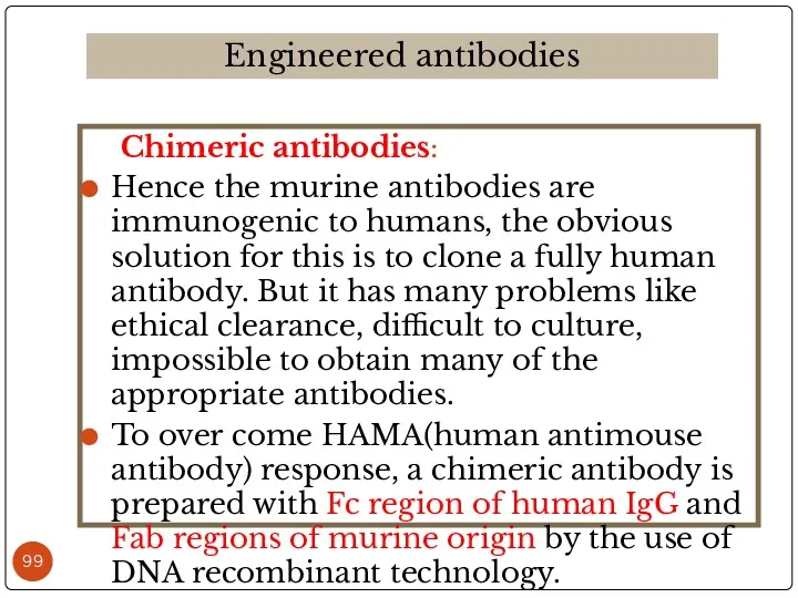
- 99. Chimeric antibodies: Hence the murine antibodies are immunogenic to humans, the obvious solution for this is
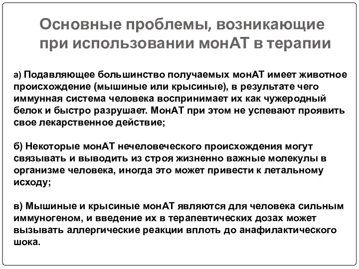
- 100. Основные проблемы, возникающие при использовании монАТ в терапии а) Подавляющее большинство получаемых монАТ имеет животное происхождение
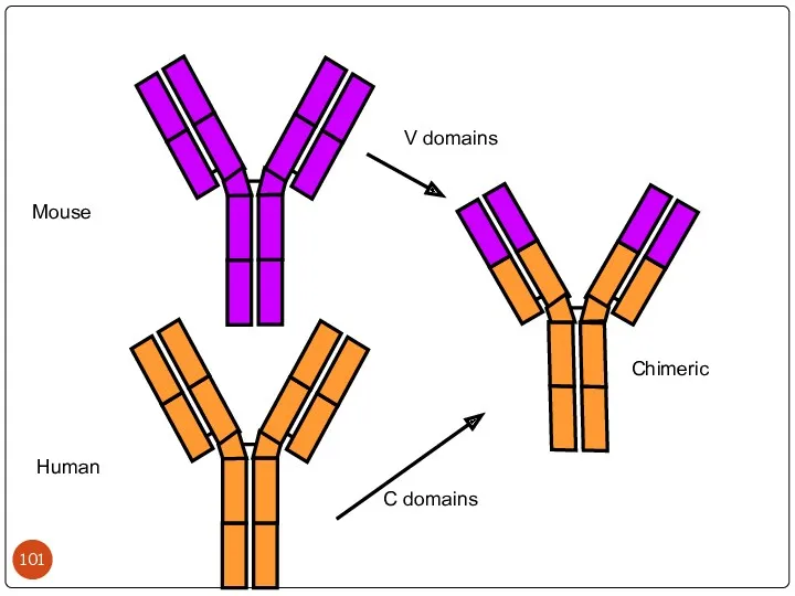
- 101. Mouse Human Chimeric V domains C domains
- 102. Химерные МКА
- 103. Humanized antibodies: Though chimeric antibodies elicit less HAMA response than murine antibodies, they are still immunogenic
- 104. Hypervariable (HVR) or complementarity determining regions (CDR) Comparisons of the amino acid sequences of the variable
- 105. Mouse Human Humanised hypervariable framework
- 106. Bispecific antibodies: These are specific to two types of antigens. They are constructed by r.DNA technology.
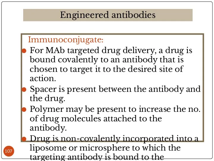
- 107. Immunoconjugate: For MAb targeted drug delivery, a drug is bound covalently to an antibody that is
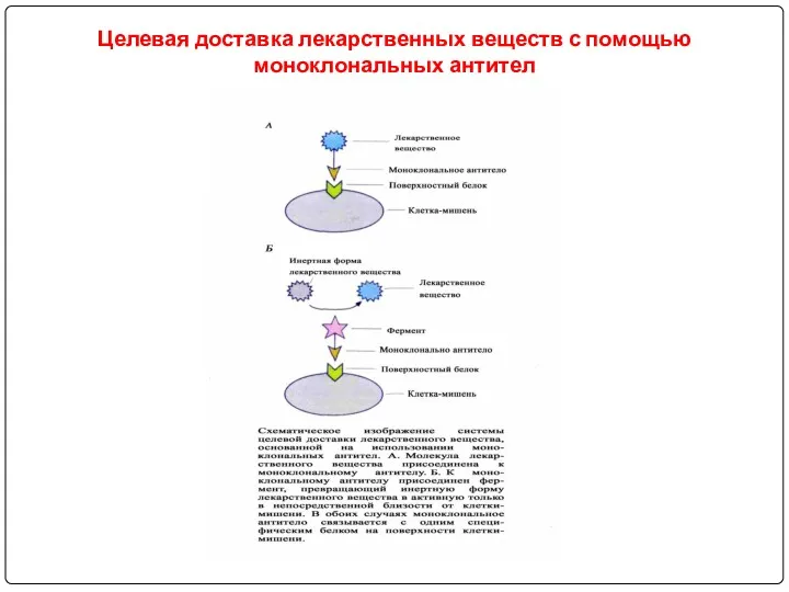
- 108. Целевая доставка лекарственных веществ с помощью моноклональных антител
- 112. Principle involved: As several classes of the drugs lack specificity for diseased cells, they show their
- 113. It is the natural in-vivo distribution pattern of the drug delivery system. It is determined by
- 114. In this some characteristics of the environment are utilized for the carrying of the drug to
- 115. Active targeting is usually done by cell-specific ligands. These are specific to specific cell types. But
- 116. Toxin conjugates (immunotoxins) EX: diphtheria toxin, Ricin have been conjugated to the tumor specific antibodies Ricin
- 117. Drug immunoconjugates: Agents like chlorambucil, methotrexate and doxorubicin are conjugated with tumor specific antibodies. Ex: doxorubicin-BR96
- 118. They are homogenous in nature. They are specific to a particular antigen with a particular epitope.
- 119. Cell Depletion Rituxan, Campath (naked) Myelotarg (drug) Zevalin, Bexxar (radioisotope) Blocking receptors Herceptin Attacking vasculature Avastin,
- 120. Препараты МкАТ, используемые при лечении онкологических болезнях
- 121. Препараты МкАТ, используемые при лечении аутоиммунных заболеваний
- 122. Препараты МкАТ, используемые в трансплантологии
- 124. Скачать презентацию
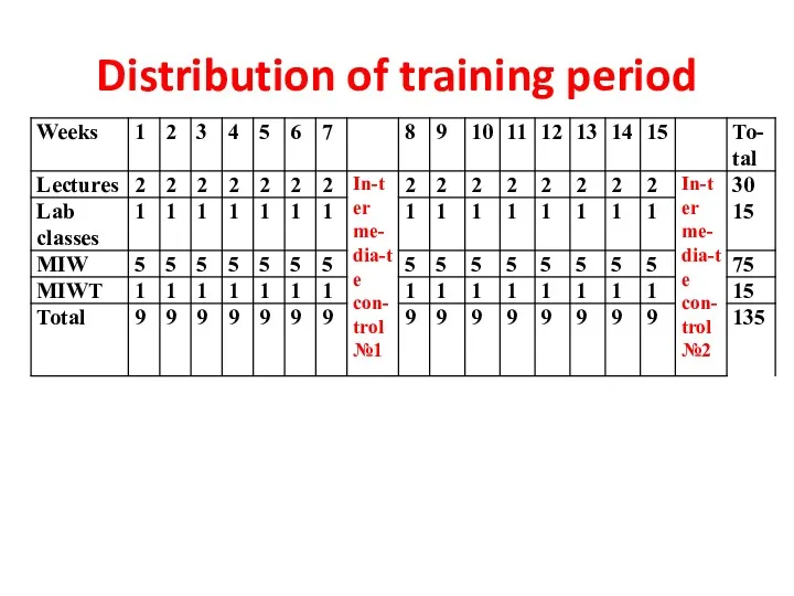
Distribution of training period
Distribution of training period
Course objectives is:
to familiarize Masters with new developments and achievements of
Course objectives is:
to familiarize Masters with new developments and achievements of
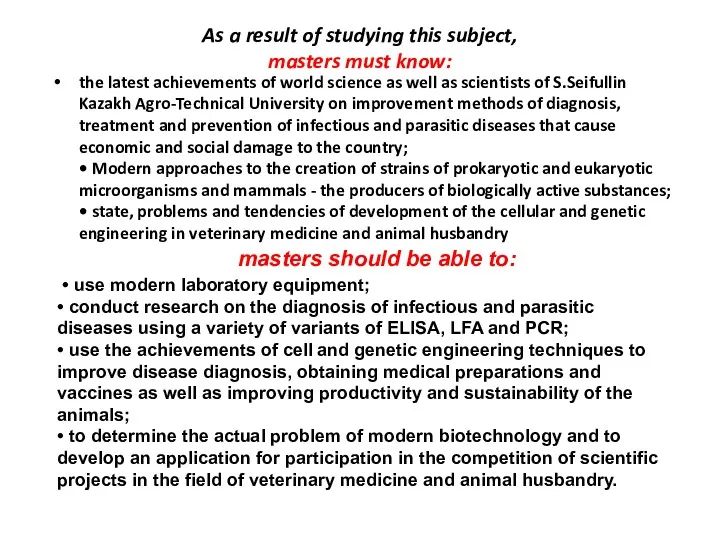
As a result of studying this subject,
masters must know:
the latest
As a result of studying this subject,
masters must know:
the latest
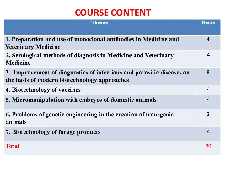
COURSE CONTENT
COURSE CONTENT
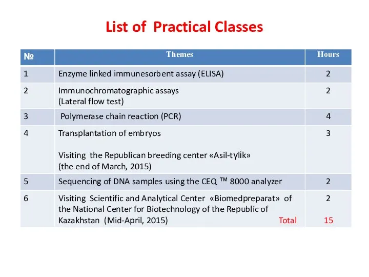
List of Practical Classes
List of Practical Classes
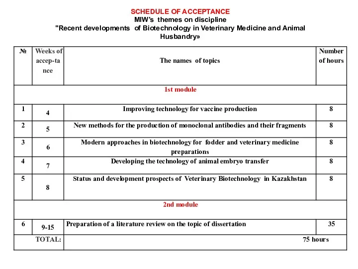
SCHEDULE OF ACCEPTANCE
MIW’s themes on discipline
"Recent developments of Biotechnology in
SCHEDULE OF ACCEPTANCE MIW’s themes on discipline "Recent developments of Biotechnology in
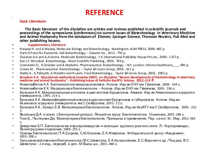
REFERENCE
Basic Literature:
The Basic literature of the discipline are articles and
REFERENCE
Basic Literature: The Basic literature of the discipline are articles and
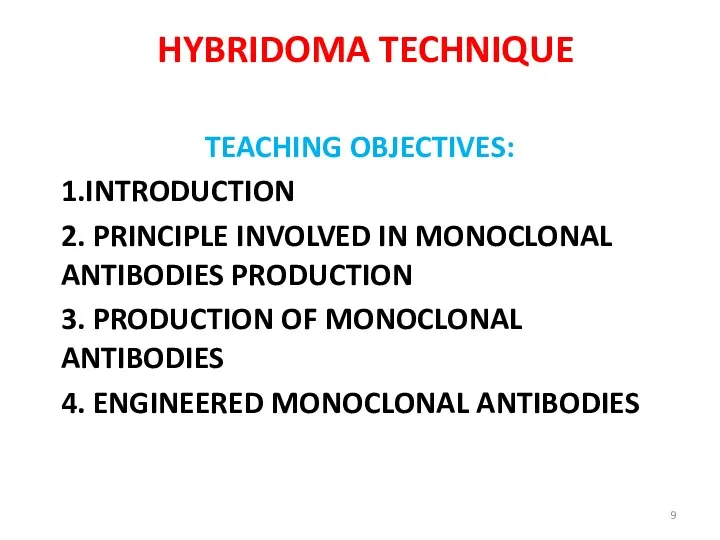
HYBRIDOMA TECHNIQUE
TEACHING OBJECTIVES:
1.INTRODUCTION
2. PRINCIPLE INVOLVED IN MONOCLONAL ANTIBODIES PRODUCTION
3. PRODUCTION OF
HYBRIDOMA TECHNIQUE
TEACHING OBJECTIVES:
1.INTRODUCTION
2. PRINCIPLE INVOLVED IN MONOCLONAL ANTIBODIES PRODUCTION
3. PRODUCTION OF
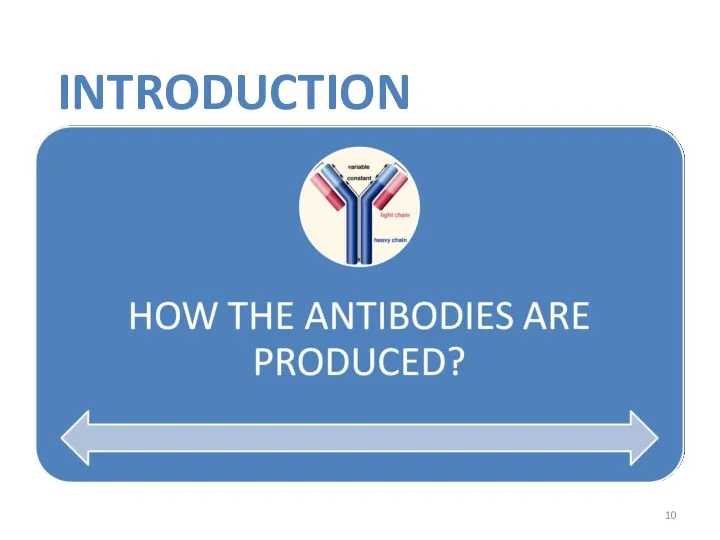
INTRODUCTION
INTRODUCTION
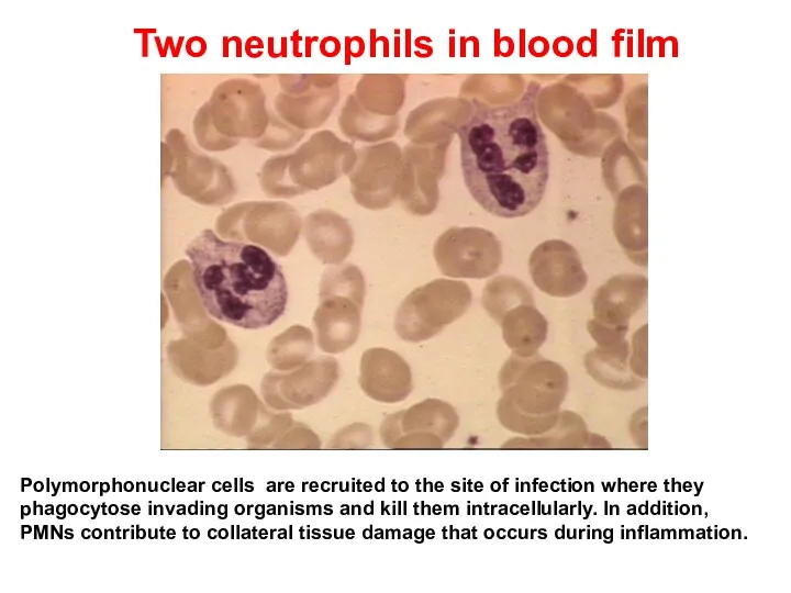
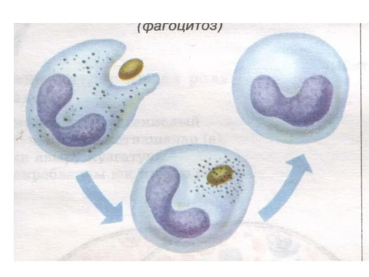
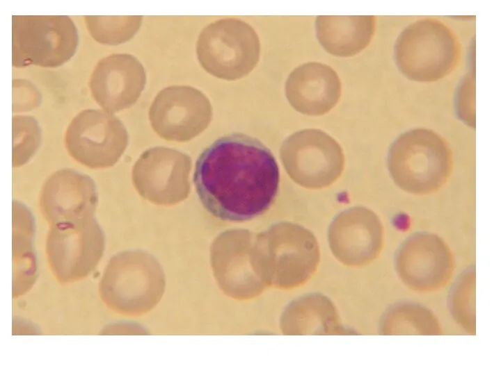
Two neutrophils in blood film
Polymorphonuclear cells are recruited to the site
Two neutrophils in blood film
Polymorphonuclear cells are recruited to the site
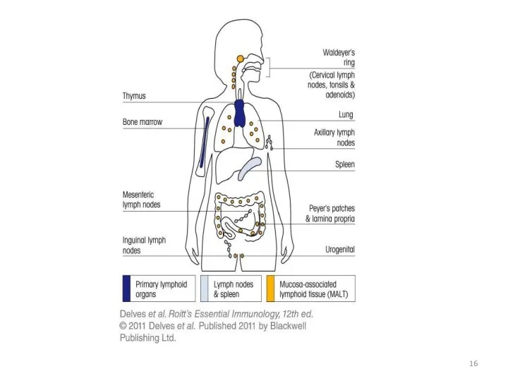
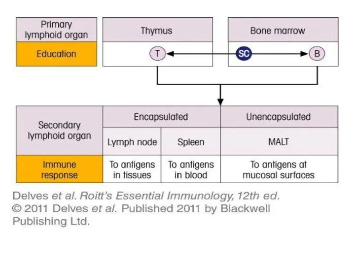
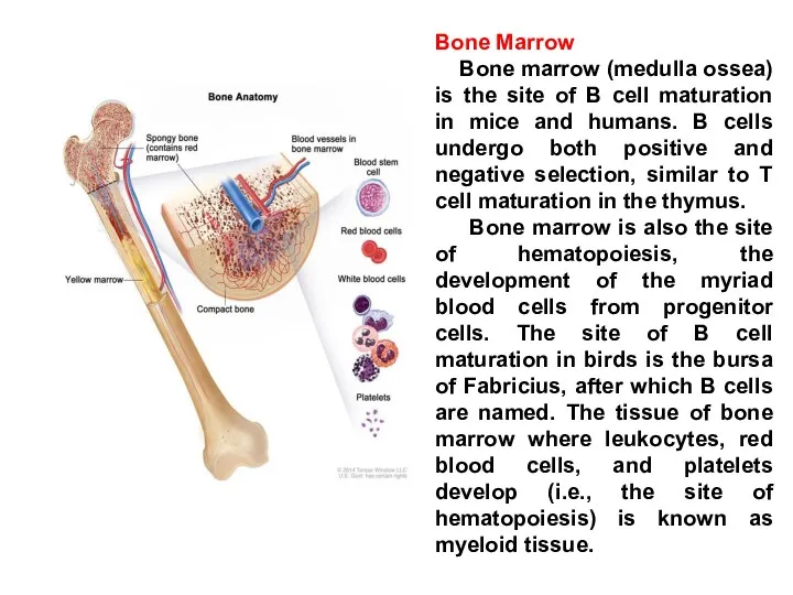
Bone Marrow
Bone marrow (medulla ossea) is the site of B
Bone Marrow
Bone marrow (medulla ossea) is the site of B
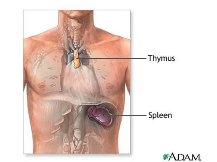
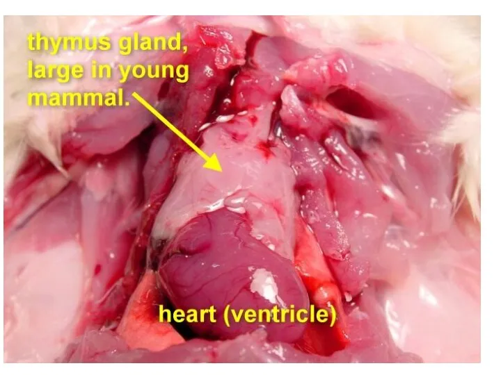

The thymus is a two-lobed organ overlying the upper part of
The thymus is a two-lobed organ overlying the upper part of
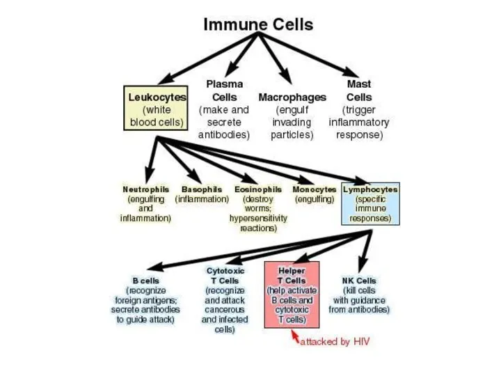
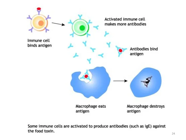
Antibodies are produced by a specialized group of cells called B-Lymphocytes.
When
Antibodies are produced by a specialized group of cells called B-Lymphocytes.
When
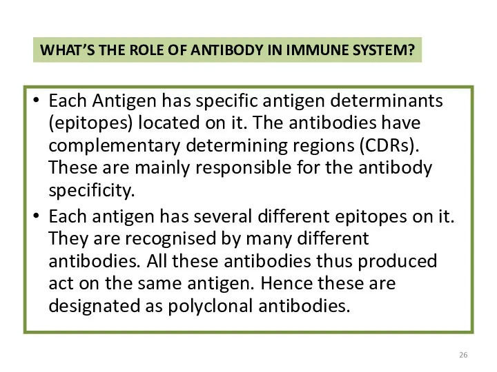
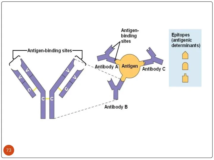
WHAT’S THE ROLE OF ANTIBODY IN IMMUNE SYSTEM?
Each Antigen has specific
WHAT’S THE ROLE OF ANTIBODY IN IMMUNE SYSTEM?
Each Antigen has specific
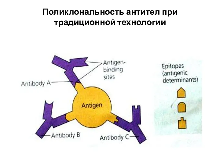
Поликлональность антител при традиционной технологии
Поликлональность антител при традиционной технологии
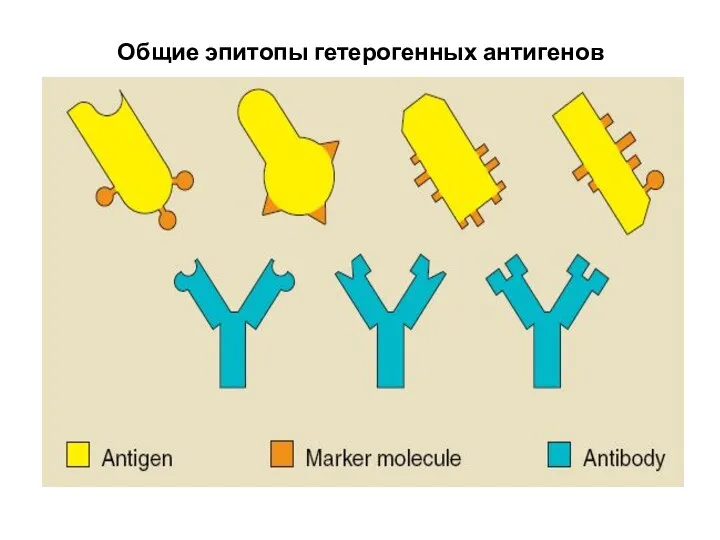
Общие эпитопы гетерогенных антигенов
Общие эпитопы гетерогенных антигенов
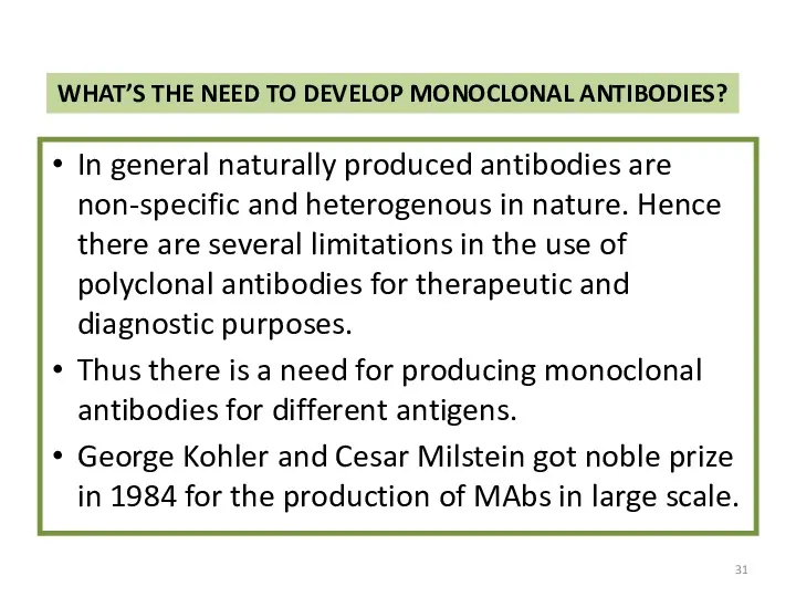
WHAT’S THE NEED TO DEVELOP MONOCLONAL ANTIBODIES?
In general naturally produced antibodies
WHAT’S THE NEED TO DEVELOP MONOCLONAL ANTIBODIES?
In general naturally produced antibodies
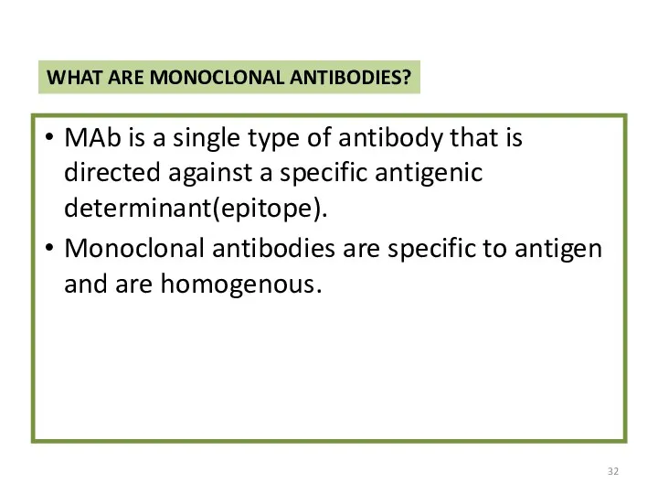
WHAT ARE MONOCLONAL ANTIBODIES?
MAb is a single type of antibody that
WHAT ARE MONOCLONAL ANTIBODIES?
MAb is a single type of antibody that
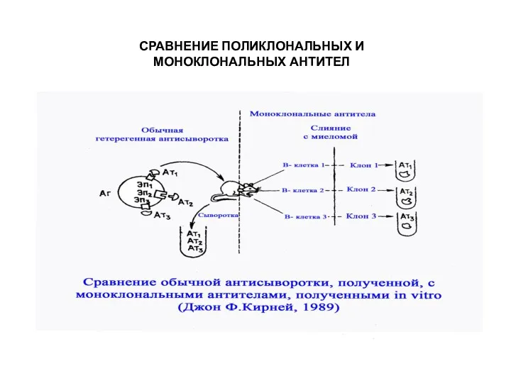
СРАВНЕНИЕ ПОЛИКЛОНАЛЬНЫХ И МОНОКЛОНАЛЬНЫХ АНТИТЕЛ
СРАВНЕНИЕ ПОЛИКЛОНАЛЬНЫХ И МОНОКЛОНАЛЬНЫХ АНТИТЕЛ
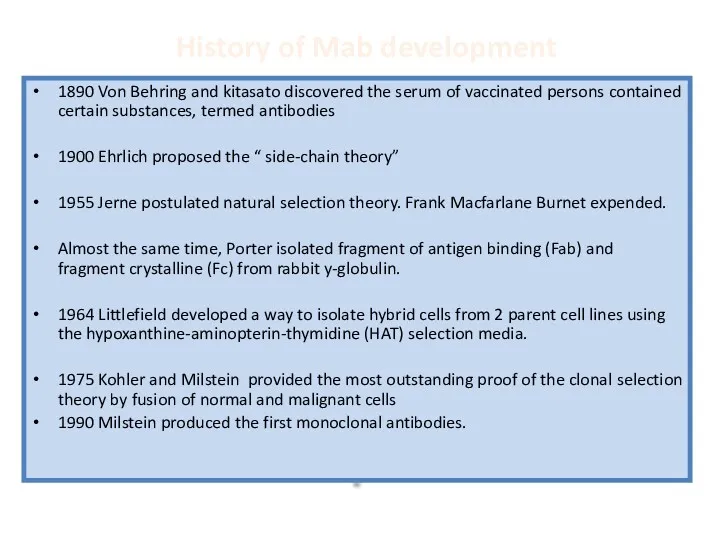
History of Mab development
1890 Von Behring and kitasato discovered the serum
History of Mab development
1890 Von Behring and kitasato discovered the serum
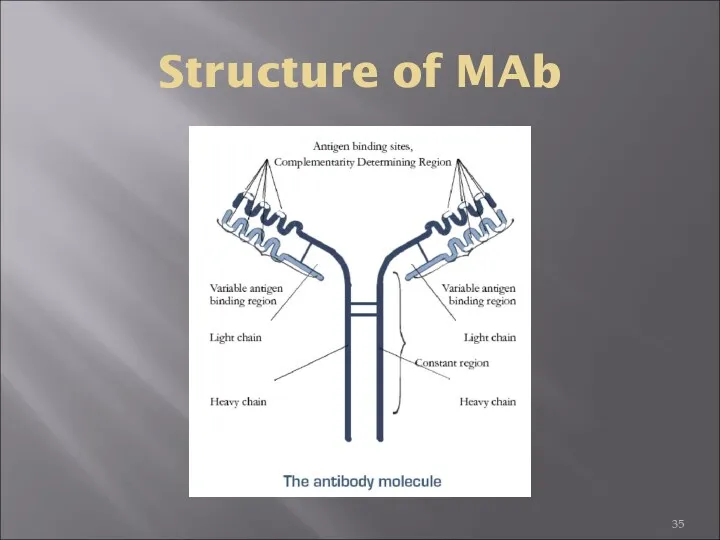
Structure of MAb
Structure of MAb
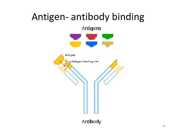
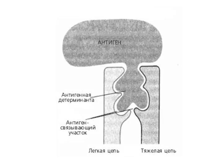
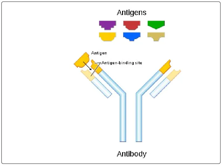
Antigen- antibody binding
Antigen- antibody binding
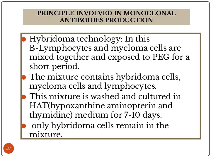
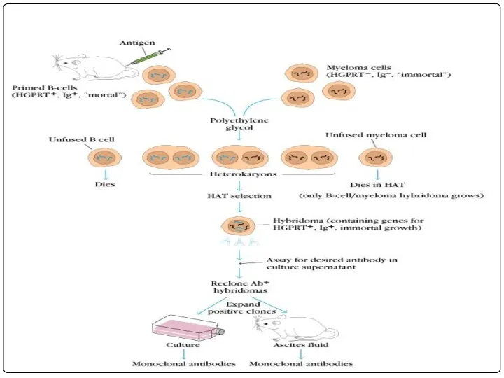
Hybridoma technology: In this B-Lymphocytes and myeloma cells are mixed together
Hybridoma technology: In this B-Lymphocytes and myeloma cells are mixed together
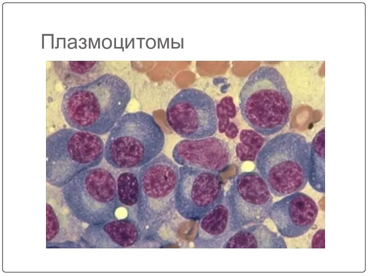
Плазмоцитомы
Плазмоцитомы
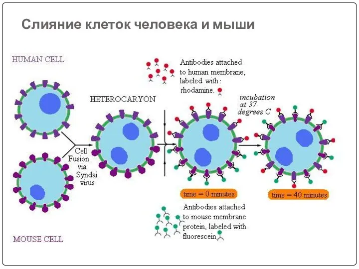
Слияние клеток человека и мыши
Слияние клеток человека и мыши
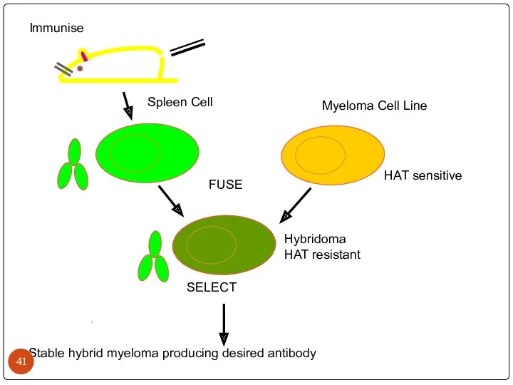
Immunise
Spleen Cell
Myeloma Cell Line
FUSE
HAT sensitive
Hybridoma
HAT resistant
Stable hybrid myeloma producing desired
Immunise
Spleen Cell
Myeloma Cell Line
FUSE
HAT sensitive
Hybridoma
HAT resistant
Stable hybrid myeloma producing desired
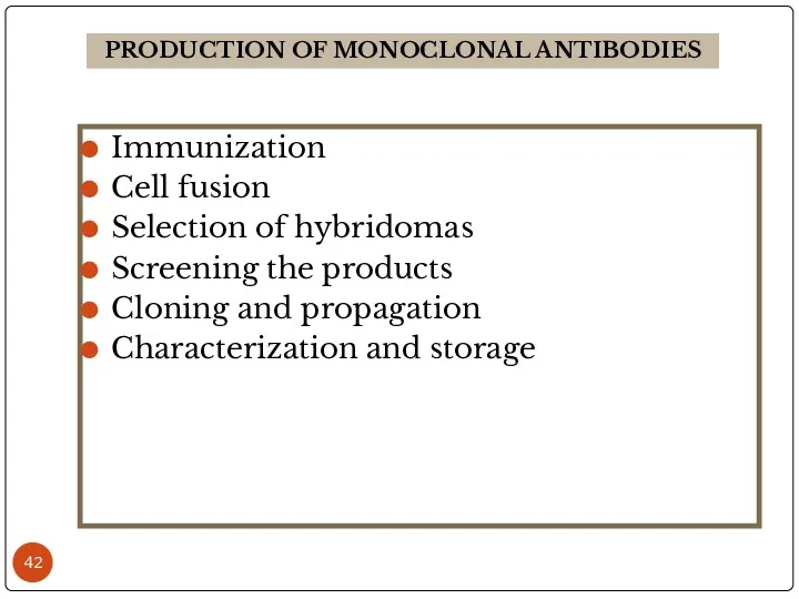
Immunization
Cell fusion
Selection of hybridomas
Screening the products
Cloning and propagation
Characterization and storage
PRODUCTION OF
Immunization
Cell fusion
Selection of hybridomas
Screening the products
Cloning and propagation
Characterization and storage
PRODUCTION OF
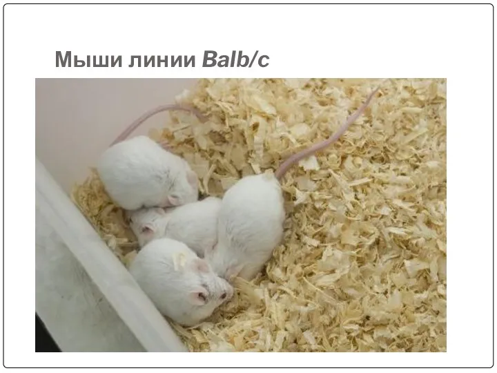
Мыши линии Balb/c
Мыши линии Balb/c
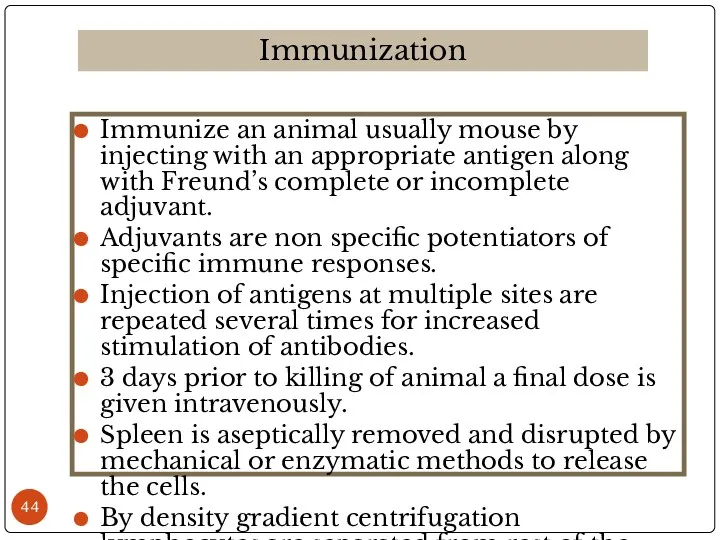
Immunize an animal usually mouse by injecting with an appropriate antigen
Immunize an animal usually mouse by injecting with an appropriate antigen
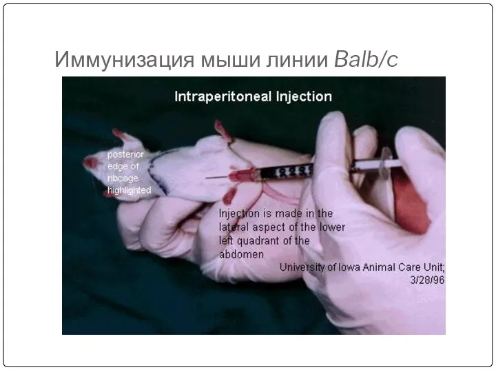
Иммунизация мыши линии Balb/c
Иммунизация мыши линии Balb/c
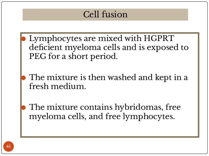
Lymphocytes are mixed with HGPRT deficient myeloma cells and is exposed
Lymphocytes are mixed with HGPRT deficient myeloma cells and is exposed
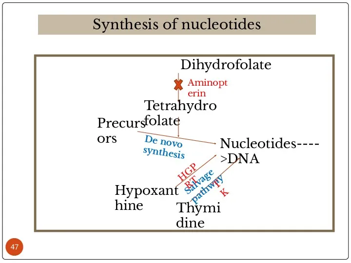
Dihydrofolate
Synthesis of nucleotides
Tetrahydrofolate
Precursors
Nucleotides---->DNA
Hypoxanthine
Thymidine
De novo synthesis
Salvage pathway
Aminopterin
HGPRT
TK
Dihydrofolate
Synthesis of nucleotides
Tetrahydrofolate
Precursors
Nucleotides---->DNA
Hypoxanthine
Thymidine
De novo synthesis
Salvage pathway
Aminopterin
HGPRT
TK

The above mixture is cultured in HAT medium for 7-10 days.
The above mixture is cultured in HAT medium for 7-10 days.

Среда RPMI-1640
Среда RPMI-1640
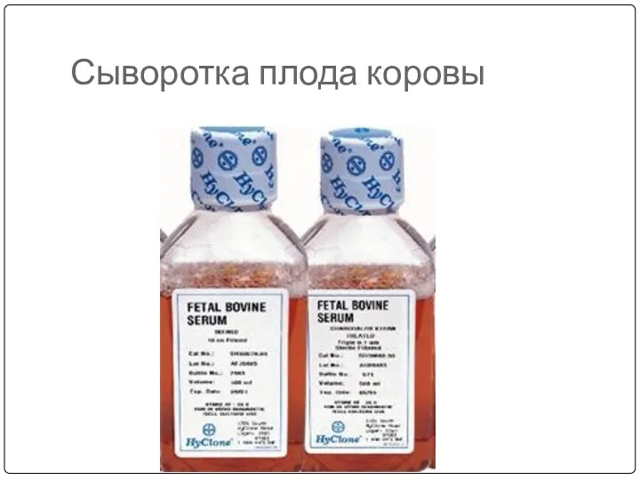
Сыворотка плода коровы
Сыворотка плода коровы
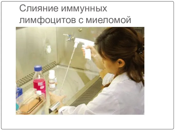
Слияние иммунных лимфоцитов с миеломой
Слияние иммунных лимфоцитов с миеломой
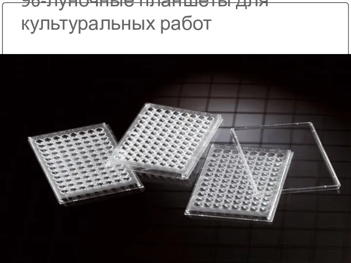
96-луночные планшеты для культуральных работ
96-луночные планшеты для культуральных работ
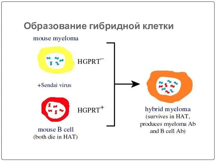
Образование гибридной клетки
Образование гибридной клетки
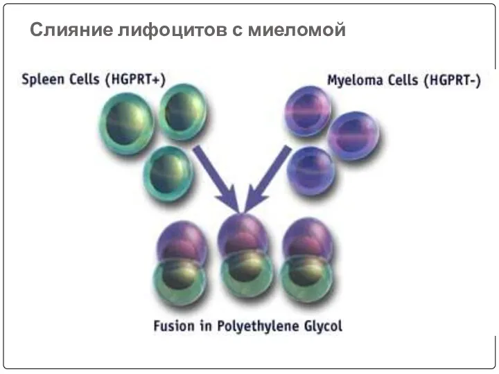
Слияние лифоцитов с миеломой
Слияние лифоцитов с миеломой
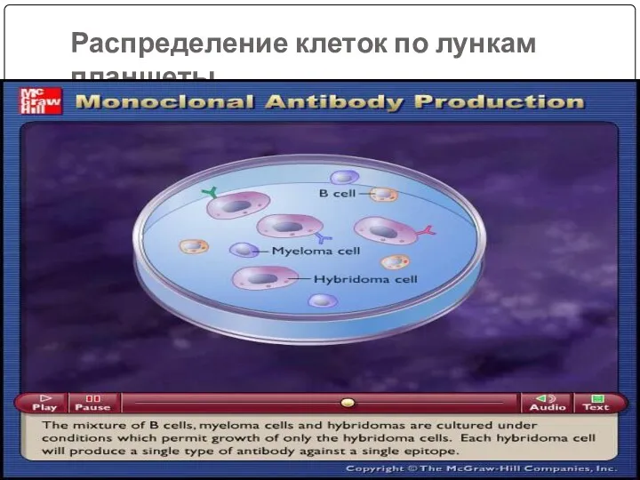
Распределение клеток по лункам планшеты
Распределение клеток по лункам планшеты
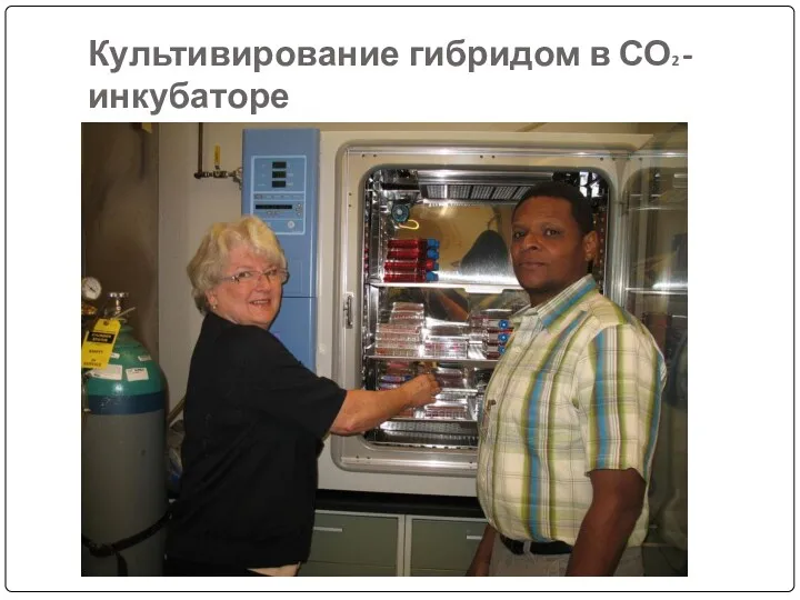
Культивирование гибридом в СО2 -инкубаторе
Культивирование гибридом в СО2 -инкубаторе
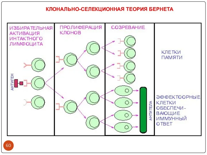
КЛОНАЛЬНО-СЕЛЕКЦИОННАЯ ТЕОРИЯ БЕРНЕТА
КЛОНАЛЬНО-СЕЛЕКЦИОННАЯ ТЕОРИЯ БЕРНЕТА
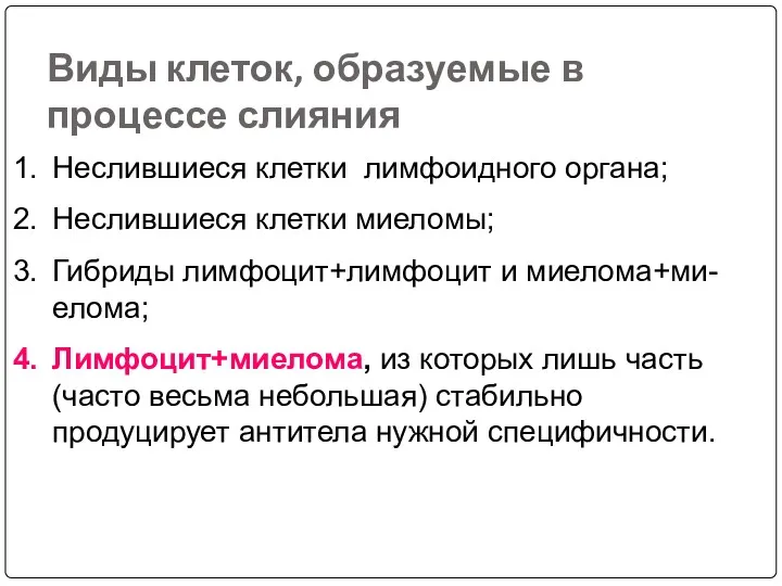
Виды клеток, образуемые в процессе слияния
Неслившиеся клетки лимфоидного органа;
Неслившиеся клетки миеломы;
Гибриды лимфоцит+лимфоцит и миелома+ми-елома;
Виды клеток, образуемые в процессе слияния
Неслившиеся клетки лимфоидного органа;
Неслившиеся клетки миеломы;
Гибриды лимфоцит+лимфоцит и миелома+ми-елома;
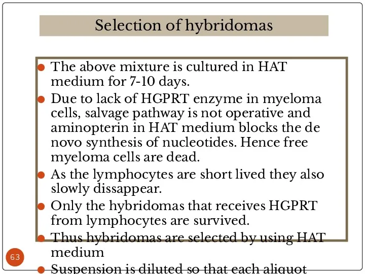
The above mixture is cultured in HAT medium for 7-10 days.
The above mixture is cultured in HAT medium for 7-10 days.
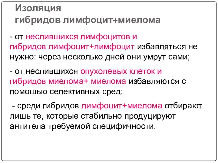
Изоляция гибридов лимфоцит+миелома
- от неслившихся лимфоцитов и гибридов лимфоцит+лимфоцит избавляться не нужно: через несколько дней они
Изоляция гибридов лимфоцит+миелома
- от неслившихся лимфоцитов и гибридов лимфоцит+лимфоцит избавляться не нужно: через несколько дней они
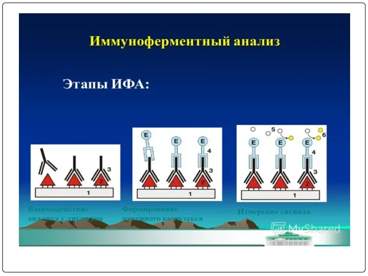
Screening is done for antibody specificity.
For this we need to test
Screening is done for antibody specificity.
For this we need to test
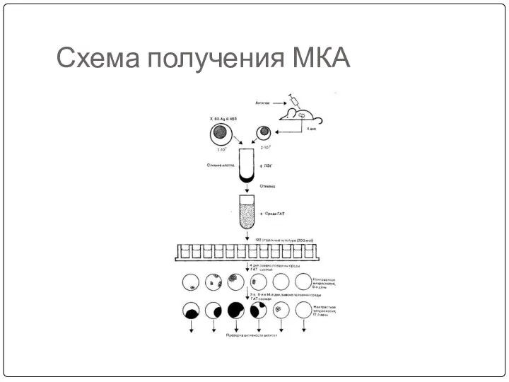
Схема получения МКА
Схема получения МКА
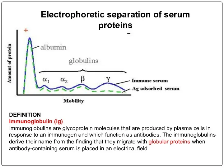
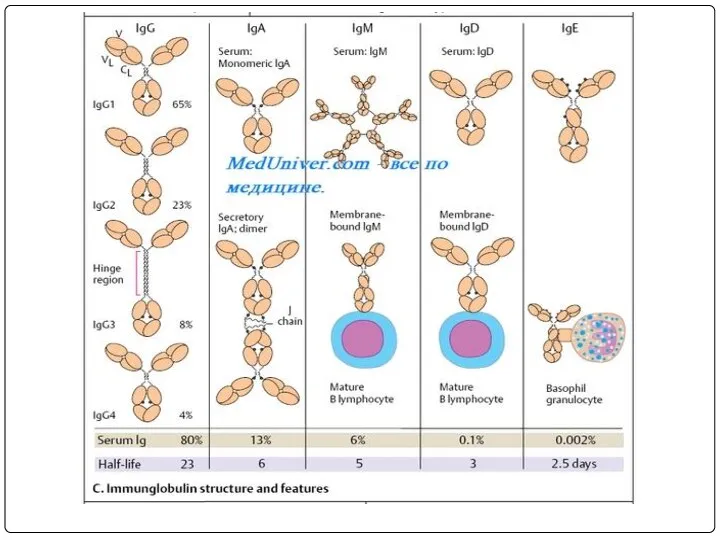
Electrophoretic separation of serum proteins
DEFINITION
Immunoglobulin (Ig)
Immunoglobulins are glycoprotein molecules that are
Electrophoretic separation of serum proteins
DEFINITION
Immunoglobulin (Ig)
Immunoglobulins are glycoprotein molecules that are
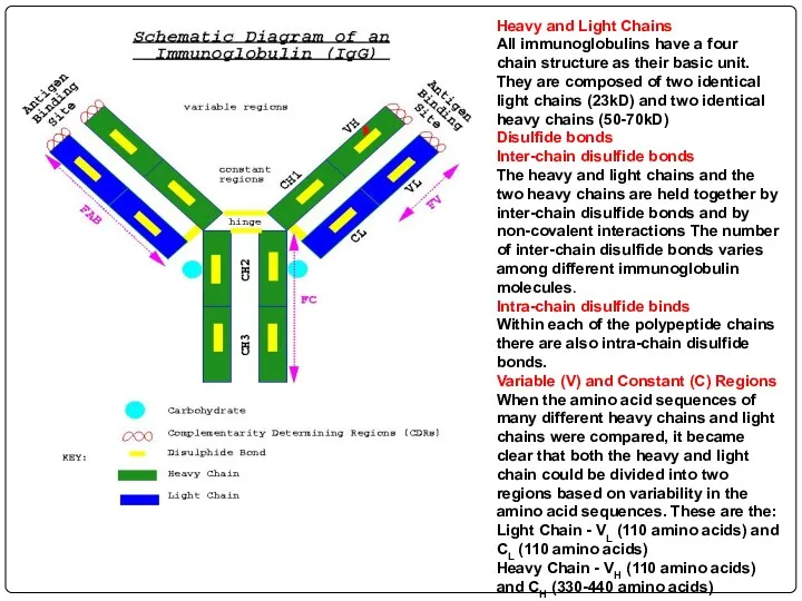
Heavy and Light Chains
All immunoglobulins have a four chain structure as
Heavy and Light Chains All immunoglobulins have a four chain structure as
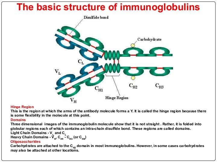
The basic structure of immunoglobulins
Hinge Region
This is the region at which
The basic structure of immunoglobulins
Hinge Region This is the region at which
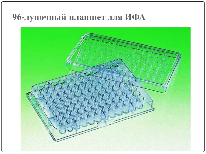
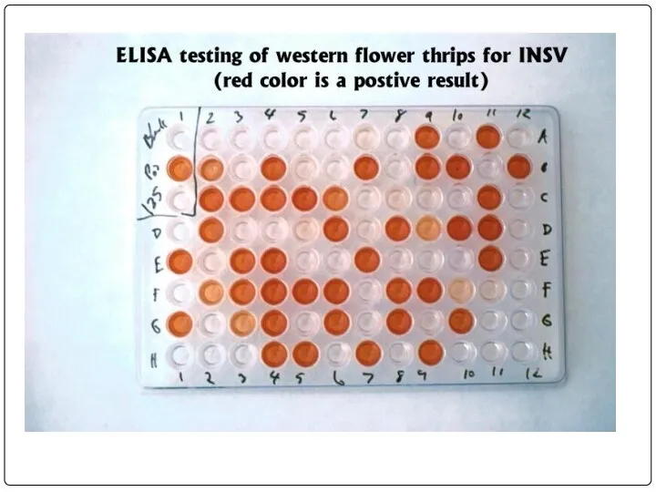
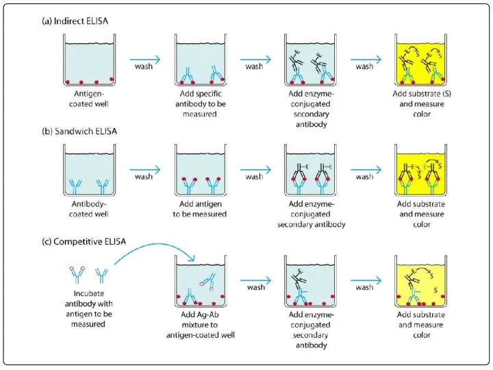
96-луночный планшет для ИФА
96-луночный планшет для ИФА
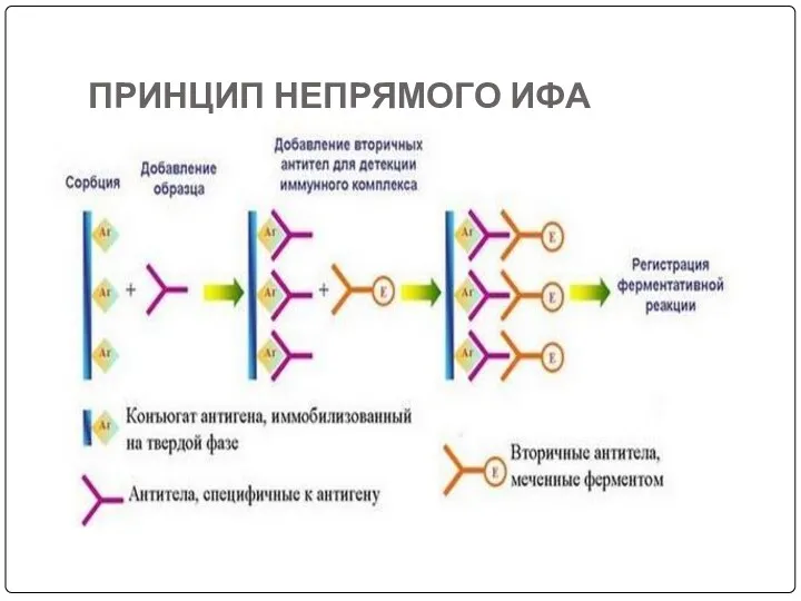
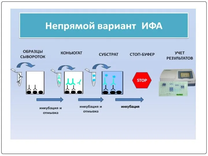
ПРИНЦИП НЕПРЯМОГО ИФА
ПРИНЦИП НЕПРЯМОГО ИФА

Спектрофотометр для ИФА
Спектрофотометр для ИФА
The single hybrid cell producing the desired antibody are isolated and
The single hybrid cell producing the desired antibody are isolated and
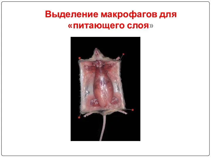
Выделение макрофагов для «питающего слоя»
Выделение макрофагов для «питающего слоя»
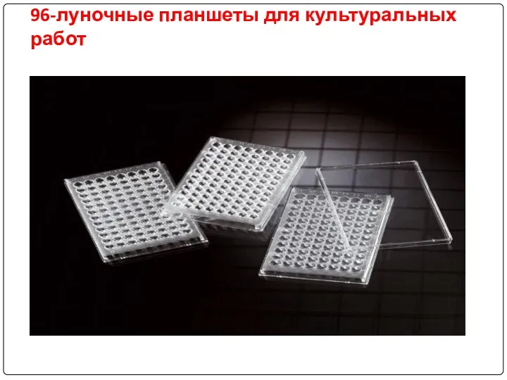
96-луночные планшеты для культуральных работ
96-луночные планшеты для культуральных работ
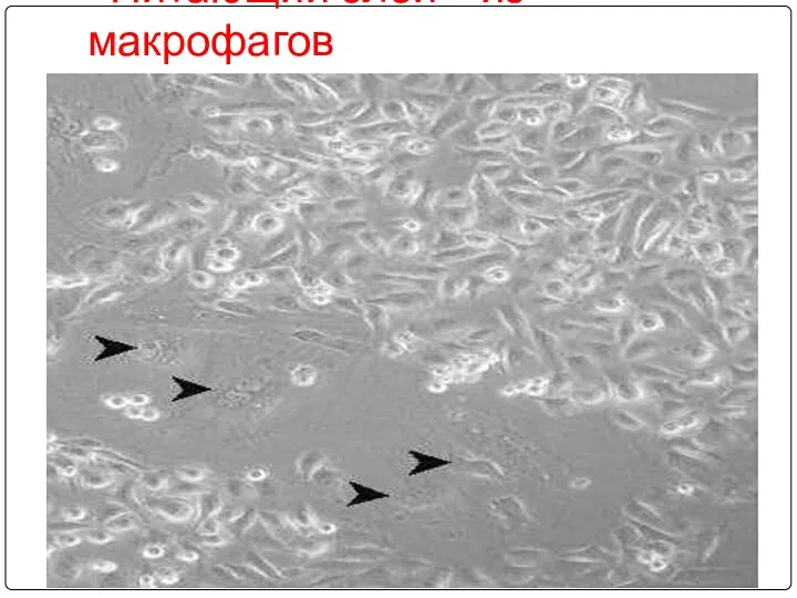
«Питающий слой» из макрофагов
«Питающий слой» из макрофагов
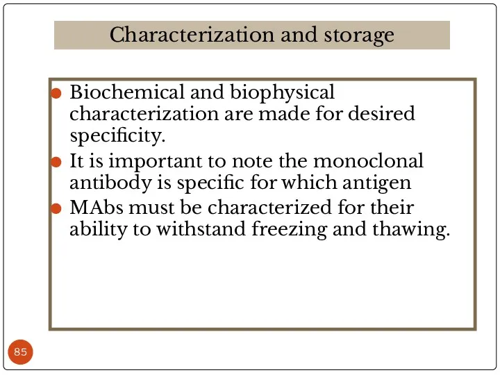
Biochemical and biophysical characterization are made for desired specificity.
It is important
Biochemical and biophysical characterization are made for desired specificity.
It is important
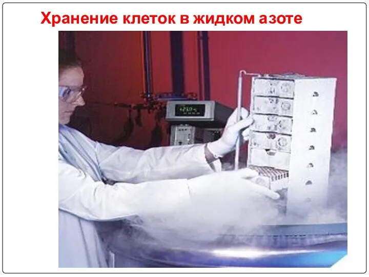
Хранение клеток в жидком азоте
Хранение клеток в жидком азоте
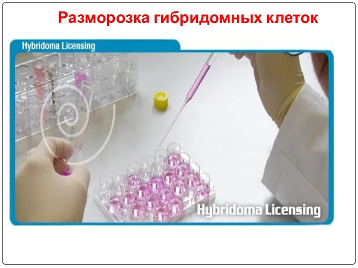
Разморозка гибридомных клеток
Разморозка гибридомных клеток
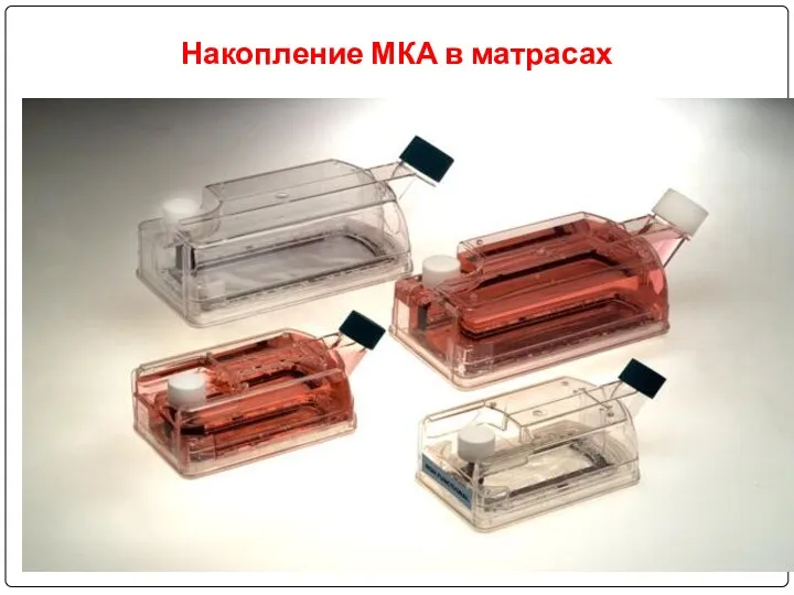
Накопление МКА в матрасах
Накопление МКА в матрасах
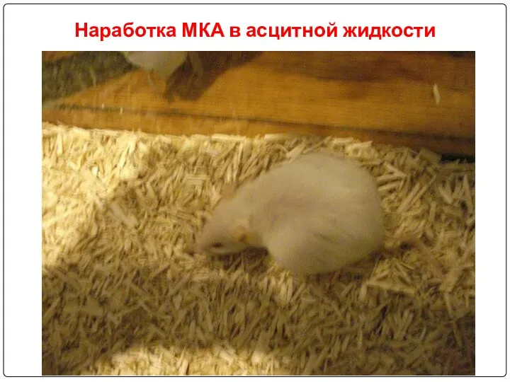
Наработка МКА в асцитной жидкости
Наработка МКА в асцитной жидкости
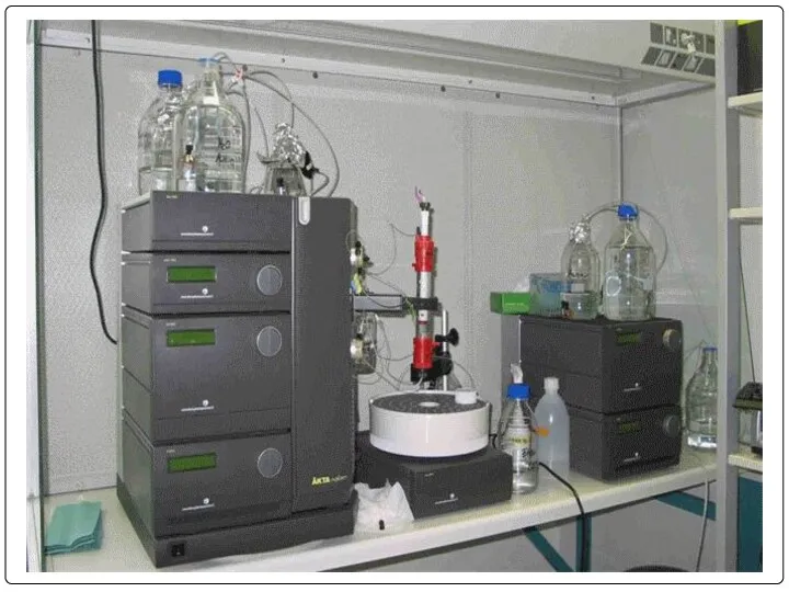
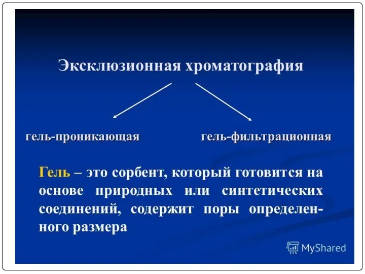
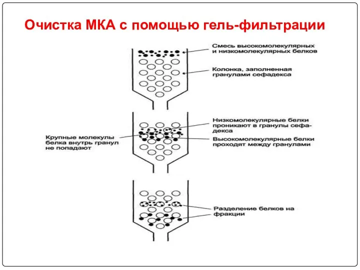
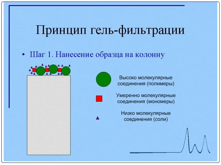
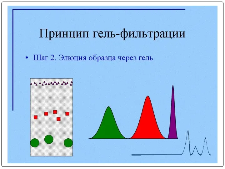
Очистка МКА с помощью гель-фильтрации
Очистка МКА с помощью гель-фильтрации
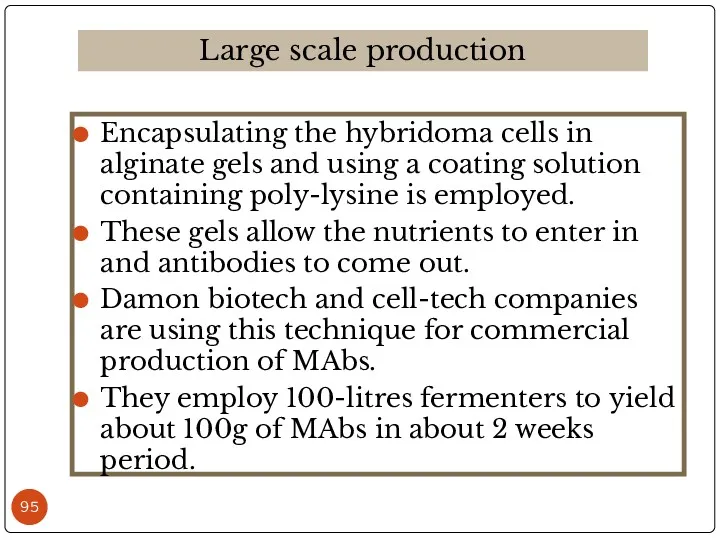
Encapsulating the hybridoma cells in alginate gels and using a coating
Encapsulating the hybridoma cells in alginate gels and using a coating
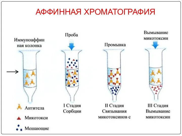
АФФИННАЯ ХРОМАТОГРАФИЯ
АФФИННАЯ ХРОМАТОГРАФИЯ
MAbs derived from mouse are murine derivatives. As they are not
MAbs derived from mouse are murine derivatives. As they are not
Chimeric antibodies:
Hence the murine antibodies are immunogenic to humans, the
Chimeric antibodies:
Hence the murine antibodies are immunogenic to humans, the
Основные проблемы, возникающие при использовании монАТ в терапии
а) Подавляющее большинство получаемых
Основные проблемы, возникающие при использовании монАТ в терапии
а) Подавляющее большинство получаемых
Mouse
Human
Chimeric
V domains
C domains
Mouse
Human
Chimeric
V domains
C domains
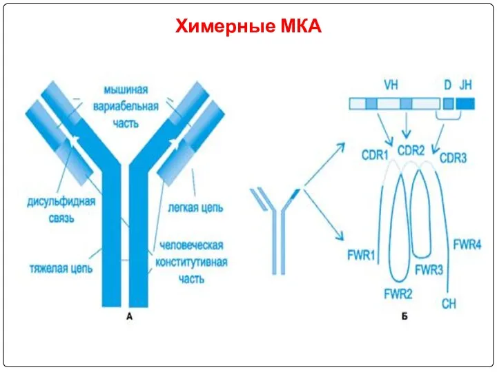
Химерные МКА
Химерные МКА
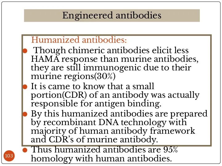
Humanized antibodies:
Though chimeric antibodies elicit less HAMA response than
Humanized antibodies:
Though chimeric antibodies elicit less HAMA response than
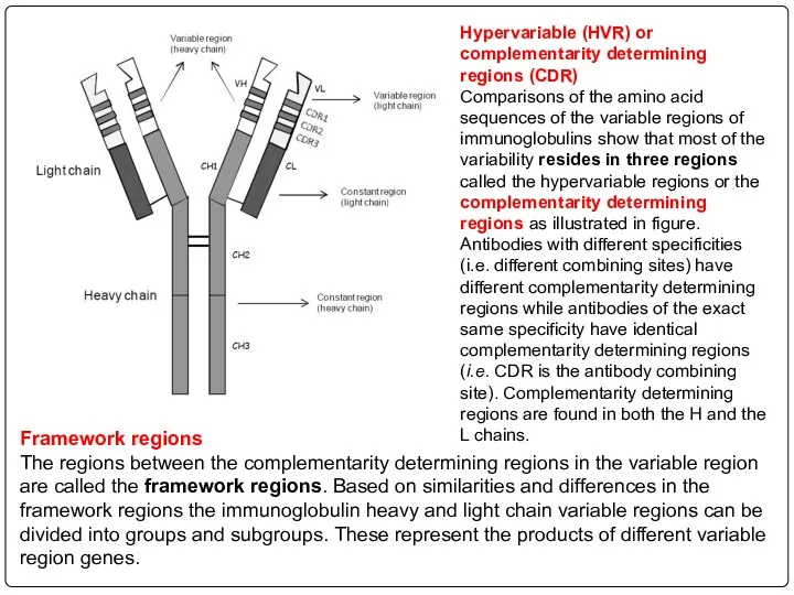
Hypervariable (HVR) or complementarity determining regions (CDR)
Comparisons of the amino acid
Hypervariable (HVR) or complementarity determining regions (CDR)
Comparisons of the amino acid

Mouse
Human
Humanised
hypervariable
framework
Mouse
Human
Humanised
hypervariable
framework
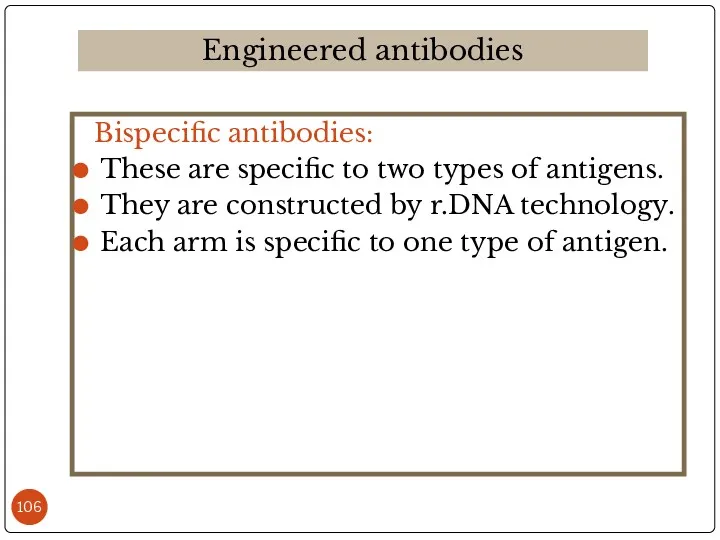
Bispecific antibodies:
These are specific to two types of antigens.
They are
Bispecific antibodies:
These are specific to two types of antigens.
They are
Immunoconjugate:
For MAb targeted drug delivery, a drug is bound covalently
Immunoconjugate:
For MAb targeted drug delivery, a drug is bound covalently
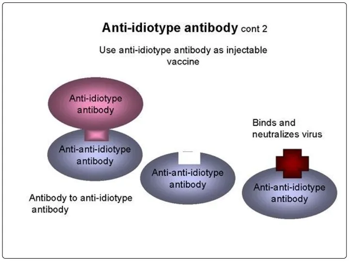
Целевая доставка лекарственных веществ с помощью моноклональных антител
Целевая доставка лекарственных веществ с помощью моноклональных антител
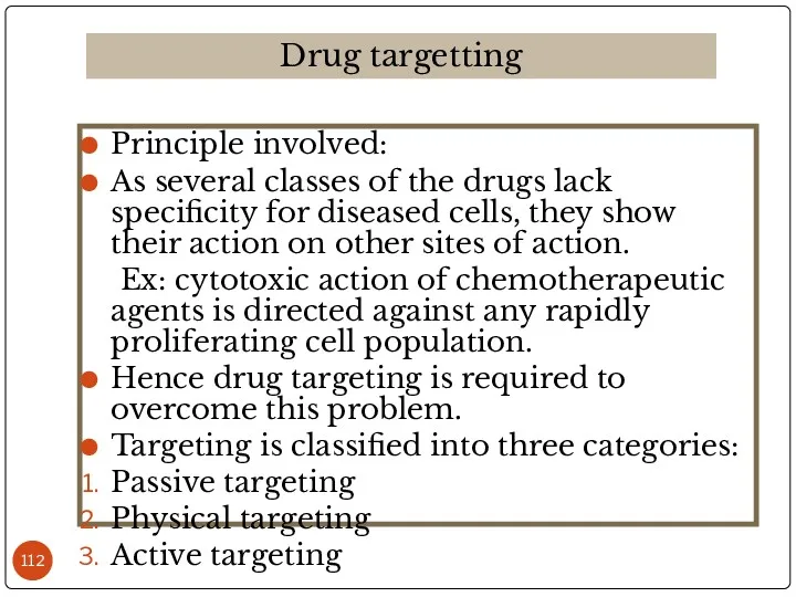
Principle involved:
As several classes of the drugs lack specificity for diseased
Principle involved:
As several classes of the drugs lack specificity for diseased
It is the natural in-vivo distribution pattern of the drug delivery
It is the natural in-vivo distribution pattern of the drug delivery
In this some characteristics of the environment are utilized for the
In this some characteristics of the environment are utilized for the
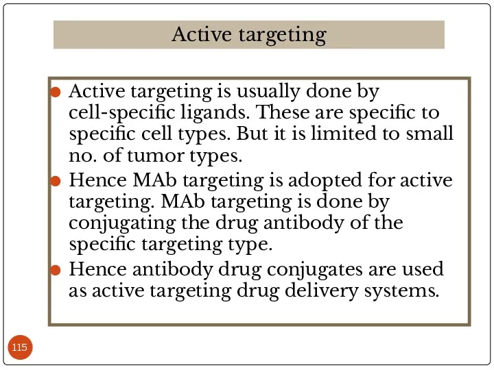
Active targeting is usually done by cell-specific ligands. These are specific
Active targeting is usually done by cell-specific ligands. These are specific
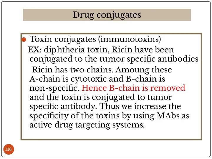
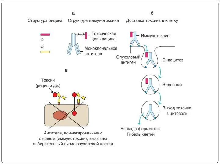
Toxin conjugates (immunotoxins)
EX: diphtheria toxin, Ricin have been conjugated to
Toxin conjugates (immunotoxins)
EX: diphtheria toxin, Ricin have been conjugated to
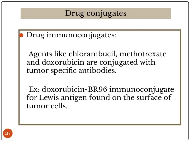
Drug immunoconjugates:
Agents like chlorambucil, methotrexate and doxorubicin are conjugated with
Drug immunoconjugates:
Agents like chlorambucil, methotrexate and doxorubicin are conjugated with
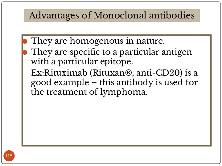
They are homogenous in nature.
They are specific to a particular antigen
They are homogenous in nature.
They are specific to a particular antigen
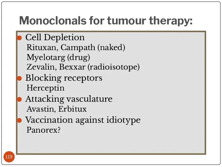
Cell Depletion
Rituxan, Campath (naked)
Myelotarg (drug)
Zevalin, Bexxar (radioisotope)
Blocking receptors
Herceptin
Attacking vasculature
Avastin, Erbitux
Vaccination against
Cell Depletion
Rituxan, Campath (naked)
Myelotarg (drug)
Zevalin, Bexxar (radioisotope)
Blocking receptors
Herceptin
Attacking vasculature
Avastin, Erbitux
Vaccination against
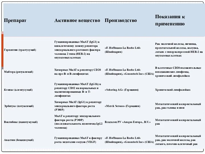
Препараты МкАТ, используемые при лечении онкологических болезнях
Препараты МкАТ, используемые при лечении онкологических болезнях
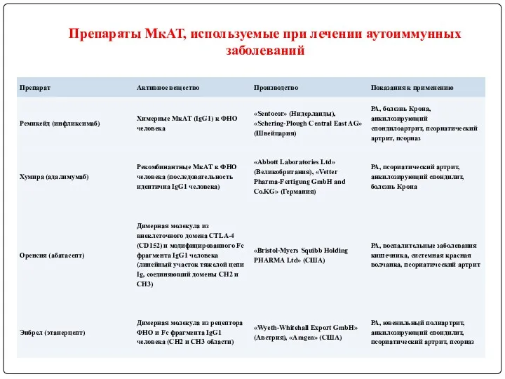
Препараты МкАТ, используемые при лечении аутоиммунных заболеваний
Препараты МкАТ, используемые при лечении аутоиммунных заболеваний
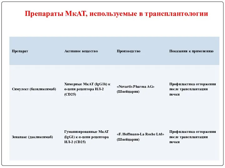
Препараты МкАТ, используемые в трансплантологии
Препараты МкАТ, используемые в трансплантологии
 Первая помощь ожогах
 Характеристики основных видов твердых лекарственных форм
 Биоревитализация. Базовая процедура в эстетической медицине
 Malaria
 Значение физических упражнений для формирования системы опоры и движения
 Иммунопатологические состояния
 Диагностика, лечение и профилактика заболеваний, сопровождающихся легочной гипертензией, во всех возрастных группах
 Өлім. Өлім белгілері. Танатогенез. Терминальді жағдай, өлімнен кейінгі өзгерістер
 Гигиенические требования к хирургическим отделениям больниц
 Современные ингаляционные анестетики
 Группы крови. Переливание крови
 Патология внешнего дыхания
 Лекарственные препараты
 Внутрибольничные инфекции
 Неотложные состояния в акушерстве и гинекологии на догоспитальном этапе
 Диетология. Влияние витамина В1 на аппетит
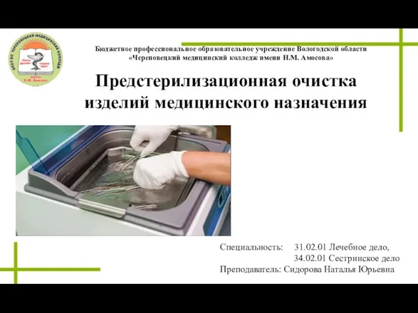 Предстерилизационная очистка изделий медицинского назначения
 Ультразвуковая диагностика при подозрении на опухоль щитовидной железы
 Неонатальный период. Доношенный новорожденный. Тема 3
 Сестринское обследование - первый этап сестринского процесса
 Тісжегінің таралуы мен белсенділігін бағалау. Пародонт тіндерінің зақымдалу белсенділігі мен таралуын бағалау
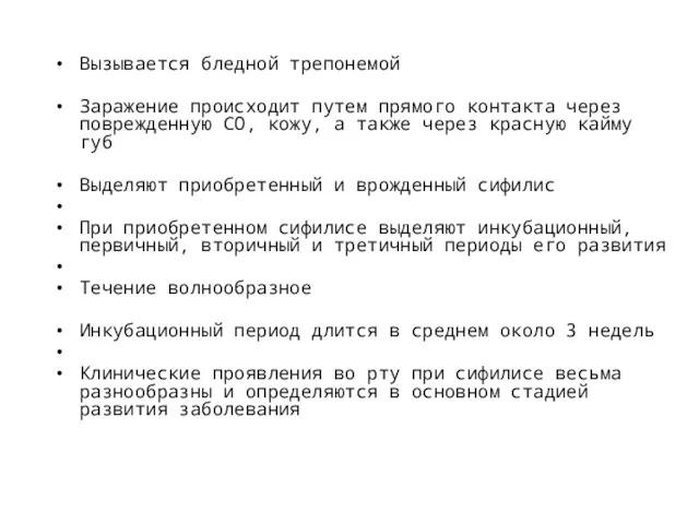 Сифилис, туберкулез
 Патофизиология эндокринной системы
 Синдром диссеминированного внутрисосудистого свертывания крови (ДВС-синдром) у беременных
 Лечебная физическая культура при травмах и заболеваниях центральной нервной системы
 Контроль качества в здравоохранении
 Туляремия дами
 Острая печеночная недостаточность