- Liver diseases

Содержание
- 2. The morphologic alterations that cause liver failure fall into 4 categories: Hepatic dysfunction without overt necrosis
- 3. Hepatosis It is metabolic liver dysfunction, that is characterized by dystrophic changers of hepatocytes without inflammation
- 4. Massive hepatic necrosis The mechanism is direct toxic damage to hepatocytes or a variable combination of
- 5. Hepatitis This term is used to describe the presence of inflammation in the liver and to
- 6. Hepatotropic viruses infect primarily hepatocytes. The known hepatotropic viruses include: hepatitis A virus (HAV), hepatitis В
- 7. HBV and HCV are mostly often transmitted by the parenteral route: - blood transfusions (high risk
- 8. CLINICAL SYMPTOMS 1. Asymptomatic infection (serologic evidence only) 2. Acute hepatitis a. Anicteric b. Icteric 3.
- 9. Acute Viral Hepatitis Sporadic attacks of acute hepatitis caused by all of the hepatotropic viruses are
- 10. 1. Liver is enlarged, reddened liver; greenish if cholestatic. 2. Hepatocyte injury: swelling (ballooning degeneration) -
- 11. 4. Regenerative changes: hepatocyte proliferation 5. Sinusoidal cell reactive changes: - Accumulation of cellular debris in
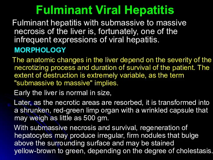
- 12. Fulminant Viral Hepatitis Fulminant hepatitis with submassive to massive necrosis of the liver is, fortunately, one
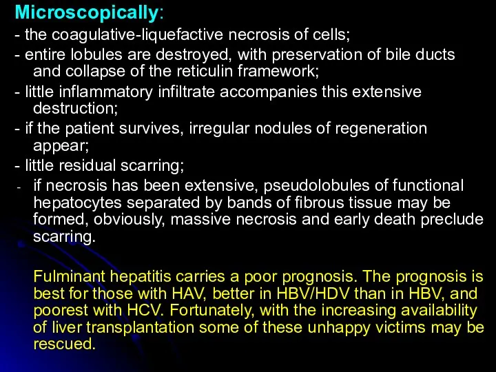
- 13. Microscopically: - the coagulative-liquefactive necrosis of cells; - entire lobules are destroyed, with preservation of bile
- 14. Chronic Hepatitis Chronic hepatitis is defined as the continuation of hepatic inflammation and necrosis for longer
- 15. CPH is characterized clinically by: - persistence of elevated levels of liver aminotransferase enzymes (AST and
- 16. In contrast, in chronic "lobular" hepatitis: the portal tracts are only mildly inflamed and random hepatocytes
- 17. Chronic Active Hepatitis In contrast to the previous condition, САН is characterized by progressive destruction of
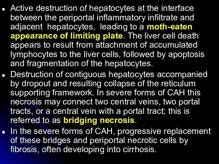
- 18. Active destruction of hepatocytes at the interface between the periportal inflammatory infiltrate and adjacent hepatocytes, leading
- 19. AUTOIMMUNE CHRONIC HEPATITIS Chronic hepatitis unrelated to known viral agents or toxins is frequently ascribed to
- 20. Liver cirrhosis Cirrhosis is a result of advanced liver disease. It is characterized by replacement of
- 21. According to cause: Alcoholic liver disease (ALD) Alcoholic cirrhosis, Non-alcoholic steatohepatitis (NASH), Viral hepatitis (B, C),
- 22. According to morphology: Micronodular (less then 3 mm), Macronodular (more then 3 mm), Mixed, Septal According
- 23. CLINICAL SYMPTOMS Cirrhosis has many possible manifestations. These signs and symptoms may be either as a
- 24. Spider angiomata or spider nevi are vascular lesions consisting of a central arteriole surrounded by many
- 25. Hypogonadism, a decrease in sex hormones manifest as impotence, infertility, loss of sexual drive, and testicular
- 26. Portal hypertension Liver cirrhosis increases resistance to blood flow and higher pressure in the portal venous
- 28. Скачать презентацию
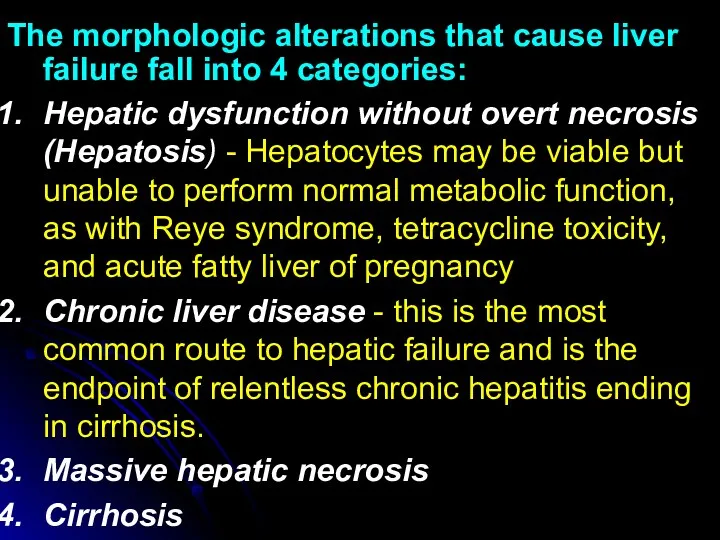
The morphologic alterations that cause liver failure fall into 4 categories:
Hepatic
The morphologic alterations that cause liver failure fall into 4 categories:
Hepatic
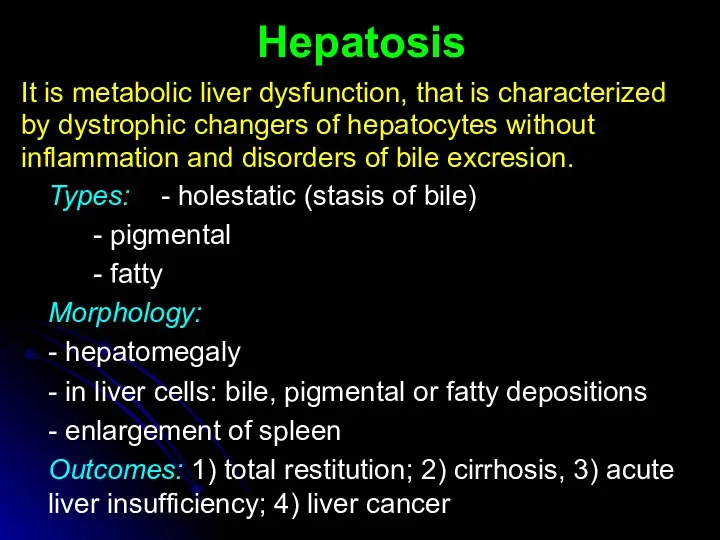
Hepatosis
It is metabolic liver dysfunction, that is characterized
by dystrophic changers of
Hepatosis
It is metabolic liver dysfunction, that is characterized
by dystrophic changers of
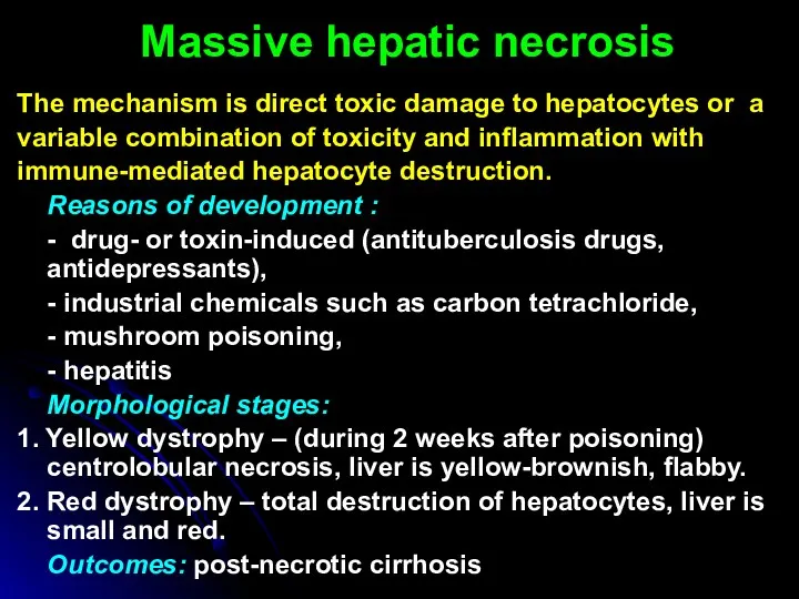
Massive hepatic necrosis
The mechanism is direct toxic damage to hepatocytes or
Massive hepatic necrosis
The mechanism is direct toxic damage to hepatocytes or
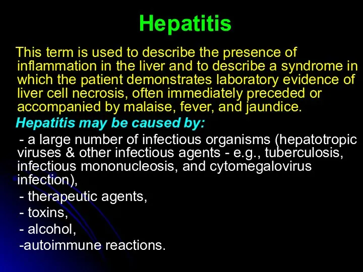
Hepatitis
This term is used to describe the presence of inflammation
Hepatitis
This term is used to describe the presence of inflammation
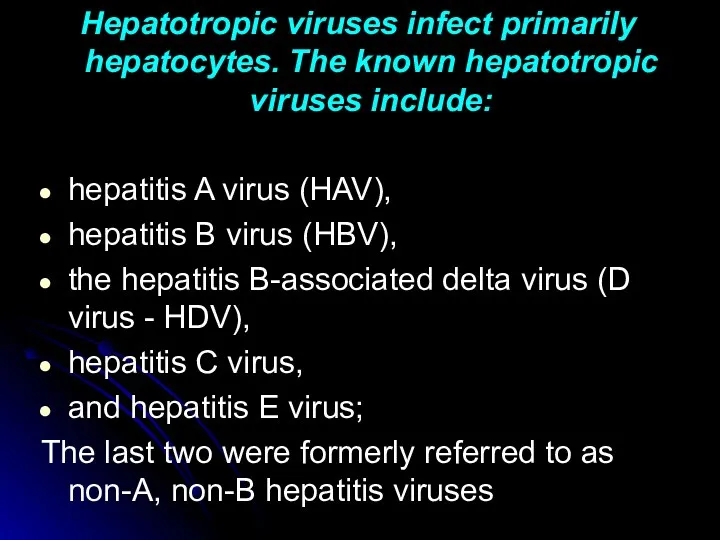
Hepatotropic viruses infect primarily hepatocytes. The known hepatotropic viruses include:
hepatitis A
Hepatotropic viruses infect primarily hepatocytes. The known hepatotropic viruses include:
hepatitis A
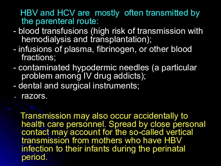
HBV and HCV are mostly often transmitted by the parenteral
HBV and HCV are mostly often transmitted by the parenteral
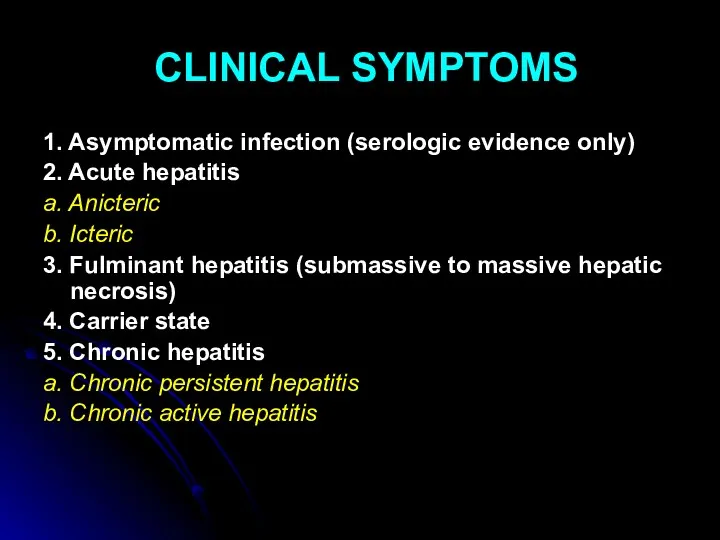
CLINICAL SYMPTOMS
1. Asymptomatic infection (serologic evidence only)
2. Acute hepatitis
a. Anicteric
b. Icteric
3.
CLINICAL SYMPTOMS
1. Asymptomatic infection (serologic evidence only)
2. Acute hepatitis
a. Anicteric
b. Icteric
3.
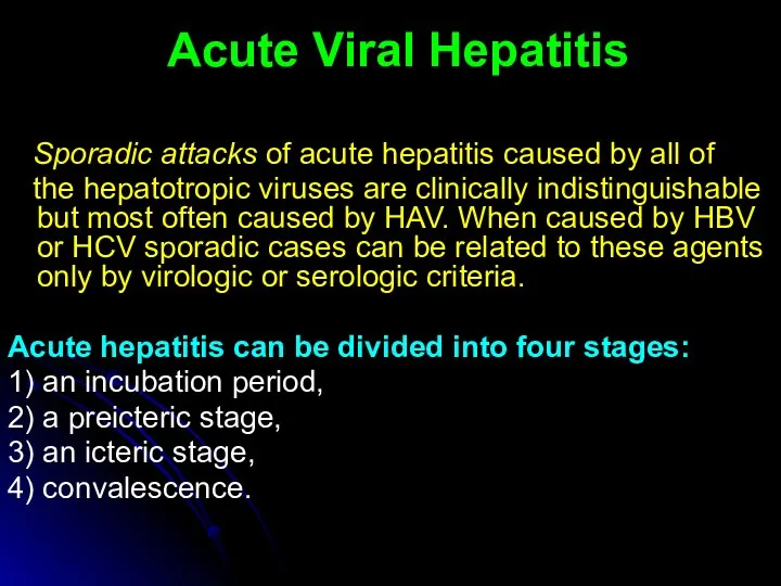
Acute Viral Hepatitis
Sporadic attacks of acute hepatitis caused by
Acute Viral Hepatitis
Sporadic attacks of acute hepatitis caused by
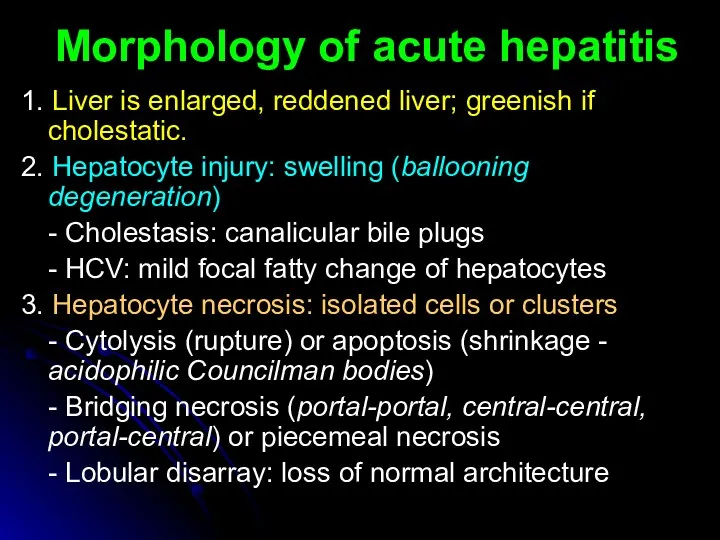
1. Liver is enlarged, reddened liver; greenish if cholestatic.
2. Hepatocyte
1. Liver is enlarged, reddened liver; greenish if cholestatic.
2. Hepatocyte
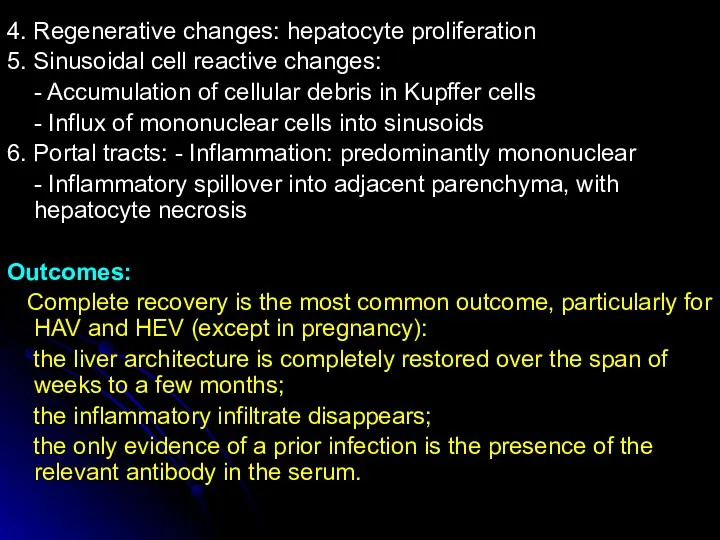
4. Regenerative changes: hepatocyte proliferation
5. Sinusoidal cell reactive changes:
- Accumulation of
4. Regenerative changes: hepatocyte proliferation
5. Sinusoidal cell reactive changes:
- Accumulation of
Fulminant Viral Hepatitis
Fulminant hepatitis with submassive to massive necrosis of
Fulminant Viral Hepatitis
Fulminant hepatitis with submassive to massive necrosis of
Microscopically:
- the coagulative-liquefactive necrosis of cells;
- entire lobules are destroyed, with
Microscopically:
- the coagulative-liquefactive necrosis of cells;
- entire lobules are destroyed, with
Chronic Hepatitis
Chronic hepatitis is defined as the continuation of
Chronic Hepatitis
Chronic hepatitis is defined as the continuation of
CPH is characterized clinically by:
- persistence of elevated levels of
CPH is characterized clinically by:
- persistence of elevated levels of
In contrast, in chronic "lobular" hepatitis: the portal tracts are
In contrast, in chronic "lobular" hepatitis: the portal tracts are
Chronic Active Hepatitis
In contrast to the previous condition, САН is
Chronic Active Hepatitis
In contrast to the previous condition, САН is
Active destruction of hepatocytes at the interface between the periportal inflammatory
Active destruction of hepatocytes at the interface between the periportal inflammatory
AUTOIMMUNE CHRONIC HEPATITIS
Chronic hepatitis unrelated to known viral agents or
AUTOIMMUNE CHRONIC HEPATITIS
Chronic hepatitis unrelated to known viral agents or
Liver cirrhosis
Cirrhosis is a result of advanced liver disease. It is
Liver cirrhosis
Cirrhosis is a result of advanced liver disease. It is
According to cause:
Alcoholic liver disease (ALD) Alcoholic cirrhosis,
Non-alcoholic steatohepatitis (NASH),
Viral hepatitis
According to cause:
Alcoholic liver disease (ALD) Alcoholic cirrhosis,
Non-alcoholic steatohepatitis (NASH),
Viral hepatitis
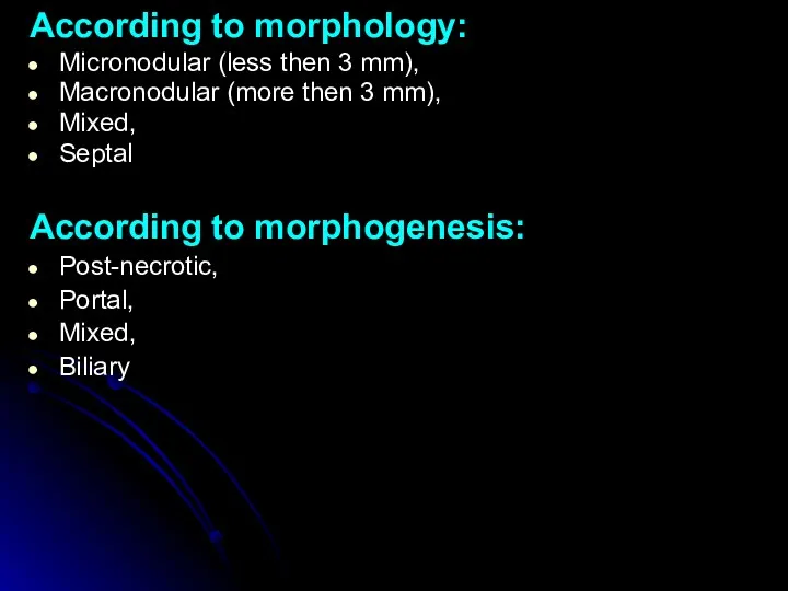
According to morphology:
Micronodular (less then 3 mm),
Macronodular (more then 3 mm),
Mixed,
Septal
According
According to morphology:
Micronodular (less then 3 mm),
Macronodular (more then 3 mm),
Mixed,
Septal
According
CLINICAL SYMPTOMS
Cirrhosis has many possible manifestations. These signs and symptoms may
CLINICAL SYMPTOMS
Cirrhosis has many possible manifestations. These signs and symptoms may
Spider angiomata or spider nevi are vascular lesions consisting of a
Spider angiomata or spider nevi are vascular lesions consisting of a
Hypogonadism, a decrease in sex hormones manifest as impotence, infertility, loss
Hypogonadism, a decrease in sex hormones manifest as impotence, infertility, loss
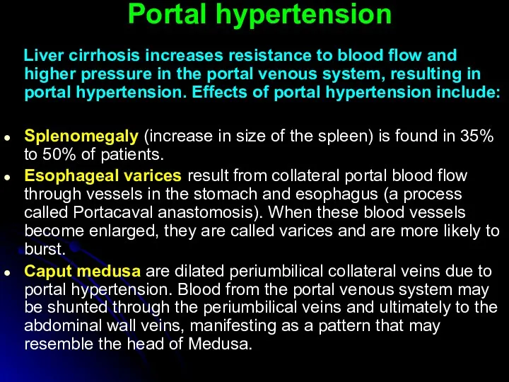
Portal hypertension
Liver cirrhosis increases resistance to blood flow and higher
Portal hypertension
Liver cirrhosis increases resistance to blood flow and higher
 Лейкокория у детей
 Методика исследования периферических и внутригрудных лимфатических узлов
 Инфекционные болезни - чума
 Наложение шин на руку
 Сердечно-легочная реанимация
 Жүктілікпен байланысты гипертензиясы Ерте токсикоздар
 Воздействие на поведение водителя психотропных, наркотических веществ, алкоголя и медицинских препаратов
 Косметические средства ухода за кожей детей при аномалиях конституции
 Diet and teen health
 Зрительный нерв. Заболевания зрительного нерва
 Реанимационная помощь
 Дәрілік заттардың әсерінің ағза қасиеттеріне тәуелділігі. Фармакологиялық әсердің дәрілік заттардың қасиеттері мен қолдану
 Antimicrobial drugs
 Снотворные и противосудорожные средства. Противопаркинсонические средства
 Предмет и задачи медицинской микробиологии. История микробиологии. Медицинская микробиология XXI века
 Ошибки диагностики и лечения при сочетанной травме
 Профилактика стоматологических заболеваний в раннем детском возрасте
 Вскармливание детей грудного возраста. Раздел 1.Лекция 6
 Современные направления пластической хирургии в акушерстве и гинекологии
 Местные анестетики
 Всероссийский день трезвости
 ВКР: Анализ работы медсестры процедурного кабинета хирургического отделения стационара
 Врожденные аномалии грудной клетки
 Вреден ли фаст-фуд для нашего здоровья
 Здоровый образ жизни и профилактика основных неинфекционных заболеваний
 Қазіргі заманғы денсаулық сақтау, әлемдік тенденция, дамыған елдер
 Иммуногенез бұзылуының морфологиясы. Тимустың, шеткері лимфалық тіндердің иммунгенез бұзылғандағы өзгерістері
 Жүкті студенттердің медико-әлеуметтік аспектілері